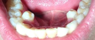
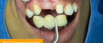
Number of teeth in animals. Animal teeth (part 3)
Behind the incisor teeth in pigs, dogs, stallions, and geldings there are fangs - dentes canini - conical teeth for capturing and holding food. Large and small cattle and most mares do not have tusks. Behind the canines are the molars. The first 3-4 molars are called premolars - dentes premolares. Behind them are grinding teeth - molars - dentes molares. Between the incisive teeth and premolars there is an interalveolar toothless edge of the lower jaw, covered with gum - gingiva. The toothless edge is significant in length in large and small cattle and horses. In stallions and geldings, the toothless edge is located from the canine to the premolar. The first lower premolar in pigs is separated from the second by a significant interdental space (diastema). The location and number of teeth in the arcades are usually shown in numbers in the formulas of one half of the oral cavity: the upper arcade is in the numerator, the lower one is in the denominator. The number of incisors is shown first, then canines, premolars and molars. The numbers are preceded by the first letter of the name of the teeth according to the international nomenclature. For brevity, these letters are often not written. Table 7 shows the formulas of primary and permanent teeth of the main types of domestic animals.
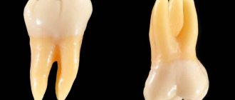
The molars of domestic animals and the first premolar in pigs and dogs do not have milk predecessors, therefore the number of milk molars is less than that of permanent teeth. Age-related changes in teeth are manifested in the timing of the eruption (appearance) of milk teeth in the oral cavity, then when they grow and are replaced by permanent teeth , growth and wear of permanent teeth. Milk teeth - incisors, canines, premolars have a crown covered with milky-whitish enamel, smooth, with a shine. The roots are short (Fig. 226, 227, 228).
When teeth are worn, the shape of the rubbing (closing, chewing) surface of the teeth and their inclination in the arcades change, which is used to determine the age of animals. Tables 8 and 9 indicate the timing of tooth changes. They differ in animals of different breeds: in early maturing breeds, teeth are replaced earlier than in late maturing ones. The growth and change of teeth reflects the state of metabolism in the animal’s body. In immaturely born animals, such as puppies and kittens, the eruption of baby teeth occurs a few weeks after birth. In mature births (calves, lambs, foals), milk teeth erupt before birth or in the first week after birth.
Teeth numbers. How dentists count teeth: arrangement and numbering principles
Each dental unit has its own functions, depending on its structure and location on the jaw. Thus, incisors are designed for biting off pieces of food, canines help to hold hard food and “tear off” stubborn pieces, premolars are needed for primary processing, and dense “plump” molars are designed for thoroughly chewing and grinding food. The order of the teeth in the mouth, accordingly, will be as follows: on each jaw there are four incisors, two canines, four premolars (two on each side) and six molars (three on each side).
In dentistry, tooth numbers are assigned according to their location on the jaw and function. Since the incisors and molars are located symmetrically on the right and left, the count starts from the middle of the row, that is, from the central incisors and further to the right and left. If we divide this row into two halves and start counting on one of them, we get: two incisors (teeth numbers 1 and 2), a canine (3rd number), two premolars (4th and 5th), three molar (6th, 7th and 8th, the last one being the wisdom tooth). These are the names and numbers of teeth that are universally accepted in dentistry.
If you have a problem similar to that described in this article, be sure to contact our specialists. Don't diagnose yourself!
Why you should call us now:
But if you simply say “sixth tooth,” then how can you understand whether it is upper or lower, and on which side of the jaw – left or right – it is located? To eliminate confusion in this matter, it is customary to designate a person’s teeth by numbers, indicating the segment of the jaw on which they are located. The segments are considered as follows: the upper right is the first and is indicated by a ten in front of the number of a particular tooth (for example, the 11th is the central right upper incisor, the 16th is the upper right molar, next immediately after the premolars, etc.), the upper left - twenty. Accordingly, the lower left is designated by thirty, and the lower right by the number 40. That is, the segments of the jaws are numbered clockwise, this makes it much easier to remember the order and, if necessary, count.
Thus, the notorious 48th number indicates the location of the wisdom teeth in the lower right segment of the jaw. And insisting on removing the 48th dental unit, the dentist simply indicates what number the wisdom tooth has in the lower right, and does not inform the patient about the supernumerary teeth that came from nowhere in the mouth.
Reasons for creating the formula
Why is numbering of row elements necessary and how does it work when performing dental work? Each tooth has its own structural features, as well as a range of tasks performed. Some elements are involved in the process of biting off large pieces, others grind and crush foods for further better absorption in the body. Only wisdom teeth, which are inherited from ancient ancestors, do not take part in the process of chewing food. Due to the heat treatment of foods and the consumption of softer foods, the size of the jaw of modern humans has decreased, and wisdom teeth have ceased to perform their functions.
However, doctors strongly advise caring for eights, treating them if necessary, like other elements. Wisdom teeth prevent the development of bite pathologies and can be used for prosthetics if, for example, sevens are missing.
The dental formula must be entered into the patient’s outpatient record in order to minimize the risk of errors during further treatment. In dentistry, it is customary to number the dentition from the central part of the jaws. Concise markings allow clinicians to quickly complete the information in the chart so that it is understandable to another dentist or dentist. Doctors cannot use individual calculation schemes, as this will complicate the work of another specialist and change the information in the patient’s hospital record.
And there is also a special formula with which parents can determine the required number of milk units in the baby’s mouth. It looks like this: N = n - 4, where N is the number of milk elements in a child, and n is the baby’s age in months. The result calculated using the formula does not always coincide with the actual number of elements erupted in the child. Mismatches in the formula are not a reason to panic. Each child’s body is individual and minor deviations from the norm are acceptable. This calculation formula only applies to children under 2 years of age.
Symptoms that require consultation with a doctor include:
- complete absence of teeth in the oral cavity in a child older than 12 months;
- the presence of less than 10 teeth in the mouth of a 3-year-old child;
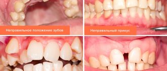
- non-compliance with the pattern of eruption of milk elements (for example, if the canines or premolars erupt first, and not the incisors);
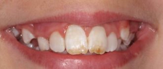
- defects on the enamel.
Mammal teeth
The teeth of mammals differ from the teeth of other vertebrates in a number of extremely significant properties.
Firstly, they are heterogeneous, but are divided into incisors, fangs and molars, which in turn are divided into true, or large molars, and false, or small molars (only in toothed whales all teeth are of the same type - in the form of sharp cones, which represents a secondary phenomenon). The incisors (incisivi) are used for biting food and are chisel-shaped. In the upper jaw they always sit only in the premaxillary bones. The fangs (canini), the number of which is no more than one in each half of the upper and lower jaws, have the shape of a sharp cone and are used to grab, hold and kill prey, while the molars serve to grind it. The small molars (praemolares), which are located in front of the large molars, undergo replacement (baby teeth are replaced by permanent ones), while the large molars (molares) are permanent and, having grown, do not change throughout the life of the animal. In addition, small molars often differ from large molars in a less complex shape and smaller size. Thus, the teeth of mammals, in contrast to the teeth of all other classes, are differentiated and serve not only for holding, but also for grinding food. In different groups of mammals, due to their way of life, teeth have a very diverse structure: rodents, for example, do not have fangs, but the incisors are especially large; In carnivores, on the contrary, the incisors are poorly developed, while the fangs are very large.
Dental formula
Dental formula
— a brief description of the dental system of mammals and other heterodont tetrapods, written in the form of special notations.
When recording a dental formula, use abbreviated names for the types of teeth of the heterodont dental system: I
(lat.
dentes incisivi
) - incisors;
C
(lat.
d. canini
) - fangs;
P
(lat.
d. premolares
) - premolars, or small molars, or premolars;
M
(lat.
d. molares
) - molars, or large molars. The abbreviated name of the type of teeth is followed by an indication of the number of pairs of teeth in this group: in the numerator - the upper jaw and in the denominator - the lower jaw.
Sample of a dental formula (using the example of a person):
This entry means: two pairs of incisors (I), one pair of canines (C), two pairs of molars (P) and three pairs of molars (M).
In addition to these main types of teeth, representatives of some groups of mammals have types that are characteristic only of them. These are the intermediate
(lat.
d. intercalares
,
in
) teeth of shrews, corresponding, presumably, to poorly differentiated incisors, premolars and, probably, canines, and
large premolar
(lat.
d. praemolares prominantes
,
PmP
) teeth of chiropterans, located between premolars and molars.[ 1]
The dental formula is widely used in the taxonomy of vertebrates when compiling characteristics of groups of various ranks, from orders to subfamilies and even genera, since it allows a compact presentation of the main characteristics of the dental system.
In practical dentistry, such designations are rarely used, and the teeth of a person’s jaws are simply numbered from incisors to molars (from 1 to 8).
All teeth are divided into 4 sectors (counterclockwise):
- The teeth of the upper jaw are on the right (respectively, the central incisor is 11, the second incisor is 12, the canine is 13, the first premolar is 14, the second premolar is 15, the first molar is 16, the second molar is 17, the third molar or wisdom tooth is −18).
- Upper jaw teeth on the left (21, 22, 23, 24, 25, 26, 27, 28, similar to the right side).
- Lower jaw teeth on the left (31, 32, 33, 34, 35, 36, 37, 38).
- Lower jaw teeth on the right (41, 42, 43, 44, 45, 46, 47, 48).
For children's teeth, a similar numbering is used from 51 to 85, or they are indicated in Latin numerals.
Examples of dental formulas of permanent teeth of some mammals
| View | Latin name | Squad | Family | Dental formula |
| Koala | Phascolarctos cinereus Goldfuss, 1817 | Two-incisor marsupials | Koalas | |
| Kutora | Neomys fodiens Schreber, 1776 | Insectivores | Shrews | |
| Ushan | Plecotus auritus Linnaeus, 1758 | Chiroptera | Leather | |
| White hare | Lepus timidus Linnaeus, 1758 | Lagomorpha | Zaitsevy | |
| Wolf | Canis lupus Linnaeus, 1758 | Predatory | Doggystyle | |
| Cat | Felis catus Linnaeus, 1758 | Predatory | Felines | |
| Horse | Equus caballus Linnaeus, 1758 | Odd-toed ungulates | Equine | |
| Boar | Sus scrofa Linnaeus, 1758 | Artiodactyls | Pork | |
| Elk | Alces alces Linnaeus, 1758 | Artiodactyls | Deer | |
| Cattle | Bos taurus taurus Linnaeus, 1758 | Artiodactyls | Bovids | |
| Mouse | Mus musculus Linnaeus, 1758 | Rodents | Mouse | |
| Elephant | Elephas maximus Linnaeus, 1758 | Proboscis | Elephantids |
Dog teeth: 1, 2, 3 - incisors, 4 - canines, 5 - premolars, 6 - molars
Incisors
(lat.
Dentes incisivi
) - teeth whose function is to bite off food.
The incisors are relatively sharp and located at the front of the jaw. In humans, in the upper and lower jaws there are two central and two lateral incisors, of which the two central ones in the upper jaw are larger. Together with the canines, the incisors form the front teeth. In different species of mammals, the incisors have undergone various transformations during evolution. In elephants, for example, the upper incisors form tusks. In rodents and lagomorphs, incisors are of particular importance and are one of the main distinguishing features of these orders. Ruminants have no incisors in the upper jaw, and the opposite obstacle to the incisors of the lower jaw is the palate.
Fangs
- cone-shaped teeth that serve to tear and hold food.
In humans and other mammals, the canines are located between the incisors and molars.
In addition to participating in the digestive system, fangs are also used by animals for defense.
Venomous snakes have fangs that are hollow inside and are used to release venom from their venom glands.
Premolar
,
small molar tooth
- (lat.
premolar
,
dentes premolares
) - one of two teeth located in the dentition of adults on both sides of the jaws behind the fangs in front of the large molars.
Molars
, better known as
molars
, are teeth that serve mainly for the primary mechanical processing of food.
Teeth
, present in the vast majority of mammals, are solid structures that develop from special connective tissue (mesoderm) cells - odontoblasts and consist mainly of calcium phosphate (apatite), i.e. chemical composition is very similar to bones. However, calcium phosphate crystallizes and combines with other substances in different ways, so that the result is the formation of various dental tissues - dentin, enamel and cementum. The tooth is primarily made up of dentin. (Elephant tusks, and therefore ivory, are solid dentin; the small amount of enamel that initially covers the end of the tusk is quickly worn away.) The cavity in the center of the tooth contains a “pulp” that feeds it, made up of soft connective tissue, blood vessels and nerves. Typically, the protruding surface of the tooth is at least partially covered with a thin but extremely hard layer of enamel (the hardest substance in the body), which is formed by special cells - ameloblasts (adamantoblasts). The teeth of sloths and armadillos are devoid of it; on the teeth of the sea otter (sea otter) and spotted hyena, which have to regularly chew hard shells of mollusks or bones, its layer, on the contrary, is very thick. The tooth is attached to the cell on the jaw using cement, which in terms of hardness occupies an intermediate position between enamel and dentin. It may also be present inside the tooth itself and on its chewing surface, such as in horses. Mammalian teeth are generally divided into four groups according to their function and location: incisors, canines, premolars (molars, false molars, or premolars), and molars (molars). The incisors are located in the front of the mouth (on the premaxillary bones of the upper jaw and, like all teeth of the lower jaw, on the dentary bones). They have cutting edges and simple conical roots. They serve mainly to hold food and bite off parts of it. Fangs (who have them) are usually long rods pointed at the end. As a rule, there are four of them (2 upper and 2 lower), and they are located behind the incisors: the upper ones are in the front part of the maxillary bones. Fangs are used mainly for inflicting penetrating wounds in attack and defense, holding and carrying food. Premolars are located between the canines and molars. Some primitive mammals have four of them on each side of the upper and lower jaw (16 in total), but most groups during evolution have lost some of their false root teeth, and in humans, for example, there are only 8. Molars, located in the back of the jaws, along with premolars are combined into a group of cheek teeth. Its elements can vary in size and shape depending on the feeding pattern of the species, but usually have a wide, ribbed or tuberous chewing surface for crushing and grinding food. In piscivorous mammals, such as toothed whales, all teeth are almost identical, approaching a simple cone in shape. They are used only for catching and holding prey, which is either swallowed whole or pre-torn into pieces, but not chewed. Some mammals, notably sloths, toothed whales and the platypus, develop only one set of teeth throughout their lives (in the platypus this is only present during the embryonic stage) and are called monophyodonts. However, most animals are diphyodont, i.e. they have two changes of teeth - the first, temporary, called milk teeth, and the permanent one, characteristic of adult animals. Their incisors, canines and premolars are completely replaced once in a lifetime, and molars grow without milk precursors, i.e. in fact, they are a late developing part of the first change of teeth. Marsupials occupy an intermediate position between monophyodonts and diphyodonts, since they retain all the milk teeth except the removable fourth premolar. (In many of them, the third cheek tooth corresponds to this, since one premolar was lost during evolution.) Since the teeth are homologous in different species of mammals, i.e. identical in evolutionary origin (with rare exceptions, for example, river dolphins have more than a hundred teeth), each of them occupies a strictly defined position relative to the others and can be designated by a serial number. As a result, the dental set characteristic of a species can be easily written down in the form of a formula. Since mammals are bilaterally symmetrical animals, this formula is compiled only for one side of the upper and lower jaws, remembering that to calculate the total number of teeth it is necessary to multiply the corresponding numbers by two. The expanded formula (I - incisors, C - canines, P - premolars and M - molars, upper and lower jaws - numerator and denominator of the fraction) for a primitive set of six incisors, two canines, eight false roots and six molars is as follows:
(*2 = 44, total number of teeth).
However, an abbreviated formula is usually used, which indicates only the total number of teeth of each type. For the above primitive dental set, it looks like this:
<="" div="" pagespeed_url_hash="3521456244″> For a domestic cow that lacks upper incisors and canines, the entry takes the following form:
<="" div="" pagespeed_url_hash="3451387543″> and for a person it looks like this:
<=»» div=»» pagespeed_url_hash=»3495707974″> Since all types of teeth are arranged in a constant order - I, C, P, M - dental formulas are often further simplified by omitting these letters. Then for a person we get:
<="" div="" pagespeed_url_hash="3086021326″> Some teeth that perform special functions during evolution can undergo very dramatic changes. For example, in the order of carnivores (Carnivora), i.e. in cats, dogs, etc., the fourth upper premolar (designated P4) and the first lower molar (M1) are larger than all other cheek teeth and equipped with sharp, blade-like cutting edges. These teeth, called carnivores, are located opposite each other and act like scissors, cutting the meat into pieces that are more convenient for the animal to swallow. The P4/M1 system is a distinctive feature of the order Carnivora, although other teeth may serve its function. For example, the milk set of Carnivora does not contain molars, and only premolars (dP3/dP4) are used as carnivores, and in some representatives of the extinct order Creodonta two pairs of molars, M1+2/M2+3, served for the same purpose.
TYPES OF TEETH. GRABING
TYPES OF TEETH. CUTTING
TYPES OF TEETH. PLANTERS
TYPES OF TEETH. COMPRESSING
TYPES OF TEETH. CRUSHING
TYPES OF TEETH. GRINDING
Teeth anatomy. Anatomy of the teeth of the upper and lower jaw
Dental buds are formed in the fetus already in the first trimester of pregnancy, during the 7th week of development. At the same time, at the site of future alveolar processes, the epithelial tissue thickens and, forming a symmetrical arc, grows into the depths of the mesenchyme. Subsequently, secondary plates are formed under it, located perpendicularly.
In the tooth buds, meanwhile, tooth enamel begins to form from epithelial cells. As the dental plate grows, the enamel organs appear in front and become separated from it. It is then that the components of the future tooth are formed.
With normal dental anatomy in humans, the epithelium is transformed into enamel, and the mesenchymal tissue forms dentin and pulp, and a cement shell appears that protects the tooth root. The rudiments themselves remain in the alveolar processes, awaiting the time of their eruption.
Based on their structural parts, teeth are usually divided into crown, neck and root:
- the crown is the visible part that is located above the gum and is directly involved in grinding food;
- the neck is the part located inside the gum, not covered with enamel, but protected by cement;
- the root is hidden in the alveolus, connecting the teeth with the bone tissue of the jaw, and through the canal of which nerves and blood vessels run into the tooth cavity.
The cavity itself is filled with soft tissue, penetrated by many nerve and vascular endings, and is called pulp.
The main part of the dental tissue consists of dentin, which is located around the pulp and is protected from damage by tooth enamel on the crown and cement in the neck and root area.
Upper jaw teeth
The crown of the tooth is spade-shaped.
The lateral surfaces gradually converge towards the neck. The vestibular surface is convex and often has the shape of a rectangle. In young people it is wavy, the waves go longitudinally and seem to divide the vestibular surface into three parts, forming three bends along the cutting edge.
With age, the waviness of the vestibular surface of the crown and cutting edge disappears (erases), and it becomes smooth. The crown is wider at the cutting edge and narrower at the neck of the tooth, the medial angle of the cutting edge is straight, the distal one is slightly rounded.
The outer line of the incisor is rounded on the mesial side, and somewhat concave on the distal side. The oral surface is concave and has the shape of a triangle with the apex directed towards the neck of the tooth. In the upper third there is a tubercle.
In young people, the palatine tubercle is divided into several small tubercles. The approximal surface has the shape of a triangle with the apex facing the cutting edge. The line of the neck of the tooth (enamel-cementum border) is curved.
The labial surface is convex only in the upper half (closer to the neck), its half, going to the cutting edge, is flattened. Lateral incisor of the upper jaw The lateral incisors are smaller in size than the central incisors, their shapes vary significantly.
The crown of the tooth is spade-shaped. The lateral surfaces of the crown are almost parallel. The crown of the lateral incisor is smaller than the central one in all dimensions (shorter and narrower by about 1 mm). The medial angle of the lateral incisor is more rounded than that of the central one.
The vestibular surface is convex (and the narrower the crown of the tooth, the more pronounced it is) and has the shape of a triangle with the apex facing the neck of the tooth. With a relatively wide crown, its shape is the same as that of the central incisor, that is, flattened in the lower part of the crown. In the dentition, the neck of the lateral incisor is located somewhat distally compared to the cutting edge.
Maxillary canine
The canine, located distal to the lateral incisor, forms the angle of the dental arch - the transition from cutting teeth to chewing teeth. In the dentition, the crown of the canine is slightly deflected vestibularly and, accordingly, protrudes from the arch of the dentition. The shape of the crown is cone-shaped, its anteroposterior size is larger at the base, and its transverse size is larger at the middle.
When examining the canine crown from above, its mesial-distal curvature is clearly outlined. The vestibular surface is convex and... has a vaguely defined longitudinal ridge, better visible at the cutting edge; the roller divides the labial surface into two unequal parts: the smaller - medial and the larger - distal.
The oral surface is narrower than the vestibular one, slightly convex and, like the vestibular surface, has a longitudinal ridge running from the neck to the cutting tubercle. The roller divides the surface into two parts - medial and distal. There are often indentations on either side of it.