Dystopia represents incorrect tooth placement in the dentition, as well as beyond its boundaries. This manifests itself mainly in the displacement of teeth in the direction of the cheeks or deep into the oral cavity and even simply in the rotation of the tooth around its axis. There are cases when teeth begin to grow in the second row or even on the roof of the mouth. Such an anomaly greatly affects the dentition: it causes discomfort, leads to crowding of teeth and malocclusion, and can also injure the tissues of the tongue and cheeks. Most often, the upper canines are subject to displacement, due to their late development and growth (at 9-12 years), the lower “eights” or, as they are also called, wisdom teeth, as well as the incisors.
Types of dystopia
- medial (manifests in the removal of teeth forward)
- distal (displacement deeper into the mouth, closer to the tongue)
- supraposition and infraposition (when teeth grow above and, accordingly, below the established norm)
- Tortoposition (rotation of the tooth in the opposite direction)
- Transposition (the order of location in the dental line is disrupted)
Incorrect position of the teeth interferes with oral hygiene procedures, as a result - the development of caries, as well as the occurrence of inflammatory processes, both on the dystopic tooth and on the teeth growing nearby. An equally important problem is bad breath. In addition to the fact that the aesthetic appearance of the dentition is disrupted, problems arise with breathing, chewing food and swallowing, and speech is also impaired.
What is a dystopic tooth?
Dystopia is an abnormal position of the crown in the dental arch. It is characterized by a displacement of the molar or incisor in the direction of the cheek, palate, or by turning it around its axis (tortoposition).
The main cause of dystopia is the problematic growth of a wisdom tooth, for example, when it is located horizontally and puts pressure on neighboring molars. The crown can also change position and go beyond the socket after severe mechanical trauma.
Dystopia in 99% of cases leads to malocclusion. It can also cause permanent injury to the oral mucosa, problems with breathing, swallowing and chewing.
Main causes of dystopia
- Heredity
- Disturbances in dental development during the embryonic period
- The appearance of extra teeth (hyperdontia)
- Discrepancy between the sizes of permanent and baby teeth
- Eruption of wisdom teeth with irregularities
- Dental injuries (dislocations caused by a blow or fall)
- Premature removal of baby teeth
If dystopia is not treated, problems with the digestive system, respiratory system, and serious diction problems may occur. It is best to carry out treatment before the bones of the facial skeleton are formed, then the treatment procedure will be faster and more effective. As a rule, treatment of dystopia in adults occurs by removing interfering teeth or through surgery - a complex procedure, but very necessary, especially when it comes to wisdom teeth and delayed canines. An experienced dental surgeon in Minsk will always perform high-quality surgery to save your teeth and a beautiful smile.
Symptoms and diagnosis
The owner of dystopic teeth does not feel entirely confident in society and is embarrassed by his smile, so as not to advertise the anomaly. In addition, this phenomenon negatively affects human health.
A malocclusion occurs, which is associated with eruption anomalies . Correct speech and chewing function are impaired, and this in turn leads to problems with the gastrointestinal tract.
A person suffering from dystopia develops a large amount of plaque and stones, and all because it becomes difficult to properly care for the oral cavity.
It also often provokes the formation of periodontal pockets in which food accumulates. This can lead to inflammatory processes.
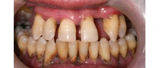
Photo: dystopia of wisdom teeth in the picture
A person suffering from dystopia can diagnose this disease himself. But in order to carry out therapeutic measures, it is necessary to contact a general practitioner or orthodontist.
During a clinical examination, the dentist will prescribe the necessary procedures for final diagnosis and determination of the position of dystopic teeth in the gums.
Before starting treatment, the following measures must be taken::
- orthopantomography , that is, it is necessary to take a picture of the jaw in order to see the condition of the roots, bone tissue, and determine the location of the diseased tooth in relation to the others;
- creating a jaw model from plaster , for which the necessary measurements are taken, subsequently used for treatment;
- teleradiography , thanks to which the ratio of the sizes of the jaw and teeth is measured;
- classification of the patient’s bite according to ICD-10, which allows for an assessment of defects.
Based on the results of the research, the dentist decides on the treatment of the patient.
Treatment methods for dystopia
- Orthodontic treatment using braces (used in the early stages, when there is room in the dentition for a dystopic tooth, as well as in case of trauma to the teeth and gums)
- Removal of a crooked tooth (in the presence of a cyst, inflammation of the gums, and also if the tooth interferes with treatment and the normal development of the rest of the dentition)
You can detect dental dystopia yourself, but only a specialist can make an accurate diagnosis. If an incorrectly grown tooth does not have a strong impact on the dentition, and also does not disturb the aesthetics of the smile, it will be enough to just slightly polish it. But, if the problem is much more serious, professional treatment will be needed. The Family Dentistry Center provides dental treatment under general anesthesia or, in other words, through intravenous sedation. While you are in medicated sleep, doctors with many years of experience will perform all necessary operations, painlessly and without causing discomfort. Don’t delay your visit to the doctor - remember, the health of your teeth is in your hands!
Anomalies in the position of individual teeth can lead to occlusal aberrations and cause internal disorders of the temporomandibular joint (TMJ). The asymptomatic abnormal position of the third molars in combination with the aesthetic optimum of the smile raises doubts in the patient about the advisability of removing such teeth and requires the dentist to objectively justify the need for this intervention [5].
The examination of patients with internal disorders of the TMJ includes an instrumental stage. Magnetic resonance imaging (MRI) of the TMJ is the “gold standard” in the assessment of intra-articular disorders [1]. MRI examination of the TMJ is carried out in the positions of habitual occlusion and maximally open mouth. For stable fixation of the lower jaw (MF) and maximum reduction of motion artifacts during functional studies, it is optimal to use a mouth retractor in the form of a threaded cone made of amagnetic material [3] .
Long-term disorders of the biomechanics of the joint in the absence of adequate treatment lead to the development of secondary osteoarthritis, manifested by deformation of the articular surface of the head of the joint and changes in the spongy substance (subchondral osteosclerosis, cystic restructuring, fatty involution). Such changes are more reliably and more completely visualized with multislice computed tomography (MSCT) or dental volumetric tomography (DVT) [1, 2].
Functional diagnostics in dentistry (orthodontics) is aimed at an integrated approach to solving clinical problems. To understand the biomechanics of the joint and occlusion, it is necessary to study the articulatory movements of N.Ch. The trajectories of these movements are individual and can change with anomalies in the occlusion of the dentition, pathological processes in the TMJ and muscles of the maxillofacial region, and postural disorders. To conduct a study of the biomechanics of the joint and assess the influence of the occlusal factor on the articulation of the LF and internal structures of the TMJ for the purpose of diagnosis and control of treatment, electronic axiographs, kinesiographs, and MPI diagnostics are used [4].
Clinical experience indicates that the development and progression of articular disc dislocations is to a certain extent associated with changes undergone by the dentofacial apparatus. In this regard, a comprehensive examination, including visualization of the TMJ, study of the biomechanics of the joint and disorders of functional occlusion, seems to us absolutely necessary when treating patients with this pathology.
Often in one patient we find several factors that are potential causes of articular disc dislocation. It is not always possible to identify the leader among them. It would be optimal to influence all possible causative factors and links of pathogenesis, but in practice this is often impossible to fully implement. In such cases, it seems advisable for us to focus treatment efforts on eliminating the most obvious cause. In this regard, I would like to share the experience of treating our patient with internal TMJ disorder.
Patient D
., 30 years old, applied to the orthodontic clinic of the Omsk State Medical Academy on July 2, 2014 with complaints of pain in the right ear when chewing, limited mouth opening with a feeling of mechanical obstruction in the area of the right TMJ. She became acutely ill: 3 weeks before treatment, for no apparent reason, in the morning there was a sudden restriction in opening her mouth, after which pain appeared. The patient did not notice any sound phenomena in the TMJ either before or after the onset of the disease. I contacted my local dentist with these complaints. The prescribed treatment (nimesulide orally, compresses with a 25% dimexide solution) for 1 week did not bring a positive result. The patient leads a sedentary lifestyle, spends at least 5-6 hours a day at the computer, and suffers from periodic headaches.
On examination, slight asymmetry of the face is noted due to the displacement of the chin to the right from the midline. On palpation in the projection of the LF heads, no pain is observed. Excursion of the right head is somewhat limited. Joint noises are not detected during low frequency movements. The maximum interincisal distance is 30 mm. There is a deviation of the bass to the right when opening the mouth. Nomination of N.Ch. anteriorly is sharply limited, painful. The chewing muscles are painless on palpation.
Oral examination: teeth 1.8, 2.8 and 3.8 are missing. The crowns of the remaining teeth are intact.
Dental crowding of the first degree, tooth supraposition 4.8, mesial inclination of the tooth crown 4.8, normal Spee curve. Retention of 2.8, 3.8 teeth.
Study of diagnostic jaw models: an increase in the width of the dentition in the molar area by 5 mm and in the premolar area by 4 mm on the upper jaw (HF) and LF, an increase in the length of the anterior segment in the HF by 2 mm, in the LF - by 1 mm. Teleroentgenogram (TRG) analysis: interincisal angle (II) - 127°, inclination angle of the upper incisor relative to the HF plane (Max1-NSL) - 109°, inclination angle of the lower incisor relative to the LF plane (Mand1-ML) - 99.8°, Wits -number - –1.1 mm, facial index according to Hasund - 87%. Horizontal growth, protrusion of the upper and lower incisors, dental-alveolar class I, skeletal class I (Fig. 1).
Rice. 1. Points and planes used for TEG analysis.
On 07/03/14, the patient underwent MSCT: in the left TMJ in a state of occlusion, the head of the TMJ has a central position in the articular fossa with moderate subchondral sclerosis and usuration, flattening of the anterior contour. In the right TMJ in occlusion: the articular head is in retroposition, with moderate subchondral sclerosis and usuration (mainly expressed along the flattened upper contour). During a functional study: in the left TMJ - forward movement of the head extending beyond the articular tubercle up to 16 mm; in the right TMJ - the movement of the head is sharply limited to the level of the base of the articular tubercle (up to 5 mm). The masticatory muscles are symmetrical in thickness and density. Conclusion: degenerative-dystrophic changes in both TMJs according to the type of arthrosis of the I degree with retroposition and signs of limited mobility of the condyle in the right TMJ.
On 07/04/14, the patient underwent MRI of the TMJ: the MR signal from the bones of both TMJs was unchanged. No pathological changes in the structures in the left TMJ during occlusion were detected. During a functional study, the forward movement of the head extends beyond the articular tubercle by 17 mm, the intra-articular disc occupies its normal position, without signs of degenerative changes, the signal from it is normal. In the right TMJ in the position of occlusion, the intra-articular meniscus is in the position of anterior dislocation. During a functional study, the movement of the head is limited to 6 mm. Disc - with signs of degenerative changes, with thinning in the center and probable fragmentation (separation): the posterior part is displaced posteriorly, into the laminar zone, the anterior part is determined to be displaced anteriorly. No fluid is detected in the joint cavity (Fig. 2).
Rice. 2. Functional MRI of the right TMJ. Oblique-sagittal plane; pd-weighted images.
A picture of degenerative changes in the right TMJ according to the type of arthrosis of the first degree with retroposition of the head in the joint, signs of degeneration of the intra-articular disc (and its probable fragmentation), manifestations of anterior dislocation of the intra-articular disc.
According to clinical and radiological examination, the patient was diagnosed with: unreducible displacement of the articular disc of the right TMJ; secondary arthrosis.
A diet limiting hard foods is recommended. It was decided to refrain from invasive measures (arthrolavage, arthroscopy). She was referred for treatment to an orthodontist.
A functional examination of the TMJ was performed using the KAVO Arcus Digma II electronic registration system. When performing a functional test of opening and closing the mouth: articulation of the MF without taking into account dental guides, visualization of the articular trajectories of movements of the articular heads of the MF is assessed as normal. When performing a functional test of opening and closing the mouth: articulation of the MF taking into account dental guides, visualization of the articular trajectory of movements of the left articular head of the MF is assessed as normal (Fig. 3).
Rice. 3. Articular trajectory of movement of the left articular head of the LF.
When performing a similar test of the right TMJ, the articulation curve at the initial segment has a “reverse bend”, indicating the presence of an occlusal obstacle; the length of the curve is shorter than the similar curve of the opposite TMJ, which indicates a decrease in translation of the head of the TMJ on the right and the predominance of rotational movements in the initial period of mouth opening (Fig. 4).
Rice. 4. Trajectory of movement of the right articular head of the LF.
To assess the position of the articular heads in the position of habitual occlusion in relation to the central relationship of the TMJ, an MPI study was performed. The SAM articulator was preliminarily adjusted in accordance with the articulatory protocol for individual programming of the articulator (Fig. 5).
Rice. 5. SAM articulator adjustment protocol.
When assessing MPI parameters in the right TMJ, the presence of compression of the bilaminar zone was noted.
Taking into account the clinical, functional study and radiation imaging data of the TMJ, the patient was recommended to remove tooth 4.8.
After tooth extraction 1.5 months later, the patient has no complaints. On examination, the mouth opens freely, painlessly, without displacement. The maximum interincisal distance is 46 mm. No joint sounds are detected. Palpation in the projection of the LF heads and masticatory muscles is painless. A repeated functional examination of the TMJ was performed using the KAVO Arcus Digma electronic registration system. When performing a functional test of opening and closing the mouth: articulation of the LF taking into account dental guides (right TMJ), normalization of the shape of the articulation curve is noted, which indicates the absence of an occlusal obstacle; an increase in the length of the curve indicates the correspondence of the translational and rotational movements of the head of the right TMJ. MPI data: displacement of the articular heads of the mandible from the central ratio is within normal limits.
Thus, the main etiological factor was identified and eliminated, which led to a pronounced clinical effect. Complete restoration of joint function, symmetry of LF movements, as well as MPI data indicate normalization of intra-articular relationships. The patient is indicated for further orthodontic treatment, elimination of crowding of teeth, restoration of bite height, removal of teeth 2.8, 3.8.
The data presented allow us to conclude that patients with suspected irreducible displacement of the articular disc of the TMJ are recommended to undergo a comprehensive examination to verify the diagnosis (MRI, MSCT of the TMJ with a closed and open mouth), as well as to identify the probable etiological factor (methods of functional gnathic research); This clinical case confirms the influence of an anomaly in the position of an individual tooth on the development of internal disorders of the TMJ.
Treatment of tooth impaction
The dentist, as a rule, makes a decision - to save the impacted tooth or remove it.
For proper treatment, the specialist conducts a comprehensive examination of the jaw:
- to make an X-ray;
- Ultrasound;
- CT.
If a tooth with retention comes out correctly, but is hampered by a too dense layer of gum tissue, the specialist performs the following manipulations:
- excision of the gingival hood;
- freeing the tooth from part of the gum tissue that prevented eruption.
After the operation (and cutting the gums of an impacted tooth is considered a surgical operation in dentistry), the doctor monitors the growth process until its final exit, and the patient is prescribed regular medical examinations.
If an impacted tooth is considered healthy, but is positioned incorrectly, the patient is prescribed orthodontic treatment (wearing braces).
Also, if the shape is incorrect or there is a lack of tissue, artistic restoration may be prescribed (extension of the coronal part, installation of veneers/lumineers).
Consequences of retention
If you have impacted teeth, urgent qualified dental care is required. Possible complications:
- cyst formation;
- abnormal development of teeth that are located next to the impacted one;
- violation of facial geometry;
- row offset;
- root resorption.
Retention of a wisdom tooth is usually very painful. The patient's temperature rises sharply, there is a feeling of pressure on the entire jaw, and severe throbbing pain. The impacted unit is quite large in size; the rudiments, as a rule, are positioned incorrectly, which creates problems during eruption up to the impossibility of exiting the bone.