One of the most common myths when undergoing an MRI is that metal braces cause discomfort during the examination. In addition, many are convinced that by acting on the brace system, the tomograph is harmful to health. Almost every patient with braces doubts the safety of this method, as they know about the property of magnets to attract metal objects. Read further in the article about whether it is possible to do an MRI with braces and the possible consequences.
The operation of the tomograph is based on the creation of an electromagnetic field with which the hydrogen atoms present in the human body resonate. As a result of changing the fields, an image is obtained, which is decrypted using a specific program. Scientists have proven that the MRI procedure does not harm the health of wearers of braces, and stories about discomfort and harm are myths that have not received real confirmation. The magnetic field does not cause deformation of braces. The metals used in them do not change their properties under the influence of a tomograph.
What are braces and what are they?
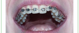
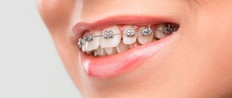
Braces are orthodontic structures that help correct bite pathologies. Treatment of dental diseases with their help is a rather lengthy process. Depending on the condition of the teeth, the patient’s age and other factors, it may take several years. During this time, the patient is not insured against other diseases that may require hardware diagnostics, including MRI.
Braces are made from various materials: metal, sapphire, ceramic or plastic. The arch of the structure, which passes through the clasps and exerts the main pressure on the dentition, is usually made of metal. As already noted, it can distort the results of the examination. For this reason, the doctor may prohibit an MRI if the patient wears braces. There is no direct contraindication to the study, but the information obtained during it cannot be trusted 100%.
However, refusal to conduct an MRI is justified only in cases where the head area is being examined. Other parts of the body can be scanned whether or not you have braces.
There are also types of metal that do not affect diagnostic results. Therefore, tomography of the brain or dentofacial apparatus can be carried out even if there are orthodontic structures in the patient’s oral cavity.
Risks of MRI diagnostics with titanium dentures
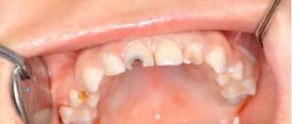
Due to the fact that prostheses implanted into the jaw bone are mostly made of paramagnetic titanium, magnetic resonance imaging with dental implants is a completely feasible and completely safe procedure. Titanium is a biologically inert material, i.e. it is not able to oxidize and release harmful substances during contact with bones, muscles and blood vessels of the human body. Of course, in medicine it is not pure titanium that is used, but its alloys with a small amount of other elements. This is done to make the material durable and light. But they also do not have any negative impact when undergoing an MRI.
By the way, titanium is comparable in strength to steel, and in lightness - to aluminum. Therefore, this metal has received recognition not only from dentists, but also from traumatologists and orthopedic surgeons.
Titanium is also used to produce abutments, gum formers, and metal arches (bases) for dentures.
MRI and braces: which metals do not affect diagnosis
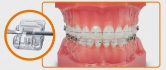
Before your MRI procedure, tell your doctor what material your braces are made of. This information can be obtained from the orthodontist who installed the structure. The physicochemical properties of various metals differ significantly. Moreover, the reaction to the magnetic field of a particular metal product may be different.
Some of them, for example, titanium structures, do not react at all to the MRI machine and the magnetic waves it creates. Also, braces containing ferromagnets - substances that have spontaneous magnetization - will not interfere with the examination.
In any case, the procedure can be interrupted if the patient feels any discomfort. Before the examination, the doctor gives him a special device with which he can signal that he is not feeling well.
Should you tell your doctor that you have dental implants?
A patient with installed dental implants must notify the doctor about this. Then the specialist makes the necessary adjustments, taking into account the location and composition of the prostheses. These settings eliminate the appearance of blurred distortions (also called field artifacts). And then the results of the examination will be clear and precise.
But if a person decides to keep silent about “non-native” teeth, then the picture will turn out blurry, because implants, although slightly, do affect the quality of the pictures. And such a careless patient will have to pay for the procedure and undergo diagnostics again. Why does the blame in this case fall on the patient? Because before the MRI, he will definitely be asked about the presence of implants and what kind of metal they are made of.
“Mom 2 years ago had an MRI of the head and blood vessels, using a high-field tomograph. And she’s had implants installed in her upper jaw for about 5 years now. And nothing, everything went fine. There are no special sensations during the procedure. Just be sure to tell the girls at the reception that there are implants before starting the procedure. They warn the doctor themselves, and then the device is reconfigured.”
Vitalina, from correspondence on the woman.ru forum.
Myths about MRI and braces
Some people believe that MRI scans performed while there are metal objects in the body can cause electric shocks. Others believe that metal structures, such as braces, become red hot and leave severe burns on (or inside) the body. There is also an opinion that orthodontic brackets lose their functionality or begin to deteriorate under the influence of the magnetic field created by the MRI machine.
All these ideas about tomography are erroneous. The examination does not cause any complications, even if it is carried out while the patient has braces. It’s just that the exposed images obtained during such a study can be treated with skepticism. The doctor will not be able to make an accurate diagnosis and prescribe treatment. However, MRI cannot cause any harm to health. Moreover, it is not capable of damaging the orthodontic structure.
Magnetic resonance imaging in the presence of a dental implant
So why wouldn't a dental implant be an absolute limitation to MRI? Here you need to understand that implants are made from metals endowed with diamagnetic and paramagnetic properties. What it is? The operation of an MRI scanner is based on the magnetizing properties of certain substances. So, para- and diamagnets react weakly to powerful magnets, such as those installed in the tomograph.
Paramagnetic materials are almost not subject to the action of a magnetic field and do not shift in it. These are the following materials:
- titanium: the main metal from which modern implants are made,
- aluminum: medical gurneys made of this metal can be brought into the room where MRI is performed,
- platinum: softer and more expensive than titanium.
Diamagnets - magnetization is not significant, it occurs in the direction of the applied field:
- hydrogen: it was the presence of this element in the human body that made magnetic resonance imaging possible,
- copper: oxidizes quickly, not suitable for implantation into the body,
- gold and silver: very soft metals.
Magnetic resonance imaging can be performed on any part of the body. If the organ being studied is located in the lower half of the body, then the implant will not affect the result in any way. And if the upper part is examined, then there may be some distortions - however, the implant itself will not move, that is, it will not be magnetized and will not break out of the body.
Alternative to MRI with braces
So, are MRIs done with braces? This method for orthodontic braces is prescribed in the following cases:
- Tomography is performed below the head area;
- braces are made of titanium or contain ferromagnets;
- The orthodontic system does not contain metal parts.
If the patient needs to examine the brain or the dental system, and he has metal braces in his mouth that are not made of titanium and without ferromagnets, the doctor prescribes alternative examination methods, for example, CT - computed tomography.
Unlike MRI, which operates on the basis of magnetic waves, CT provides scanning by exposing the body to x-rays. Computed tomography is prescribed for bruises and fractures, dental injuries, bone diseases, internal bleeding and other pathologies. The effectiveness of MRI and CT may vary depending on which organ is being examined and what disease is suspected.
When a patient requires a detailed examination of the jaw, he is usually prescribed a CT scan, since the images obtained using MRI will be overexposed in the places where the braces are. Computed tomography, no less effective than magnetic resonance imaging, allows us to examine the hard tissues of the oral cavity. However, if it is necessary to assess the condition of the soft tissues in the area of the dental system, the doctor may suggest temporarily removing the orthodontic structure. In this case, additional consultation with an orthodontist will be required.
Is it possible to do an MRI of the head with braces?
When a part of the body away from the braces is examined, no complications will arise at all. Perhaps elements made of a magnetic alloy will actually become magnetized in the field created by the tomograph and come into resonance with it. This is only dangerous because the generated image will be distorted, and the doctor will not be able to properly see the area of interest.
If the patient is scheduled to study the lumbar region of the spine or, for example, the knee joints, the braces simply will not fall into the zone of action of the constant magnetic field, and no problems will arise.
The situation is different with the study of the brain and facial area. In this case, metal braces will react to magnetic waves and increase the risk of diagnostic errors.
Reviews
In the capital, almost every medical institution has a tomograph and everyone is examined indiscriminately. When I was scheduled for pelvic diagnostics, I had braces. I thought that the procedure would be postponed until the end of treatment or the plates would be removed ahead of schedule. But the doctor convinced that the braces would not interact with the magnetic field, since it affects a completely different area.
Inna, Moscow
They ordered a head examination, and I had braces. There was no time to wait, since a dangerous diagnosis was in question. I found out from the dentist that my braces are made of plastic and there are no metal parts in them. Therefore, I did the procedure without fear and did not regret it. The primary diagnosis was not confirmed.
Egor, Krasnodar
How to prepare for an MRI of the brain?
No special, lengthy preparatory manipulations are required before the session, but a number of recommendations should be followed to ensure that the MRI of the brain is comfortable and without complications. First, be sure to warn your doctor about the presence of permanent metal implants. If it is possible to remove such parts for a while, this should be done in advance, for example, visit the dentist and remove the braces. Before going to the clinic, leave all body jewelry at home.
Secondly, take care of comfortable, loose clothing for the duration of the procedure. The diagnosis is carried out in a lying position, so items of clothing should not hinder movement (a miniskirt and skinny jeans will definitely not fit). Clothes should not have metal fasteners, buttons, belts, etc. Check your pockets for lost keys, small change, magnetic cards, etc.
Before going for an examination, you need to prepare the necessary medical documentation, including a referral from the treating specialist, a card or an extract from it, the results of other studies and previous photographs, if available.
It is better for women to avoid wearing makeup on this day, as many cosmetic and decorative products contain metallic inclusions. Before screening, each patient will have to complete a written questionnaire regarding health issues. This is required to identify cases of allergies to medications, chronic diseases that prevent the use of contrast, and other disorders that are subject to a ban on tomography.
What is the procedure?
MRI is one of the methods of modern non-invasive diagnostics. The method is based on the response of hydrogen atoms of internal tissues and organs to the influence of a powerful magnetic field. The equipment detects when hydrogen atoms change their position and transmits the image to the screen in the form of a three-dimensional picture.
The MRI equipment itself is a large cylinder with holes at both ends. Inside, the device is equipped with a movable table on which the patient lies during the examination. There are several options for tomographs:
- Open.
- Closed.
- Partially open.
In open tomographs, as a rule, very obese patients are examined, as well as patients suffering from severe claustrophobia.
The tomography procedure must be carried out completely motionless, therefore, to ensure immobility, the patient is fixed with special belts. To get a complete and detailed picture, you need to take several pictures. Each shot takes some time, and the entire procedure can last from 20 to 50 minutes.
Despite the fact that the tomography procedure is absolutely painless and does not cause harm to health, it is often undesirable to do it. Firstly, the cost of tomography is quite high, and secondly, such a study requires a certain amount of time. If the tomography shows an unreliable result for a patient with metal braces, the procedure will have to be repeated.
When is it necessary to undergo an MRI of the brain?
There are many indications for using MRI of the brain. Screening is used both as an independent primary method and as a secondary procedure that confirms or refutes data obtained by other diagnostic methods. MRI is often prescribed to determine the patient's operability in the preoperative period, as an accompanying visualization during surgery. After instrumental treatment in the head area, a secondary control examination is possible to monitor tissue recovery and regeneration after surgery.
The main indications for seeking diagnostics are:
- vascular disorders, destruction of the walls of blood channels, changes in the configuration of the network, the presence of obstacles in it - blood clots;
- head injury, bruises, penetrating and open wounds, suspected internal bleeding;
- tumor processes, uncontrolled growth of tissue structures;
- a progressive or sharp decrease in sound perception, impaired speech function in the patient;
- spread of infection, foci of inflammation;
- diagnosis of the cause of changes in mental well-being, impairment of cognitive functions (thinking, remembering);
- congenital developmental pathologies;
- manifestation of epileptic syndrome;
- chronic pain in the skull of “unexplained” etiology.
MRI or CT?
The research method using a computed tomograph (CT), just like MRI, allows you to obtain layer-by-layer images of sections of the scanned parts of the body and transfers the images to a computer monitor. Using this method, you can obtain three-dimensional models by combining all the received images into one picture.
An X-ray computed tomograph works on the principle of circular illumination of the areas under study with x-rays, which are directed perpendicular to the body. Since different tissues of the body absorb X-rays in different ways, the computer, processing the images, displays a detailed and clear picture of the examined internal tissues and organs on the screen.
The computed tomography method gives fairly clear and accurate results, but MRI is considered more effective.
Myths for patients with implants
The most common myth about magnetic resonance imaging scares most patients. An incompetent interlocutor - a work colleague or a neighbor next door - may say in horror: “What, you can’t do a tomography with implants - the prosthesis will be pulled out of the jaw by a magnet and it will stick somewhere, or the device will be damaged.” And the person will believe and refuse MRI in favor of a less informative, but more harmful examination (for example, computed tomography).
Every patient referred for magnetic resonance imaging should remember that dental implants are not a contraindication. And if you are denied such an examination, then you need to look for a clinic with modern equipment and a qualified radiologist.
Author: Vasiliev A. A. (Thank you for your help in writing the article and the information provided)
Features of undergoing MRI
MRI research is based on the study of hydrogen atoms and their behavior when exposed to electromagnetic pulses. The research design is as follows:
- A magnetic field is created using a tomography machine
- The reaction of the nuclei of hydrogen atoms behaves differently under the influence of the field
- When hydrogen atoms resonate, the field of the apparatus changes, forming an image.
- The results are read using a special program.
Doubts regarding the admissibility of a tomograph when wearing braces are based on the characteristics of the magnetic field, which can attract metal objects, and therefore the materials used in the manufacture of braces. This forces patients to refuse the procedure.
In the famous series about Dr. House there is a story about conducting a CT scan on a patient with a tattoo. During an MRI session, the tattoos became heated due to the inclusion of metal particles in the paint.