Microscope - luxury or necessity?
Harry Kahr, who was the first to use a microscope in dental treatment, briefly and clearly substantiated the principle of its necessity: “You can only treat what you can see.” With traditional treatment, the doctor’s “view” is limited to what is reflected in the dental mirror, while the microscope intended for dental treatment has a thirty-fold magnification, making the examination of the surface of the tooth and its internal structure more detailed and complete. This not only allows the doctor, who normally works almost by touch, to better see the area being treated, but also reduces the amount of living tissue removed during drilling, and also reduces to zero the risk of “looking through” a carious cavity or an uncleaned canal.
Bacteria of this disease
The cause of tooth decay is cariogenic bacteria. Some types of microflora have a destructive effect on the hard shells of teeth.
What microorganisms cause damage?
In the formation of pathology, the main role is played by bacteria and pathogens of the genus Streptococcus, species mutans, sanguis, sabrinus, viridans, salivarius, mitis and lactobacilli (Lactobacillus).
The bacteria Streptococcus mutans has the greatest destructive effect on teeth .
Dozens of species of other bacteria live in the mouth , the vital activity of which does not cause tooth decay. Some of them are enemies of streptococci, which cause caries, and destroy them.
The role of microbes in development or pathogenesis
Pathogenic microbes synthesize organic acids and other active compounds in the process of anaerobic glycolysis. These substances react with the enamel and cement of the tooth and destroy them. As a result, carious cavities are formed.
Under what conditions do bacteria develop in the oral cavity? There are several reasons for the active reproduction of cariogenic organisms on teeth:
- Imbalance of bacterial flora in the mouth.
- Diet with excess carbohydrates.
- Insufficient amount of saliva.
- Lack of fluoride in drinking water.
- Neglecting to brush your teeth, not using toothpicks and floss.
- Stress, decreased immunity.
How it happens
There are several key stages in its development :
- Plaque forms. Staphylococci form deposits that firmly adhere to the surface of enamel and cement. The deposits consist of 10% bacteria , the remaining 90% are waste products of microflora. Most often, plaque appears in fissures - recesses in the upper part of the chewing teeth, in the lateral areas, and between them.
Photo 1. A thickened layer of plaque on the lower front teeth from the back.
- A plaque is formed from thickening plaque. Once it has reached the plaque stage, it cannot simply be brushed off. The acids under the plaque destroy (demineralize) the tooth tissue, and the dentist diagnoses caries.
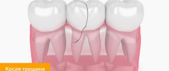
- Caries appears. Bacteria attack the hard shells of the tooth. They show brown and black pits, cracks, and grooves. The destruction process extends to dentin.
- Dentin becomes porous and fluid gets into it , which puts pressure on the tubules. The tooth aches when sweet, salty, cold or hot food enters the cavity. The carious cavity increases in size and reaches the root canal. The inflammatory process begins in the pulp. If the nerves are affected, severe toothache begins. And not only after eating, but also at rest.
If you endure the pain, the pulp dies, as do the nerve endings. The infection can spread to the periodontium , circulatory system and bone tissue.
Important! If a tooth is left untreated, caries will destroy it inside and out. form around the destroyed root .
Is it necessary to use a microscope when treating tooth canals?
The use of a microscope can be justified both in diagnosis and in the treatment of teeth and gums, equally in surgery, therapy, periodontology, dental restoration, and orthodontics. However, where the use of a microscope cannot be avoided, it is in endodontics - the treatment of dental canals. Each tooth has from one to three (in rare cases, four) canals, the diameter of which is on average 1 millimeter - a value barely visible to the naked eye. In addition, dental canals have bends and branches; a doctor can see them in detail, and therefore fully treat them, only under magnification, otherwise he will act by touch or “by the picture” - an enlarged image from an x-ray.
Eliminating bacteria living on teeth
Numerous photos and scientific videos give reason to say that it is in dental plaque that there is a huge amount of bacteria. To prevent bacteria from forming on teeth and causing discomfort, comprehensive treatment is necessary.
But treating bad breath and tooth decay will be ineffective if you do not get rid of bad habits. Everyone knows what teeth damaged by smoking look like in photos and in real life! It is smoking that you need to get rid of first of all. In addition, you should stop drinking alcoholic beverages - alcohol harms all body systems.
The next stage of treatment is, in fact, a consultation with a dentist. He will examine the plaque under a microscope and take the necessary photos. Treatment of teeth damaged by microbes and plaque occurs only on an individual basis.
For dental treatment to be effective, you must not only take the medications prescribed by your doctor, but also make sure that there are enough vitamins in the menu. Here are a few hygiene principles that also increase the effectiveness of treatment:
- You need to brush your teeth twice a day.
- It is necessary to change your toothbrush once every three months.
- Use dental floss as recommended by your doctor.
- The mouth must be rinsed after every meal.
- In order to produce a sufficient amount of saliva, it is necessary to use chewing gum without sugar, but without abusing it.
- It is necessary to regularly remove plaque from the tongue, since photographs taken under an electron microscope confirm the presence of a huge number of microbes on it.
In a clinical setting, the most effective method of getting rid of bacteria living on teeth is ultrasonic cleaning. Such treatment is carried out no more than once or twice a year.
I work as a veterinary doctor. I am interested in ballroom dancing, sports and yoga. I prioritize personal development and mastering spiritual practices. Favorite topics: veterinary medicine, biology, construction, repairs, travel. Taboos: law, politics, IT technologies and computer games.
What are the indications for dental treatment under a microscope?
If an X-ray examination shows the presence of a large number and complex structure of dental canals, the use of a microscope becomes simply necessary. Also, one hundred percent indication for the use of a microscope is retreatment of canals. Previously, when it was discovered that endodontic treatment was carried out poorly or not in full (and the signal indicating this was, as a rule, acute pain), the tooth had to be immediately removed. Today, it has become quite possible to unfill, re-treat and re-fill tooth canals, and all this is thanks to the use of a microscope. One of the common complications of endodontic dental treatment in the past was root canal perforation. This happened when a doctor, drilling “by touch,” accidentally drilled through the tooth tissue, leaving a hole inside that provoked inflammation and ultimately led to the removal of the injured tooth. Today, the risk of perforation is reduced through the use of a microscope when processing canals, and if this does occur during traditional endodontic treatment, the situation can be quickly corrected using the same dental microscope.
Another unpleasant moment, which, alas, was the norm ten or even five years ago, is the fragments of the instrument left in the canals when the nerves were removed, as well as fragments of the nerves themselves. The microscope here equally helps both to prevent the occurrence of such a problem and to eliminate it. Another undeniable breakthrough made thanks to the introduction of microscopy into widespread dental practice is the ability to treat granulomas and cysts in a tooth without removing the latter.
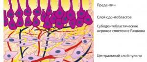
Tooth structure
The tooth is divided into two main parts : the crown , which rises above the gum, and the root , located in the periodontal canal. Between the tooth and the bone there is a layer of soft tissue called periodontium.
The hard frame is dentin . In the upper part it is covered with a thick cover made of hard enamel. On the surface of the chewing teeth there are depressions - fissures . Dentin in the cervical and root areas is surrounded by cement.
Inside the dentin there is a root canal filled with pulp, which contains blood vessels and nerves that exit the root through the apical foramina. They are connected to the neurovascular bundle. The main bundle connects to the circulatory and nervous system of the body.
In what other cases is a dental microscope used?
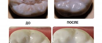
Endodontics is the most common and most justified use of such expensive equipment as a microscope, but its field of application is not limited to root canal treatment alone. It is absolutely fair to say that with a microscope is always better than without it. Even in standard caries treatment, the use of a microscope greatly increases efficiency and reliability, and also provides guarantees of long-term results. For example, detecting caries at the initial stage, when its treatment can be carried out without using a drill and removing a large amount of tissue, is almost impossible with the naked eye, especially in hard-to-reach places; A microscope provides such an opportunity.
Photo of a tooth under a microscope
Ease of use and psychological aspects
Working without a microscope, the dentist has to bend over the patient, getting close to him. He himself is not particularly comfortable working in this position, and patients often experience discomfort. The microscope solves this problem by allowing the doctor to be approximately 0.5 meters away from the patient.
In addition, modern microscopes allow images to be displayed on a screen. Some patients are simply interested in watching the operation process, while others feel calmer, seeing that nothing “terrible” is happening to their tooth.
What are the features of dental treatment under a microscope?
If you decide to use a microscope in your treatment, your appointment with the dentist will proceed as follows: firstly, you will be in a lying position and not sitting, as is the case with traditional treatment; secondly, the doctor will be behind your head or sitting almost on the same level as you to your right, and the microscope itself will be located at a distance of 20 - 25 centimeters from your face. The use of a microscope in treatment means that the doctor will work with it constantly, rather than look into it from time to time. In this case, the image from the camera equipped with the dental microscope will be transmitted to the monitor of the assistant, who controls the treatment process and provides the doctor with the necessary tools and preparations.
Progress of the procedure
Treatment using optical magnification is more comfortable for the patient. He reclines in a chair, which helps him relax better. In addition, the instruments that the doctor uses are much smaller than usual, and therefore do not cause discomfort or a gag reflex. This also allows the patient to swallow saliva normally. In addition, multiple magnification of the image allows the doctor to see the work area in great detail, and the patient does not need to open his mouth wide.
Where is it possible to undergo dental treatment under a microscope in Moscow?
Unfortunately, today not all dentists in Moscow have a dental microscope. This is due not only to the high cost of such equipment, but also to the need for specialized training for doctors to work with such equipment. Therefore, if at the diagnostic stage such complications-fraught features as branched dental canals or the possibility of a dental cyst were discovered, it is worth asking whether the dentistry of your choice allows dental treatment under a microscope. If this is not possible, you should think about looking for another clinic and a doctor with appropriate competence. Any conscientious dentist, even if he is not able to independently carry out dental treatment under a microscope, if there is an urgent need for it, will advise you to go to a dentistry that has such an opportunity.
Our specialists
- Gogia Tea Zurabovna
Dentist-therapiston Komsomolskaya
- Gracheva Natalia Alexandrovna
Dentist-therapist
on Komsomolskaya
- Bystrikova (Murashkina) Valeria Pavlovna
Dentist-therapist
on Mayakovsky
- Yarygina Larisa Borisovna
Dentist-therapist
on Kalanchevskaya