Anesthetics with a high vasoconstrictor dilution of 1:100000
In pediatric practice, they are used only for a number of surgical interventions for the purpose of hemostasis. The duration of anesthesia is 75 minutes for dental pulp and 360 minutes for soft tissues.
Such outpatient interventions include the removal of an impacted, supernumerary tooth, apperculectomy, cystectomy, plastic surgery of the frenulum and vestibule of the oral cavity, and removal of a tumor. The use of anesthetics with high dilutions of epinephrine for other types of dental interventions is unjustified and disproportionate to the volume of intervention.
Application in pediatric dentistry
The technique is popular in pediatric dentistry, as it allows you to completely anesthetize any dental procedure, creating a trusting attitude towards doctors in young patients.
The porous structure of children's primary teeth is especially susceptible to topical anesthesia. The drug easily penetrates tissue, turning off sensitive receptors even in the deep layers of the gums. This allows the method to be used as the main method of pain relief - not only for superficial procedures, but also for the treatment of deep caries of primary teeth.
ATTENTION! In pediatric dentistry, drugs with a low level of toxicity are used. Drugs such as Dicaine are contraindicated for use in patients under 10 years of age.
Anesthetics with low vasoconstrictor dilution 1:200000
Indicated for most outpatient interventions in pediatric dentistry. 4% articaine 1:200000 provides anesthesia for soft tissues for 180 minutes and dental pulp for 45 minutes, which satisfies the protocol for most outpatient interventions.
Currently, anesthetics based on 4% articaine with epinephrine 1:400000 have appeared in European countries. They provide anesthesia for dental pulp tissue for 20 minutes and soft tissue for 1 hour. This anesthetic provides the duration of anesthesia required by the doctor and a short period of “numb” tissue, which is so important for pediatric patients. Currently, these anesthetics are not certified in the Russian Federation, but work is underway to introduce them into domestic dentistry.
It is also worth noting that there is no difference in the depth of anesthesia and effectiveness between anesthetics with vasoconstrictors 1:100000, 1:200000 and 1:400000. There is a difference only in the duration of local anesthesia of the dental pulp: 25, 45 and 75 minutes, respectively. Many dentists mistakenly divide anesthetics into “strong” (1:100,000) and “weak” (1:200,000). This statement is misleading.
For short-term interventions in children with concomitant pathologies, the use of anesthetics without a vasoconstrictor is indicated. However, their use does not guarantee complete safety and does not reduce the risk of complications. It is worth noting some of their pharmacological features. Vasoconstrictors are added to the local anesthetic solution not only to increase the duration of pain relief, but also to reduce their toxicity. The fact is that all anesthetics have a vasodilating effect and undergo fairly rapid absorption into the bloodstream. The addition of a vasoconstrictor slows the absorption of the anesthetic and prolongs its action. When using an anesthetic without vasoconstrictors, this effect does not occur. The anesthetic is forced into the blood, which can lead to a toxic reaction. This complication is possible if the permissible dosage is exceeded, which is different for anesthetics with and without a vasoconstrictor. In summary, it should be noted that anesthetics without a vasoconstrictor do not affect the functioning of the cardiovascular system, are less allergenic since they do not contain preservatives, but due to accelerated absorption they are toxic and safe to use only if the dosage is observed.
Preparations used for topical anesthesia
Local topical anesthesia often contains the same types of active ingredients as injection anesthesia (lidocaine, dicaine, bumecaine, etc.), but in higher concentrations. The composition is often supplemented with antiseptics, inflammatory process blockers, and flavorings. The main forms of release are cream, gel, ointment, spray, aerosol.
The entire list of available tools can be divided according to the principle of action:
- cauterizing agents - acid-containing preparations that can seal dentinal tubules and close nerve endings from external influences;
- dehydration compounds - preparations based on carbonic acid salts dehydrate tissue, constricting blood vessels and depriving the tissue of sensitivity (more suitable for superficial manipulations such as brushing teeth and removing tartar);
- analgesic liquids with a high concentration of anesthetic compounds block the conduction of peripheral nerve fibers (the most common group of anesthetics);
- preparations of physiological effects based on strontium, fluorine, sulfides - block the transmission of impulses to nerve endings (such drugs often have an additional therapeutic effect and can be used to treat and strengthen enamel, protect against caries);
ATTENTION! Despite the safety and simplicity of the method, using topical anesthetics yourself at home is undesirable. In each individual case, the dentist selects and prescribes the drug individually, taking into account the source of pain and the personal characteristics of the patient.
Anesthetics without a vasoconstrictor
Provide varying durations of pain relief for dental tissues. In particular, 2% lidocaine provides anesthesia of the dental pulp within 5 minutes, while the rate of onset of anesthesia is also 5 minutes, which is unsatisfactory for the doctor. Therefore, the use of 2% lidocaine without a vasoconstrictor is inappropriate for dental anesthesia.
3% mepivacaine, in comparison with other anesthetics, has a less pronounced vasodilator effect, which makes it possible to use it without adding a vasoconstrictor. The anesthetic provides pain relief for 10-20 minutes, while treatment must be carried out from the 5th to 20th minute during therapeutic interventions and from the 10th to 20th during tooth extraction surgery.
Articaine 4% is currently available in the Russian Federation. This anesthetic is short-acting: anesthesia of dental pulp for 6 minutes, soft tissue for 45 minutes. Its widespread use in pediatric dentistry is limited due to its short action, which is not suitable for most interventions.
Kinds
This anesthetic is a rich agent. It is applied to a limited area of radical action. Local anesthetic effect can be obtained:
- Using medications;
- When the mucous layer cools;
- If the impact point is cauterized;
- With other physico-chemical methods of exposure;
Thanks to such methods, the degree of vulnerability is reduced. Dentists even remove teeth in this situation, especially non-molar ones. Of course, the doctor must determine the type of pain relief. More details about each type:
Cauterization
This is the very first type of anesthesia of the superficial layers of tissue. As a rule, aggressive drugs with strong effects are used. They may contain nitric or carbolic acids, zinc chloride, and silver nitrate. The technology allows you to freeze not only the periodontium, but also dentin. When applied to fabrics:
- pores narrow;
- they become clogged;
- nerve fibers become closed;
The cauterization method worked for a short time. It is not widely used, since its substances are significantly aggressive. The products used are highly toxic. They negatively affect the chewing organs and oral cavity.
Dehydration
The technology is based on reducing the vulnerability of units against the background of the inclusion of dehydrating substances in the mixture. Most often these are carbonates or bicarbonates of potassium, sodium, and magnesium. Sometimes other microelements with similar effects are used. This method is used by hygienists when performing professional teeth cleaning. It can also be used for light manipulations.
Physiological agents
This is a separate type of pain relief. The application of special mixtures helps protect the patient from discomfort during treatment. The following pastes have a physiological effect:
- Sulfidic;
- Glycerophosphate;
- Aspirin;
- Strontium;
They specifically act on the receptors of dentinal tissues. Thanks to this, the transmission of impulses is blocked, so the nerve fibers do not perceive them. They are often used by dentists in the treatment of incisors, canines, and molars with damaged enamel and pathological dentin. They should be used regularly so that the damaged structure is restored. In addition, the product has a beneficial effect on healthy areas of the masticatory organs.
Local painkillers
High-quality dental treatment often requires the use of local anesthetics. They are among the most popular substances for superficial pain relief. The remedies eliminate the vulnerability very quickly. The doctor calculates with great accuracy the time when they will act. Dentists usually use benzocaine, tetracaine, and lidocaine. These substances quickly stop the conduction of nerve endings.
Dosage
In all cases of local anesthesia, it is necessary to calculate the dosage of the administered anesthetic in terms of the child’s body weight. For articaine preparations with a vasoconstrictor, the recommended dosage is 5 mg per 1 kg of weight. Before performing local anesthesia, the child’s weight is checked with the parents. In clinical practice, it is convenient to use a table with weight and the maximum permissible dose of the administered anesthetic (Table No. 1, 2).
Table No. 1
| WEIGHT | MG | ML | CARPOOLS |
| 10 | 44 | 1.5 | 0.8 |
| 15 | 66 | 2.2 | 1.2 |
| 20 | 88 | 2.8 | 1.4 |
| 25 | 110 | 3.6 | 1.7 |
| 30 | 132 | 4.4 | 2.4 |
| 35 | 154 | 5.1 | 2.9 |
| 40 | 176 | 5.9 | 3.2 |
| 45 | 198 | 6.6 | 3.6 |
| 50 | 220 | 7.3 | 4.0 |
| Mepivacaine 3% without vasoconstrictor. Maximum dose 4.4 mg/kg. 3% solution in 1 carpule 1.8 ml (54 mg). | |||
Table No. 2
| WEIGHT | MG | ML | CARPOOLS |
| 10 | 50 | 1.2 | 0.69 |
| 15 | 75 | 1.9 | 1.0 |
| 20 | 100 | 2.5 | 1.4 |
| 25 | 125 | 3.1 | 1.7 |
| 30 | 150 | 3.7 | 2.1 |
| 35 | 175 | 4.4 | 2.4 |
| 40 | 200 | 5.0 | 2.8 |
| 45 | 225 | 5.6 | 3.1 |
| 50 | 250 | 6.2 | 3.4 |
| Articaine 4% with a vasoconstrictor. Maximum dose 5 mg/kg. 3% solution in 1 carpule 1.8 ml (72 mg). | |||
Quite often, at outpatient dental appointments we encounter children suffering from obesity and metabolic syndrome, which is largely due to changes in the nutritional culture of the population. The dosage of the administered anesthetic in these cases has some peculiarities. In particular, if the doctor is going to administer anesthesia to an overweight child, the dosage of the administered anesthetic is calculated without taking into account adipose tissue.
Intracanal anesthesia
The intracanal method of pain relief speaks for itself. Typically, intracanal anesthesia in dentistry is carried out in this way: using a drill, a hole is made in the tooth corresponding to the diameter of the needle, and the drug is injected directly into the pulp or deeper into the canal. Sometimes the carious cavity itself is used for this. If we are talking about the intraligamentary technique, then we mean the introduction of a local anesthetic solution into the space at the root of the tooth (periodontal), and the tuberal technique means the introduction of a substance into the posterior alveolar branches of the upper jaw.
Injection equipment
For local anesthesia in children, carpule syringes of various designs are used. Preference should be given to injectors intended for carrying out an aspiration test (Rabinovich S. A., Vasilyev Yu. L., Sokhov S. T., 2013; Tarasenko S. V., Kuzin A. V., Belyaeva E. A., Kurtyshov A. A., 2013). Local injection anesthesia in children carries the risk of intravascular injection of local anesthetic. This fact is explained by the high degree of vascularization of the tissues of the maxillofacial region of children. Thus, the frequency of intravascular administration of an anesthetic during mandibular anesthesia in adults is 10-15%, and in children - 20-25%. Syringes with a plunger in the form of an anchor and a corkscrew have the best technical characteristics. The choice of injection needle depends on the method of anesthesia. For conductive methods, needles with a diameter of at least 0.4 mm (27G) should be used. When conducting conduction anesthesia, 0.3 mm (30G) needles bend excessively in the tissues (deflection), which leads to the deposition of the anesthetic away from the intended end point of pain relief (Rabinovich S. A., Vasiliev Yu. L., 2011).
Needles 0.3 mm (30G) are advisable to use for infiltration anesthesia and periodontal methods of pain relief.
Do not forget that when performing local anesthesia, the needle may break off. This complication, as a rule, occurs when the child makes sudden movements: withdrawing the head, abruptly closing the mouth. In most cases, these severe complications occur when using 30G needles when performing mandibular anesthesia in children.
There is an opinion that the thinner the needle, the less painful the patient perceives the stage of puncturing the mucous membrane and advancing the needle into the tissues. This opinion can be classified as a misconception. There are studies confirming that the diameter of the needle does not affect the reduction in the degree of pain of the anesthesia (Malamed SF, 2002).
Mechanism of action of surface anesthesia
Anesthesia occurs as follows: the anesthetic enters the tissue of the crown or gums, “seeps” 1–3 mm deep and affects the terminal nerve endings - the “end” parts of the nerve fibers. As a result, the fibers temporarily lose sensitivity. The effect occurs within a few minutes and lasts up to a maximum of half an hour.
The method allows you to temporarily relieve the sensitivity of the mucous membrane of the oral cavity and gums, dentin, that is, internal hard tissues, and pulp (neurovascular bundle).
Features of pain relief for temporary teeth
Of course, the choice of pain relief method depends on the planned intervention.
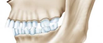
In the treatment of caries and pulpitis of temporary teeth, preference is given to infiltration methods carried out from the vestibular (buccal/labial) side, and there is no need for lingual or palatal anesthesia. This is explained by the predominance of spongy substance and a large number of nutrient openings in the structure of the jaw bones, facilitating the diffusion of local anesthetic.
In order to reduce the degree of pain of the injection and increase the level of comfort for the child, the following recommendations for local anesthesia should be followed:
- A preliminary distracting preparatory conversation with the child is necessary, during which the doctor uses terms that the child understands. In this case, the words “injection”, “syringe”, “anesthesia”, “removal”, “needle” are not pronounced.
- The child should not see (look at) the injection equipment. All instruments are presented by an assistant out of sight of the child.
- Application anesthesia should be performed so that the child does not feel the puncture stage of the mucous membrane.
- The rule must be observed: “one needle, one injection.” If the needle accidentally comes into contact with the bone tissue of the jaw, its tip is deformed; as a rule, it bends in the direction opposite to the bevel. A repeated injection with the same “dull” needle in another segment will involve applying pressure that is unpleasant for the child.
- The anesthetic should be injected slowly, 1 ml/min. Forced administration of the anesthetic leads to hydrodynamic tissue trauma, which causes pain.
- Two-stage injection of a portion of anesthetic. When performing infiltration anesthesia, the doctor initially injects a small amount of anesthetic? 0.1 submucosally, while a zone of tissue infiltration the size of a millet grain is visually determined. Next, the doctor talks with the child for 2-3 minutes.
- The subsequent administration of the main portion of the anesthetic is painless.
- The temperature of the anesthetic solution should be close to the child’s body temperature. To do this, do not store anesthetic carpules in the refrigerator. Before the injection, the doctor can “warm” the carpule in his hand or in running hot water.
The volume of injected anesthetic for infiltration anesthesia of temporary incisors and canines is on average 0.3 ml; for anesthesia of temporary molars, 0.5-0.6 ml is used. Subperiosteal anesthesia should not be performed as this will cause pain to the child and will not increase effectiveness. The needle should move submucosally towards the apexes of the roots of the teeth. In the upper jaw, when performing infiltration anesthesia, one should not insert a needle into the area of the frenulum of the upper lip, mucous strands in the area of temporary canines. These areas have an extensive network of nerves, providing hypersensitivity in these areas.
When performing an operation to remove temporary teeth of the upper jaw with a diagnosis of periodontitis, various combinations of infiltration methods are used. In the upper jaw, when removing incisors and canines, infiltration vestibular (0.5 ml) and incisal anesthesia (less than 0.2 ml) is used. In this case, classical conduction anesthesia with the advancement of the needle into the incisive canal is not carried out, but a certain amount of anesthetic is injected on the side of the incisive papilla, until signs of ischemia appear. If possible, painful incisal anesthesia should be avoided, which can be replaced with topical anesthesia or by inserting a needle into the vestibular interdental papilla and then moving it to the palatal side with a continuous supply of anesthetic.
The choice of dilution of the vasoconstrictor depends on the expected duration of the intervention and the presence of concomitant pathology in the child. As a rule, the duration of treatment for a child does not exceed 20-30 minutes.
When anesthetizing the first and second temporary molars, infiltration vestibular (0.5-0.6 ml) and palatal (0.2 ml) anesthesia is used. Palatal anesthesia can be replaced by the above-described approach through the interdental papilla. To reduce the degree of pain of local anesthetic injection into the hard palate, preliminary application anesthesia should be used.
When removing temporary teeth of the lower jaw with a diagnosis of periodontitis, a combination of conduction and infiltration methods is used. When removing incisors and canines, infiltration vestibular (0.3 ml) and lingual (less than 0.2 ml) anesthesia is used. Lingual infiltration anesthesia is performed in the area of the transition of the attached gum to the mobile mucosa of the floor of the mouth. The anesthetic is injected into the submucosal layer; the needle is not advanced to prevent intravascular injection. You can anesthetize the lingual mucosa by inserting a needle into the interdental papilla from the vestibular side and then moving it towards the lingual mucosa. When removing temporary first molars of the lower jaw, the above-described technique is used with the only difference that 0.5 ml of anesthetic is injected from the vestibular side of the tooth being removed. In the presence of local inflammation, it is necessary to perform conduction mental anesthesia. When removing the second temporary molars of the lower jaw with a diagnosis of periodontitis, either mandibular anesthesia (1 ml) or a combination of mental anesthesia (0.6 ml) and lingual infiltration (0.2 ml) is performed.
During the period of physiological change in occlusion, the removal of temporary teeth for orthodontic indications has some features. The most common mistake is removing a “moving” tooth under application anesthesia. The depth of application anesthesia is 2-3 mm of the mucous membrane, and the roots of the tooth may not be completely resorbed, and their removal will be painful. Application anesthesia can be used as an independent method only if the tooth is fixed by mucous membrane on one edge. In other cases, infiltration anesthesia should be performed. The anesthetic is administered before signs of gum ischemia appear; its volume is minimal.
Recommendations for administering anesthesia
When it is not possible to achieve a sufficient analgesic effect, you can repeat the procedure again. Each tooth has its own degree of pain perception, so one dose may not completely numb the tooth. Each time the dosage is selected individually, even for the same patient.
Before surgery and dental treatment, you need to prepare, especially if anesthesia will be used:
- Do not drink alcohol on the eve of a doctor’s visit;
- it is permissible to take herbal-based sedatives if you are afraid of visiting the clinic;
- If you are menstruating, you should, if possible, reschedule your visit to the dentist.
Application anesthesia can only be used by a doctor in a clinical setting, since pain relief drugs can have side effects, and improper use of medications inevitably leads to overdose or other problems. When using aerosols, you must remember that the drug gets on the oral mucosa and is sprayed quite strongly, which makes it difficult to correctly determine the dosage.
Features of pain relief for permanent teeth
Treatment of permanent teeth in children certainly requires pain relief. The choice of pain relief method depends primarily on the child’s age and degree of development. Difficulties in pain relief usually arise in the treatment of mandibular molars, which is due to varying degrees of development of the cortical layer of the bones of the facial skeleton. In most cases, when treating uncomplicated caries of permanent teeth of the lower jaw in children under 12 years of age, infiltration anesthesia is sufficient. In children over 12 years of age, infiltration anesthesia is effective only in the area of incisors and premolars.
To treat the canines of the lower jaw in children over 12 years of age, mandibular anesthesia or mental anesthesia should be performed, the volume of injected anesthetic is 0.6-1 ml. For the treatment of first molars with short-duration interventions (15-20 minutes), infiltration buccal anesthesia is effective, with the volume of injected anesthetic being 0.6-1 ml. Infiltration anesthesia of the second molars of the lower jaw is not so effective; in most cases, complete anesthesia of the pulp tissue is possible only with the use of conductive mandibular anesthesia.
There are some peculiarities in conducting conduction methods of anesthesia of the lower jaw. First of all, their implementation must be justified from the point of view of nosology and the method of treatment chosen by the doctor. If possible, conduction methods of anesthesia should be avoided and preference should be given to safer infiltration techniques.
There are studies confirming that the diameter of the needle does not affect the reduction in the degree of pain of the anesthesia.
Mandibular anesthesia in children of different ages is carried out according to different intraoral landmarks. This is due to the different position of the mandibular foramen in relation to the occlusal plane of the teeth.
In children under 6 years of age, the mandibular foramen is located below the occlusal surface, so the needle is inserted at the level of the occlusal surface. Accordingly, in children 6-10 years old, the needle is inserted 5 mm above the occlusal surface, and only in children over 10 years old, mandibular anesthesia is carried out by analogy with adults. When performing mandibular anesthesia, an aspiration test should be performed in all cases to reduce the risk of intravascular injection of anesthetic. The anesthetic should be administered slowly.
Mental anesthesia is carried out taking into account the period of development of the dental system. Compared to adults, the mental foramen in children is located significantly anteriorly: in children under 4 years old - in the area of temporary canines, in children 4-6 years old - in the area of the first temporary molar.
Other methods of pain relief. In addition to conduction and infiltration methods of pain relief, which have already become traditional, periodontal and intraosseous techniques are now becoming increasingly widespread in dentistry. However, the question of the safety of their use remains open. In particular, when performing intraligamentary anesthesia, the distribution of the anesthetic passes through the cancellous substance of the jaw bones, reaching the periapical region. The vasoconstrictor has a long-term vasoconstrictor effect (Brannstrom M., Lindskog S., Nordenvall KJ, 1984; Tagger E., Tagger M., Sarnat H. 1994), the safety of which has not been studied for teeth during their formation.
Intraosseous anesthesia is not indicated for use in pediatric dentistry. There is a danger of injury to the tooth buds and the negative impact of the vasoconstrictor on the growth zones of periapical tissues. Also, the vibration and noise of the intraosseous injector can frighten the child or, even worse, provoke sudden movements that can lead to injury from the rotating elements. The author does not recommend intraosseous anesthesia in pediatric practice, although there are studies in the literature about their “highest” effectiveness in children (Sixou JL, Rogier ME, 2006).
If possible, conduction methods of anesthesia should be avoided and preference should be given to safer infiltration techniques.
However, the above methods cannot be completely denied in the treatment of permanent teeth with formed apices. In particular, the effectiveness of intraseptal, intraosseous and intraligamentary anesthesia in the treatment of mandibular molars is high (Medvedev D.V., Petrikas A.Zh., Tazova O.E., 2010; Petrikas A.Zh., Yakupova L.A., Medvedev D.V., 2010).
These techniques can be used as an independent method of pain relief for short interventions lasting 15-20 minutes. They can also be used additionally if the initial anesthesia is insufficient: treatment of acute pulpitis of the mandibular molars (Anisimova E.N., Rabinovich S.A., Butaeva N.T., 2013; Makeeva I.M., Erokhin A.I., Kuzin A.V., 2012), incorrect choice of anesthetic, the effect of which has expired.
Infiltration anesthesia
This is one of the most common methods of anesthesia in modern dentistry. There are two types of infiltration anesthesia: direct and indirect. Direct anesthesia is injected directly under the mucous membrane near the teeth that require treatment, and acts at the injection site. Indirect spreads to surrounding tissues and covers a larger area, while its prevalence depends on the type of surrounding tissues. For example, on the upper jaw the alveolar process is more porous, while on the lower jaw it is denser, which means that the effect of infiltration anesthesia on the upper jaw will be more effective.
Tuberal anesthesia
Tuberal anesthesia is so named in connection with the injection site - the tubercles of the upper jaw, which are called tuber in Latin. The posterior alveolar nerves are located here, innervating the area of the alveolar ridge from the third to the first molar. This type of anesthesia is the most dangerous in terms of possible complications due to individual differences in the structure of this area and the location of nerves and blood vessels in it. There are intraoral and extraoral methods of administering tuberal anesthesia. It is believed that the intraoral method is more likely to cause injury, while the external method is safer and it is also easier to ensure adequate antiseptic treatment of the surface before injection.
Ultrasound anesthesia
When performing anesthesia, it is very important to choose the right injection site, since a mistake can lead to serious complications. This is especially true for conduction anesthesia, where the drug must be in close proximity to the nerve, but the needle cannot touch it. Ultrasound successfully helps determine the site of anesthetic injection. Under the control of an ultrasound machine, it is possible to calculate down to the millimeter the location of the needle and its proximity to the nerve, thereby ensuring the most effective and safe anesthesia of the desired area.
Computer anesthesia
Computer anesthesia allows dentists to calculate more accurate doses of the drug, administer it at the desired speed, pre-selected by the computer, and painlessly guide the syringe needle, which has a special cutting edge. Anesthesia administered with high precision can last longer - up to 40 minutes, and visual and audio signals provided by the device allow the doctor to position the needle as needed, without the risk of tissue damage, introducing anesthetic into the bloodstream or placing the needle too far from the nerve.