Dentists use various classifications of caries to determine the extent of its development. One of the oldest such systems is the scale created by the American scientist Greene Black. With its help, doctors assess the condition of teeth with carious lesions and determine the method of treatment. Let's take a closer look at the features of this classification.
In this article
- What is caries: main characteristics of the disease
- Black caries classes
- Other types of caries
- Diagnostics
Dentistry techniques are constantly being improved, but the basis for modern scientific developments are theories developed in the 19th and 20th centuries. For example, many dentists in their practice are guided by Black’s classification, which helps determine the degree of development of caries and methods of its treatment.
In 1986, American practicing dentist Green Vardiman Black developed a scale that allows you to assess the condition of a patient’s teeth with carious lesions. With the help of his classification, it became easier for doctors to make a diagnosis. Previously, dentistry was not considered as a science. After Black's scientific research, the idea of it changed, and it began to develop faster.
The American scientist created not only a classification of caries, but also the first foot-operated drilling machine. In addition, he developed a filling formula that is still used today. However, the most famous of all the developments of the dentist is the scale of carious lesions based on symptoms and the extent of the disease. Let's tell you how Black's classification differs.
What is caries: main characteristics of the disease
First, a few words about caries. This disease is characterized as a slow-moving pathological process that destroys hard dental tissues - dentin. It often occurs due to excessive proliferation of cariogenic microbes in the oral cavity. This is the main reason, but there are other factors that can lead to the development of this pathology. Among them:
- disruption of the salivary glands;
- anatomical features of the jaw;
- poor nutrition;
- genetic predisposition;
- systemic diseases;
- weak immunity.
The main provoking factor that increases the risk of developing caries is poor oral hygiene. The worse a person takes care of his teeth, the less often he brushes them, the more often he develops carious lesions.
The disease manifests itself in the following symptoms:
- increased sensitivity of teeth;
- the appearance of white and dark spots on the enamel;
- visible damage: chips, cracks;
- pain even in the absence of external stimuli;
- bad breath.
These are common signs of the disease. Each stage of the pathological process has its own symptoms. Black's classification of caries allows one to determine the degree of pathology based on individual symptoms. After this, it is easier for the doctor to prescribe treatment.
What other systems exist?
Black's classification is topographical; dentistry uses several more ways to divide types of caries into features:
Universal classification ICD 10
ICD 10 is a generally accepted and unified classification of diseases that applies to all human organs, including teeth. The classification of caries according to this system is described in detail here.
Histological
Involves sorting according to histological characteristics, i.e. the conclusion is made based on what tooth tissue is affected: enamel, dentin or cement. The classification includes 3 corresponding varieties:
- Enamel caries.
- Dentin caries
- Cement caries.
According to the clinical course
Using diagnostic methods and analysis of patient complaints, the doctor determines the nature of the disease:
- Spicy.
- Chronic.
According to the depth of the lesion
The main method that helps to choose an approach to treatment. For example, medium caries lesions of the contact surface of the tooth (type 2 according to Black). There are 4 types:
- At the spot stage.
- Surface.
- Average.
- Deep.
In relation to the condition of the pulp
A conclusion is made about the involvement of the pulp in the process of tooth destruction.
- Simple.
- Complicated.
By number of teeth affected
How many teeth are affected by caries at the time of the patient’s visit to the dentist.
- Single.
- Multiple.
- Generalized.
If you find an error, please select a piece of text and press Ctrl+Enter.
Tags caries treatment
Did you like the article? stay tuned
Previous article
Pathological causes of burning tongue and proper treatment
Next article
Gentle and effective treatment of periodontitis with a smart device Vector
Black caries classes
Black's classification includes 6 degrees of caries development, which the scientist divided into classes. The stage is determined by the location of the carious lesion and the symptoms that accompany it. The scientist himself developed only 5 classes, and the 6th was subsequently included in the system by WHO. Today, the classification consists of 6 classes, for each of which a corresponding treatment model has been developed.
Class 1 caries
Class 1 caries is awarded to a disease in which carious lesions are localized in the recesses located on the chewing side of the teeth, or fissures. This type of caries also affects the inside of the incisors and canines. In rare cases, in class 1, the disease spreads to the molars.
Class 1 caries is treated with a filling. In this case, it is necessary to exclude the possibility of its edges chipping, since it will have to be applied to the chewing surface. To prevent damage to the filling, during preparation the doctor reduces the bevel of the enamel by placing a dense composite material on it. In order to create a cavity in which it is placed, a cone-shaped bur is used, due to which the recess for the filling takes the shape of a fissure.
To close cavities of the 1st class of caries, a chemical composite is used, which is applied along the base of the cavity to lay in the pulp area. In some cases, a light-curing composite material is used, which hardens under the influence of a special lamp. This composite is applied in oblique layers. Both options allow you to create a filling that fits tightly to the tooth, eliminating the possibility of it chipping.
Class 2 caries according to Black
Class 2 caries is accompanied by damage to the contact or chewing surface of teeth that are located in the same jaw row. Often the lesion is localized between two molars or premolars. Treatment of class 2 caries according to Black is carried out according to the following algorithm:
- opening of a carious cavity;
- cleansing of necrotic tissue;
- formation of a hole for a filling;
- laying composite material;
- finishing treatment.
Using a special matrix and wooden wedges, the dentist slightly displaces the diseased tooth to ensure a tight fit of the filling. Sometimes caries destroys it so much that the distance to the neighboring tooth increases greatly. In this case, the filling will overhang. To exclude this option, the carious tooth is wrapped in a matrix and brought closer to the one next to it. Clear fixation of the composite is carried out using an adhesive composition.
Class 3 caries
Class 3 caries according to Black describes carious destruction of the contacting areas of two adjacent incisors or canines. The disease does not affect the edges and corners of the teeth. With this development of pathology, it is important not only to eliminate carious cavities, but also to restore the aesthetic appeal of the incisors. They fall into the smile zone, so any damage to them immediately becomes noticeable. In this regard, for caries of class 3, conventional cement fillings, amalgams or cast inlays are not used.
During treatment, the doctor removes necrotic tissue, and then selects material of a suitable color. As a rule, a composite of two shades is used - transparent and white. To ensure an imperceptible transition from one to another, the enamel is overlapped gradually.
Class 4 caries
Class 4 caries according to Black is characterized by damage to the contact surface of the canines and incisors. Unlike the previous type of pathology, the carious process moves to the crown and corners.
According to Black, this form of the disease develops due to the progression of class 3 caries or chipping of the incisor as a result of mechanical trauma.
Class 4 caries is treated as follows:
- eliminating the cause of the disease;
- removal of necrotic tissue;
- installation of composite material;
- restoration of the appearance of enamel.
If the incisor is destroyed by less than ⅓, a composite restoration is performed. If half of the tooth is damaged, a veneer is selected, and if more than ½ of the incisor is damaged, a crown is installed.
Class 5 caries according to Black
Class 5 caries according to Black is tooth destruction in the cervical zone, that is, at the gum itself. In addition, sometimes carious cavities are observed on the roots of molars. Pathology can occur in any part of the jaw. It occurs as a result of the proliferation of cariogenic microbes, erosion of hard tissues, enamel hypoplasia and other pathological processes.
When treating class 5 caries, correction of the soft tissue margin is required. After this is done, a temporary filling is applied to protect the pulp. Composite is usually used as a therapeutic material. However, in case of large-scale tooth damage, it is more advisable to establish a composite isomeric composition. If the carious process has damaged the enamel in the smile area, the dentist uses helio-curing materials of appropriate shades.
Class 6 caries
Class 6 of caries according to Black’s classification corresponds to the formation of carious cavities on the crowns of incisors and canines. This type of disease also includes lesions of the fissures of canines and chewing teeth. Such defects arise due to abrasion of the enamel with subsequent destruction of dentin. As a rule, class 6 caries is accompanied by malocclusion. Sometimes it is the result of incorrectly installed prostheses.
When treating a disease, it is necessary to identify its cause in order to avoid relapse. If a change in bite height is not required, the dentist simply prepares the diseased tooth and applies composite material. Otherwise, the doctor increases the height of the bite through orthopedic treatment and covering the tooth with a crown. If the enamel in the smile area is damaged, veneers are installed.
Black's caries classes are not the only classification; there are others. Let's describe them briefly.
Other types of caries
Types of caries according to the form of occurrence:
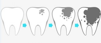
- Acute: progresses quickly, within 10-14 days the pathology goes through almost all stages - from a white spot to dentin destruction.
- Chronic: develops slowly, the enamel is destroyed gradually, turning dark brown. This disease develops in cases where acute caries is not treated, but for some reason the factor that provoked it has been eliminated.
- Multiple: occurs on several teeth or in different places of one tooth.
- Secondary: formed under the filling.
Secondary and multiple caries can occur in acute or chronic form.
Types of caries by localization:
- Cervical. A carious lesion covers a tooth at or under the gum.
- Interdental. The pathological focus is localized on two teeth at the point of their contact. Often such caries occurs on the incisors.
- Fissure. A carious cavity forms on the chewing surface. Usually develops on molars and premolars.
This classification partially overlaps with the types of caries according to Black, but the second is more informative.
The topographic system, which takes into account the depth of tooth damage, involves dividing caries into the following types:
- Spot stage. A small white spot appears on the enamel, indicating demineralization of this area and the beginning of destruction.
- Superficial lesion. Caries has not yet reached the dentin, but damage can already be noticed during an external examination.
- Average degree. Cariogenic microorganisms have destroyed part of the enamel and transferred to hard tissues.
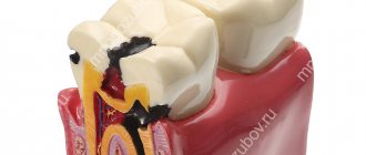
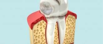
- Deep defeat. Dentin destruction begins. The pulp is protected from the pathological focus only by a thin layer of hard tissue. At this stage there is a risk of developing periodontitis or pulpitis.
If caries has progressed to pulpitis or other dental pathology, it is called complicated.
There are other types of the disease in question, for example, tooth root caries. This is one of the most complex forms of the disease, which occurs due to the formation of plaque or stone near the gums in large quantities. Microbes do not just destroy enamel, they penetrate into the gum pockets and penetrate under the mucous membrane. When treating a pathology, the doctor first cleans out the plaque, and only after that proceeds to the standard filling steps.
Caries of primary teeth stands out separately. They get sick even more often than indigenous children, because children eat a lot of sweets. Despite the fact that such teeth are temporary, they also need to be treated, otherwise there is a risk of the carious process spreading to the root of the new tooth. The treatment regimen is classic.
The essence of the system
Black's classification of carious lesions is a system for dividing the disease into certain classes depending on the location of the destroyed area of hard tissue and the coverage of certain elements of the jaw row.
Despite the fact that this scale was developed more than a hundred years ago and does not include secondary and root caries, its use is widespread in modern dental practice.
Dr. Black identified 5 main classes of caries, to which another degree of development of the disease was later added.
The purpose of creating this classification was to select the optimal method of therapy - selection of a suitable material for filling and a method of preparing the affected surface.
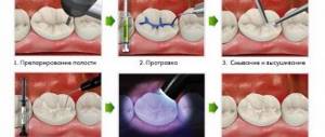
The topography of types of cavities according to Black is well demonstrated in the following diagram:
Diagnostics
Modern methods for diagnosing dental diseases make it possible to accurately determine the type of caries, its degree, causes and other parameters. Often, it is enough for the dentist to conduct an external examination and interview the patient to assess the nature of the pathology. However, in some cases, these methods are not enough to reveal the full picture. The dentist has the following types of research at his disposal:
- Probing. With its help, the depth of the carious cavity and the density of tissues affected by the disease are examined. In addition, the probe allows you to examine the pulp.
- Percussion is a method of tapping on a tooth, helping to determine the presence of cavities inside the tooth, for example, after filling and relapse.
- Vital coloring. Demineralized areas of tooth enamel are stained with dye for 3 minutes. It shows in several shades the degree of distribution of the carious lesion in different areas.
- Thermal test. The aching tooth is irrigated with a stream of cold water to cause a painful reaction. Using this method, the localization of the pathological process is determined.
- Radiography. With the help of this study, hidden carious lesions are identified, their size and depth are assessed.
- Electroodontometry (ED) is a test for the excitability of the pulp, which can react to external stimuli differently depending on the condition: normal, inflammation or necrosis.
- Luminescent method - examination of teeth under an ultraviolet lamp. Carious lesions under UV rays differ in color from healthy tissue.
A doctor can detect and treat tooth decay at any stage. However, depending on the time of detection and the degree of pathology, the treatment process can be quick or lengthy, simple or complex. The cost of the procedure also depends on the complexity of the procedure. Dentists advise coming for an examination every six months, even if there are no signs of the disease, since some forms of caries can be asymptomatic for a long time.