Healthy gums are no less important for any person than strong teeth. Healthy gums have a pale pink color and a clear texture without any bumps or swelling. Various pathological formations in the mouth alarm most people, and rightly so. Any growth on the gum should not be ignored. What exactly such neoplasms in the mouth may indicate, their causes and possible methods of treatment, we will consider below.
What is meant by a growth on the gum?
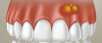
When people talk about a growth on the gum, they usually mean a cyst, which often appears for no apparent reason. If the growth does not hurt when pressed, then it is called epulis (or supragingival). If you open it, a loose mass is released from the growth or liquid contents flow out. Without timely therapy, the formation will sooner or later open up on its own, transforming into a small tumor with a hole on the surface, from which the fistulous tract goes into the thickness of the cyst. From the resulting fistula, ichor or pus is periodically released. At the same time, the patient’s general condition also suffers: he is bothered by headaches, loss of vitality, and the lymph nodes near the source of infection (jaw, neck or ear) are usually enlarged and painful.
What does a growth on the gum mean?
The formation of a cyst on the gum occurs at any age. Fortunately, such a neoplasm is not always a sign of a serious pathology.
Most often, an unpleasant growth on the gum appears from a wound formed after an infection enters the oral cavity.
This process can especially often manifest itself in children, since parents can only dream of a child observing strict hygiene. Most often, growths on the surface of the gums appear in children during teething. At this moment, all the main factors are present that contribute to the penetration of infection into the gum cavities: putting dirty hands or objects into the mouth, reduced immunity and wounds in the gum mucosa. By putting various objects into the mouth, the child thereby tries to relieve pain and itching at the site of tooth eruption by massaging the gums.
Reasons why tooth fragments remain in the gums
Pieces of teeth remain in the following cases:
- Removal of wisdom teeth or multi-rooted teeth of complex configuration. If the tooth is severely damaged or the enamel is thinned, the walls may crack when it is removed. A small piece goes unnoticed by the doctor, and fragments measuring 1-3 mm are often not visible even on a control x-ray. In such cases, everything depends on the professionalism of the dentist - the more experience, the less likely it is that after tooth extraction there are fragments left in the gums.
- Crushing of an adjacent tooth during extraction. Sometimes, during the operation, a nearby tooth may be damaged due to fragile enamel or incorrect actions of the dentist. A small fragment gets into the fresh socket of the extracted tooth, and if it is not removed in time, it becomes a cause of discomfort, pain or suppuration.
- The crown crumbled, and the remains of the tooth were overgrown with gum. This applies to pulpless teeth - a living nerve would not allow the patient to delay a visit to the dentist. Most often this happens when the old filling is destroyed, and caries gradually eats up the crown part, reaching the roots. Remains of a tooth root overgrown with gums become a breeding ground for infection for neighboring units, negatively affecting the gums.
Important!
If you see a dentist in time, you can save on prosthetics or implantation. Today, doctors restore a tooth, even when a minimal amount of tissue from the crown of the tooth remains and the root is untouched by caries. The remainder of the tooth root is more vulnerable to pathogenic bacteria - it lacks integrity, and the microflora penetrates deep into the tooth very quickly. A weak, fragile and carious root cannot serve as a support for an artificial crown and must be removed.
Appearance and types of growth
Externally, the gingival neoplasm looks like it has grown and extended beyond the gum. The growth on the gum most often looks like a small, dense, bright red tumor.
Typically, gingival growth is benign and does not exceed 2-3 mm in size.
It begins with minor inflammation (usually after microtrauma), but then the inflammatory process thickens and increases.
There are three types of epulis: angiomatous, fibrous and giant cell.
Angiomatous formation is more common in children 5-10 years old. This type of cyst is rough to the touch and has a red tint. This growth is soft to the touch and often bleeds when pressed. This epulis can not only grow quickly, but also form again after its removal.
The fibrous growth is identical in color to the surrounding gum. It is usually dense and grows very slowly and insignificantly. If you press on such a formation, it will not cause pain or bleed.
Giant cell epulis is lumpy, elastic, with a red-blue color. It can form both from the gum mucosa and from the alveolar process of the bone. This type of growth can be significant in size, which leads to constant injury and bleeding.
Elimination of a single gum recession in the anterior part of the lower jaw
A. M. Avanesov , Doctor of Medical Sciences, Professor, Deputy Dean of the Faculty of Medicine of RUDN University, Head of the Department of General Dentistry
K. A. Avanesov , general dentist, assistant at the Department of General Dentistry of RUDN University
K. S. Pchelkina student of the Faculty of Medicine of RUDN University
Gum recession is a fairly common pathology that can cause tooth loss, aesthetic defects and functional disorders. The causes of recessions are varied: for example, anatomical features such as crowding of teeth, small vestibule of the oral cavity. Poor hygiene can also contribute to the loss of soft tissues, for example, traumatic teeth brushing techniques, bad habits. Occlusal overloads and defects caused by poor-quality treatment, such as overhanging edges of fillings, crowns, and veneers, cannot be ruled out. Moreover, these factors can occur either individually or in combination, which significantly worsens the clinical situation and requires complex treatment.
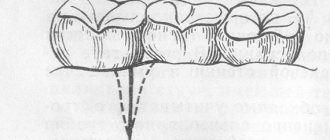
It is with great regret that it is worth noting that this very interesting topic has not received due attention at the dental faculties of Russian medical universities; for this reason, one can often find either ignoring the problem (Fig. 1), or an irrational approach to eliminating gum recession, such as covering the exposed root with a composite material where soft tissue plastic surgery is clearly required.
Rice. 1. Advanced state of the pathological process
Such an irrational “composite” approach ultimately only aggravates the clinical situation by disturbing biological parameters (Fig. 2).
Rice. 2. An example of frequent irrational treatment of recessions with composite
In this case, the overhanging edges of composite fillings compress soft tissues, promoting retention of the microbial plaque, which initiates a local inflammatory process that triggers the mechanism of alveolar bone lysis.
Using the example of our article, we want to demonstrate, especially to young specialists taking their first independent professional steps, that solving this problem is quite within the capabilities of a general dentist, namely general dental practice, such a popular and sought-after specialization, which is developing so vigorously in modern Russian dentistry.
A patient came to the Department of General Dentistry of RUDN University with complaints of exposed gums, pain, bleeding during brushing and eating in the area of the 4.1 tooth (Fig. 3, 4).
Rice. 3. Initial clinical picture
Rice. 4. Initial clinical picture
The clinical diagnosis was made based on the clinical examination: gum recession, Miller class III, in the area of 4.1 teeth.
The clinical study protocol included an instrumental examination, determination of the level of hygiene, photographing the initial clinical situation (a mandatory option for all manipulations performed at the department) using a SLR digital camera equipped with a macro lens and a macro flash, a control targeted X-ray image (Fig. 5), on in which the loss of alveolar bone between teeth 4.1 and 3.1 is clearly visible, which is clearly visible even without a photograph, judging by the characteristic architectonics of the gingival papilla. The lack of bone support significantly aggravates the situation and makes it impossible to eliminate this recession 100%. Despite the presence of local pathological changes, tooth 4.1 does not have pathological mobility and does not have a pathological periodontal pocket; slight bleeding is detected when probing in the area of the causative tooth from the vestibular side.
Rice. 5. X-ray control of the bone defect
A clear deficiency of keratinized attached gingiva is determined. On the lingual side there are minor dense, low-mineralized dental deposits, which predominantly grow towards the incisal edge. The main reason for the recession in this situation is orthodontic problems that have not been corrected in a timely manner in the form of crowding of teeth and excessive vestibular inclination of the 4.1 tooth against the background of a thin gum biotype; it is for this reason that the treatment will be complex and carried out jointly with the orthodontist, after consultation with whom the decision was made before starting orthodontic treatment to surgically eliminate recession.
After analyzing the clinical examination data, we decided to eliminate the recession in two stages. Why was this step-by-step option chosen, rather than a one-time option? The fact is that with the existing biotype of the gum (namely, keratinized attached gum) and the deficiency of soft tissues necessary for plastic surgery, it will be problematic to carry out immediate removal, and this may cause the appearance of unwanted additional defects at nearby donor sites. It is for this reason that at the first surgical stage we decided to increase the volume of attached keratinized gum using a connective tissue gingival graft, which already at the second stage will allow us to safely perform a modified plastic surgery using the sliding flap method, previously proposed by Grupe and Warren in 1956.
All surgical procedures were performed under infiltration anesthesia. A full-thickness connective tissue gum graft was obtained from the donor site of the upper jaw on the left (Fig. 6), in the area of the 2.6 tooth (Fig. 7); while the receiving bed was being prepared, the graft was placed on a gauze pad moistened with saline solution.
Rice. 6. Operation. Donor site
Rice. 7. Full-thickness connective tissue gingival graft.
Pathologically changed tissues from the edge of the recession were excised with a V-shaped incision, with a deviation of 0.5 mm from the edge of the defect, then a subperiosteal tunnel was formed using a rasp, where the graft was placed and fixedly fixed with non-resorbable suture material to the surrounding soft tissues (Fig. 8 ), the middle part of the graft remained open, since it was not possible to completely cover the graft with local tissues due to their deficiency and the risk of possible scarring.
Rice. 8. Connective tissue gum graft is fixed
Solcoseryl adhesive dental paste was applied to the suture line. After the work with the receiving bed was completed, the defect at the donor site in the area of the 2.6 tooth was closed using a collagen bandage “Digestol” produced by OJSC “Zelenaya Dubrava” (Russia). In the postoperative period, the patient was recommended to independently apply the dental adhesive paste “Solcoseryl” to the donor and recipient sites; it was recommended to dissolve 2 tablets of the antiseptic drug “Lizobakt” daily, 4 times a day, for 8 days. The postoperative period was uneventful; the patient complained of minor pain in the donor site, which is typical for this type of traumatic manipulation.
At the control examination on the 10th day after surgery (Fig. 9), the sutures were removed and necrosis of the open middle part of the graft was clearly visible, which, unfortunately, occurs quite often, however, the part of the graft that was placed in the tunnel and, therefore, it was protected and received diffuse nutrition and remained viable. The donor site was actively epithelialized (Fig. 10).
Rice. 9. Control after 10 days, necrosis of the open part of the connective tissue gum graft
Rice. 10. Epithelization of the donor site on the 10th day
After another 10 days, we observed complete engraftment of the graft and its revascularization (Fig. 11, 12).
Rice. 11. Control 20 days after surgery
Rice. 12. Healing of the donor site 20 days after surgery
A follow-up examination after 4 months (Fig. 13) demonstrates that the task set at the first stage has been fully completed.
Rice. 13. Control 4 months after transplantation
Now, having the volume of soft tissue necessary for plastic surgery, we implemented the second stage of the intervention, namely, we performed plastic surgery with a displaced sliding flap according to the classical technique proposed by the above authors. Briefly, with a scalpel No. 15, using a V-shaped incision (marked with green lines), the wound surface necessary for healing was obtained, then, retreating 1 mm downwards from the marginal edge of the gum in the area of 4.2 teeth, an L-shaped incision was made (marked with yellow lines), after After detachment of the complete non-split flap, an additional releasing incision (marked with a blue line) was made in order to move and fix the flap without tension (Fig. 14).
Rice. 14. Schematic stages of flap formation
Note that before the operation, the surface of the root of the 4.1 tooth was treated with EDTA gel to disinfect the root dentin and create additional retention points for fixation of soft tissues. The displaced flap is fixedly fixed with synthetic monofilament suture material, size 6.0, including with the help of wrapping sutures on teeth 4.1 and 3.1, for the best stabilization (Fig. 15).
Rice. 15. The flap is moved and fixed, the open area of bone is visible
A small open area of bone is covered with a biological collagen bandage “Digestol” to prevent necrosis (Fig. 16, 17).
Rice. 16. Biological dressing “Digestol” was applied
Rice. 17. Fixation of the flap, wrapping sutures
The patient is recommended to apply the dental adhesive paste "Solcoseryl" to the postoperative area, as well as take the antiseptic drug "Lizobakt" according to the above regimen. The postoperative period (Fig. 18) was uneventful; the patient, unlike the first stage, did not show any complaints at all.
Rice. 18. Condition 10 days after plastic surgery
After 11 days, the sutures were removed (Fig. 19), complete healing of the surgical wound and epithelization of the open area of the bone in the area of the 4.2 tooth were observed.
Rice. 19. Stitches removed
A follow-up examination after 2 months (Fig. 20) demonstrates that the task of eliminating recession has been completed, the local tissues in the area of 4.1 teeth are formed and have a healthy appearance.
Rice. 20. Follow-up examination after 2 months
The patient was recommended to undergo periodic professional removal of dental plaque (which, of course, was done before surgery as well) at the stages of the upcoming orthodontic treatment.
In conclusion, we emphasize: soft tissue plastic surgery of gingival defects, with proper planning of surgical intervention, allows you to obtain a result that not only pleases the eye, but also protects the adjacent bone tissue. Let us note once again that these manipulations significantly expand the arsenal of professional capabilities of a general dentist.
Causes
Most often, gum growths appear after some kind of trauma to the gums. This can be facilitated by either external or internal factors. Such cases may be:
- insufficient hygiene;
- pathology of the jaw bones;
- malocclusion (extra or protruding teeth, curvatures);
- removal of a tooth;
- unprofessional dental care (poor quality fillings, infection after minor operations);
- injuries or scratches to the gums;
- periodontitis;
- bad habits (smoking or alcohol);
- diseases of internal organs (usually digestive);
- infection after jaw surgery.
Is it possible to grow a tooth if only the root remains?
If the crown is broken and the root remains in the gum, the tooth can be restored. But this is not always realistic, but only under certain circumstances:
- The root is not rotten, not affected by caries.
- The length of the root is twice as long as the crown being restored.
- There are no cysts or granulomas at the root tips.
How to restore a tooth:
- Artificial crowns and inlays. For this method, it is important that the fragment of the tooth wall protrudes above the gum by at least 1 mm. First, an inlay is made from the impression, then a crown. The procedure can take up to two weeks, and the price depends on the materials chosen.
- Fiberglass post with metal-free crown. Fiberglass posts are transparent and look aesthetically pleasing, especially when it comes to teeth in the smile area. It takes from a week to ten days to make a crown, depending on the clinic’s capabilities: if the center has its own technical laboratory, it will take less time.
- Restoration with composites. The advantages of this method include comparative cheapness and short recovery time, but you need to understand that this is more of a temporary solution due to the unreliability and fragility of the materials. On average, a composite restoration lasts 5 years.
Treatment
Any growths in the mouth should be treated by a doctor.
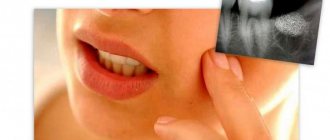
To determine the type of cyst, an x-ray method is used. If necessary, histology of the cyst tissue is also performed.
Dentists best treat cystic formations in the early stages. The most radical and fastest method is to remove the diseased tooth in the area of the damaged gum. This was the only way to treat cysts until recently. Then the cavity was completely sanitized and cleared of infected tissue.
Currently, there is no need to remove the tooth, since modern methods of washing the resulting cavity through the fistula canal with various antiseptic solutions are used. This course of treatment is very long and includes the use of general anti-inflammatory therapy and new generation antibiotics. The cystic cavity is washed until all microorganisms are removed from it. Throughout the course of conservative treatment, a special paste is injected into the root canal and the resulting cavity in the cyst to help restore bone tissue.
What to do if there are tooth remains
- After the blow. If, due to an injury, half a tooth or only the root remains, in most cases, a crown can be built up. It all depends on the condition of the teeth, the complexity of the fracture, and a number of other factors.
- After removal. Fragments of tooth fragments may remain in the socket and come out a couple of days, a week or even a month later. And they can lead to inflammation, fever, and complications. When the hole is fresh, there is a risk of confusing the remainder of the tooth after extraction with the interroot septum and, when trying to remove the fragments, damage the unhealed wound, which will lead to alveolitis. Therefore, if you have any suspicions, you should consult a doctor as soon as possible for advice.
- After complete destruction of the crown due to caries. The best solution is to pull out the remains of the tooth, then perform prosthetics or insert an implant. The longer rotten stumps remain in the gums, the more negative the consequences and the more expensive the treatment.
Is it possible to cure a growth on the gum on your own?
Of course, self-treatment can only be an aid to professional therapy. Traditional methods can only speed up wound healing after surgical or conservative drug treatment. For this, herbal infusions or decoctions are usually used (sage, chamomile, oak bark, St. John's wort, calendula, tricolor violet).
To prevent infection of a postoperative wound, soda solutions are often used, and in case of tissue swelling, a sea salt solution is used (1 tablespoon per glass of water).
Traditional healers also use ointments based on medicinal herbs (yarrow, sweet clover, calendula, tansy, sorrel, dandelion root) on the affected areas of the gums. The herbal components of the ointment are scrolled through a meat grinder and mixed with calendula oil and ichthyol ointment or Vishnevsky ointment.
When is tooth root removal required?
It is not always necessary to remove the remaining fragments after extraction: small fragments of 1-3 mm can painlessly come out on their own after a while. But if you feel sharp edges with your tongue, be sure to visit a doctor for a consultation, even if there is no pain or swelling of the soft tissues. The doctor will quickly remove the tooth fragment from the gum after removal and use an x-ray to check whether there is still a piece.
The remainder of the rotting tooth root in the gum must be removed when the crown has already crumbled due to carious lesions. Such fragments have a porous structure, on which bacterial plaque and tartar quickly form. In most cases, cysts and granulomas appear at the top of a rotten stump, leading to even greater problems with the gums.
Important!
Complex tooth extraction is often accompanied by fracture of the root apex. The splinter provokes increased bleeding, which obscures the dentist’s view. In such cases, the procedure develops according to two possible scenarios. The doctor can continue the extraction, relying on his many years of experience, or schedule a second appointment, telling the patient in detail what to do at home before the next removal. When the dentist suggests waiting until the tooth pieces come out on their own, this indicates inexperience. If a fragment remains after a tooth has been pulled out, it can cause various complications: from alveolitis to purulent inflammation.
Complications
Why can’t you treat a gum cyst yourself? Yes, because the infection in this case has already entered deep into the jaw tissue, where it is simply impossible to destroy the microbes yourself. As the disease progresses, putrefactive bacteria penetrate the dental pulp, from where they enter the bone tissue through the root canals.
Further, the process can provoke the development of a very serious and difficult to treat disease - osteomyelitis. In this case, the patient complains of severe weakness, high temperature, and enlarged lymph nodes. Osteomyelitis is especially common as a complication of gingival growth in children.
In addition, infection from the cystic cavity can spread throughout the body. After all, during inflammation, the body sends an increased flow of blood to the affected area. Dead blood lymphocytes settle in the gum cavity in the form of purulent secretion, the excess of which comes out through the fistula canal.
It is sepsis (blood poisoning) that is the most life-threatening complication of a growth on the gum.
It is important to treat any oral diseases immediately after they occur. It is in the oral cavity that the blood flow is so strong that any inflammation can instantly develop and spread through the bloodstream throughout the patient’s body.
Given the close location of the brain, a purulent process from the oral cavity can spread to this organ and lead to irreparable consequences.
Why does the gum move away from the tooth?
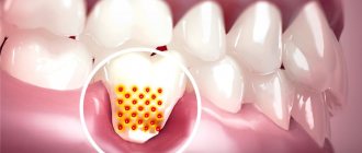
A prerequisite for the destruction of the periodontal attachment is the presence around the necks of the teeth - soft microbial plaque or supragingival tartar. The formation of dental plaque occurs due to irregular oral hygiene. In some patients with periodontitis, tartar may not be visually visible, because... when the attachment of the gum to the tooth is destroyed, it can already be localized below the level of the gum (then it is called “subgingival”).
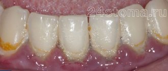
But usually the destruction of the attachment of the gums to the teeth is always preceded by a long period of chronic inflammation of the gums, during which the patient is only bothered by periodic bleeding of the gums, possibly bad breath, redness or cyanosis of the gingival margin, as well as swelling of the gingival papillae. These symptoms are characteristic of gingivitis, an inflammatory disease of the gums in which the attachment of the gum to the tooth is not yet broken.
The lack of professional treatment for gingivitis (usually patients during this period self-medicate using antiseptic rinses and toothpastes for bleeding gums) leads to destruction of the attachment of the gums to the teeth. As a result, so-called periodontal pockets (gingival pockets) are formed between the teeth and gums, and patients in this case may complain that their gums have moved away from their teeth.
Stages of gum inflammation (gingivitis and periodontitis) –
Conclusions: Figure 10 shows the differences between gingivitis and periodontitis, and that with periodontitis, the gums are not attached to the teeth to the full depth of the periodontal pockets (from 3 to 6 mm or more). Therefore, if your gum has moved away from a tooth, there is simply no point in delaying a visit to the dentist, unless, of course, your goal is to remove these teeth in the near future. And in this case, there is no method of self-medication (magic rinses or ointments for gums) that would allow you to save the situation without going to the dentist.
In the absence of professional treatment of gums by a dentist, such teeth will first become mobile, then they will begin to fan-shaped and shift in different directions (under the forces of chewing pressure). In Fig. 11-13 you can see advanced forms of periodontitis in patients who, unfortunately, turned to the dentist too late. The teeth in these photographs have a very high degree of mobility and many of them only need to be removed.
Consequences of lack of treatment -
Prevention
Prevention of the development of gingival cysts includes measures for the timely treatment of various dental problems (bad bite, untreated caries, poorly made filling, etc.).
It is also important to use the following oral care rules:
- mandatory daily brushing of teeth twice (morning and evening) with additional use of dental floss;
- rinsing the mouth after any meal (it is possible to use salt or ready-made pharmaceutical rinses, herbal decoctions, chewing gum);
- timely elimination of discomfort when wearing braces or removable dentures to prevent gum trauma;
- preventive visit to the dentist once a year to identify and treat any pathologies of the teeth and oral cavity.
Any growths that appear in the mouth should not be taken lightly. If there are growths, a person experiences discomfort and pain in the mouth, and it is difficult for him to eat normally. It is very important not to neglect the disease, so as not to get a lot of problems and troubles. Indeed, in addition to constant pain, the body is constantly poisoned by a huge number of harmful microorganisms, which leads to the development of various foci of inflammation in the body. The common expression “it will go away on its own” is completely inappropriate in this case. Any growth in the mouth should be promptly diagnosed and treated. Otherwise, serious problems cannot be avoided. Do not try to cope with tumors in the mouth yourself, as in this case self-medication may not be effective. Take care of yourself and your health!
Sources used:
- Arithmetic of periodontology. Hand tools / T.M. Elovikova. — M.: MEDpress-inform
- Stoeken, Judith E.; Paraskevas, Spiros; Van Der Weijden, Godefridus A. The Long-Term Effect of a Mouthrinse Containing Essential Oils on Dental Plaque and Gingivitis: A Systematic Review (English) // Journal of Periodontology
- Workshop on therapeutic dentistry / Ya.I. Gutner. — M.: State Publishing House of Medical Literature
How to remove tooth debris
Most often, patients ignore visiting the dentist because of the common fear of pain during extraction: the memories are still too fresh of how in the 90s, in many clinics, teeth were mercilessly pulled out without pain relief. Modern dentistry has good anesthetics that make the procedure painless. The most unpleasant sensations are an injection into the gums when frozen and a possible aching pain after the drug wears off.
There are several methods for removing residue. The choice of technology depends on where the tooth is located and the reason why the fragment appeared:
- Forceps. The doctor separates the circular ligament and soft tissue before applying forceps, tightly grasping the tooth pieces with his cheeks and removing them from the gums. Instruments may differ in shape and structure, depending on which teeth and on which jaw need to be removed. For example, straight forceps are used for incisors and canines, bayonet-shaped forceps are used for molars, and premolars are removed with S-shaped forceps. The teeth of the lower jaw are the easiest to remove: teeth with short roots and thin socket walls most often do not cause complications after extraction.
- Elevator. If the root is deep in the hole, then the forceps give way to elevators. With their help, the dentist expands the space between the root part of the tooth and the socket, while simultaneously turning the instrument along the longitudinal axis. The concave part of the cheek is used to remove the fragment that remains after tooth extraction.
- Surgical removal. When the remains cannot be removed with instruments, the surgeon performs an operation. After an injection of anesthesia, the mucous membrane is incised, the flap is peeled off and trephination or sawing out of part of the tooth with a small bur is carried out. When all parts are removed, the flap is sutured into place.