To talk about local inflammation, you must first understand what lymphadenitis is in general. This is an inflammatory process in the lymph nodes, sometimes accompanied by suppuration. It manifests itself through enlargement of one or more lymph nodes and can occur in several regions of the body at once. Clinical signs of the disease depend on the form and type of lymphadenitis: acute or chronic, regional (damage to lymph nodes of one anatomical group) and generalized (involvement of several groups of lymph nodes), serous, purulent, gangrenous, hemorrhagic, phlegmonous.
In the body, lymph nodes create a protective barrier against viruses, infections, cancer cells, participate in the formation of lymphocytes (special cells that destroy foreign microorganisms) and the production of phagocytes, antibodies (immune cells), participate in digestion and metabolic processes, distribute intercellular fluid between body tissues and lymph.
Normally, the lymph nodes are not palpable or are palpated in the form of elastic, small-sized formations that are not fused with adjacent tissues or with each other. Depending on the prevalence, the following types of lymphadenitis should be distinguished:
- local - enlargement of one lymph node in one of the areas (single cervical, supraclavicular lymph nodes);
- regional - enlargement of several lymph nodes in one or two adjacent areas (supraclavicular and axillary, supraclavicular and cervical, occipital and submandibular lymph nodes, etc.);
- generalized - enlargement of lymph nodes in three or more areas (cervical, supraclavicular, axillary, inguinal, etc.).
With axillary lymphadenitis, enlarged lymph nodes in the armpit are palpated. Symptoms of axillary lymphadenitis:
- Enlarged lymph nodes
- Painful sensations when palpating the affected area, turning the body, or moving
- Inflamed nodes are fused together with each other and adjacent tissues into one dense conglomerate
- The skin over the inflamed lymph nodes is hyperemic
- Symptoms of body intoxication appear - fever, weakness, headache, lack of appetite, feeling weak, muscle pain
Often the disease is accompanied by symptoms of the underlying disease; in addition, the patient may be bothered by:
- night sweats;
- weight loss;
- prolonged increase in body temperature;
- frequent recurrent upper respiratory tract infections;
- pathological changes on a chest x-ray;
- hepatomegaly;
- splenomegaly.
Treatment of arm lymphedema at the Innovative Vascular Center
The innovative vascular center has been developing the direction of conservative and surgical treatment of arm lymphostasis for more than 10 years, relying on the German experience of conservative treatment used by Dr. Shingale and microsurgical technology of lymphovenous anastomoses and lymph node transplantation.
We provide a full-fledged treatment and rehabilitation complex, which allows you to reduce swelling by 70-100% and control it. The course of treatment is 14 - 28 days.
Surgical treatment is used for stage 2-3 lymphedema of the arm and requires a preliminary course of conservative therapy. The experience of our clinic shows that the manifestations and progression of lymphedema can be significantly reduced and even reversed.
Specific lymphadenitis
The specific group includes L. caused by pathogens of actinomycosis, syphilis, tuberculosis, tularemia, plague, etc. For the clinical picture, diagnosis and treatment of the main types of specific L., see the articles Actinomycosis, Syphilis, Tularemia, Plague.
Tuberculous lymphadenitis
Tuberculosis lymph nodes is a manifestation of tuberculosis as a general disease of the body (see Tuberculosis). More often, especially in childhood, the period of primary tuberculosis is combined with damage to the intrathoracic lymph nodes (see Bronhadenitis). Relatively isolated damage to certain groups of lymph nodes and nodes is possible, more often in adults, against the background of old inactive tuberculous changes in other organs, when tuberculous L. is a manifestation of secondary tuberculosis. The frequency of tuberculous L. depends on the severity and prevalence of tuberculosis and social conditions. Among children, tuberculous lesions of peripheral lymph nodes, according to E. I. Guseva (1973), P. S. Murashkin (1974), etc., are observed in 11.9-22.7% of patients with active forms of extrapulmonary tuberculosis.
Tuberculosis of peripheral lymph nodes is caused mainly by Mycobacterium tuberculosis of the human and bovine type. Mycobacteria of the bovine type are usually the causative agent of tuberculous lymphadenitis in agriculture. cattle breeding districts.
The ways of spreading the infection are different. Submitted by B. P. Aleksandrovsky et al. (1936), A. I. Abrikosov (1941), F. L. Elinson (1965), V. A. Firsova (1972), Kurilsky (R. Kourilsky, 1952), etc., the tonsils can be the entrance gate of infection, when affected, the cervical or submandibular lymph nodes are involved in the process. nodes. The infection most often spreads by lymphohematogenous route from affected intrathoracic lymph nodes, lungs or other organs.
Pathomorphol, changes in the affected nodes depend on the severity of the infection, the condition of the patient’s body, the type of Mycobacterium tuberculosis and other factors. A. I. Abrikosov identifies five forms of tuberculous lesions of lymph nodes: 1) diffuse lymphoid hyperplasia; 2) miliary tuberculosis; 3) tuberculous large cell hyperplasia; 4) caseous tuberculosis; 5) indurative tuberculosis. In wedge, practice, the classification proposed by N. A. Shmelev is used, in which three forms of tuberculous L. are distinguished: infiltrative, caseous (with and without fistulas) and indurative.
With the acute onset of the disease, there is a high temperature, symptoms of tuberculosis intoxication, enlarged lymph nodes, often with pronounced inflammatory-necrotic changes and perifocal infiltration.
A characteristic sign of tuberculous L., distinguishing it from other lesions of lymph nodes, is the presence of periadenitis. Affected lymph nodes represent a conglomerate of formations of various sizes welded together. In adults, more often than in children, the onset of the disease is gradual, with less enlargement of lymph nodes and less frequent formation of fistulas due to the predominantly productive nature of inflammation.
A number of researchers associate the acute onset of the disease and the tendency to rapid formation of caseosis and fistulas with infection with the bovine type of mycobacterium tuberculosis.
The most often affected are the cervical, submandibular (Submandibular, T.) and axillary lymph nodes. The process may involve several groups of lymph nodes on one or both sides.
X-ray of the soft tissues of the neck: areas of calcification in the submandibular and cervical lymph nodes (indicated by arrows).
Diagnosis
placed on the basis of a comprehensive examination of the patient, taking into account the presence of contact with tuberculosis patients, the results of the reaction to tuberculin (in most cases it is pronounced), the presence of tuberculosis damage to the lungs and other organs. These punctures of the affected lymph play an important role in making a diagnosis. node. In the lymph nodes, calcilates can form, which are detected radiographically in the form of dense shadows in the soft tissues of the neck (Fig.), submandibular region (submandibular triangle, T.), axillary and inguinal regions. Tuberculous L. is differentiated from nonspecific purulent L., lymphogranulomatosis, metastases of malignant tumors, etc.
Treatment
determined by the nature of the damage to the lymph nodes and the severity of tuberculous changes in other organs. With an active process, first-line drugs are prescribed: tubazide, streptomycin in combination with PAS or ethionamide, prothionamide, pyrazinamide, ethambutol. Treatment should be long-term - 8-12-15 months.
In addition, streptomycin is injected (or injected) into the affected node, and bandages with streptomycin, tubazid, tibon ointment are applied. In case of severe purulent process, broad-spectrum antibiotics are prescribed. In case of caseous lesions of lymph nodes, surgical intervention is indicated against the background of a general course of anti-tuberculosis therapy (see Tuberculosis).
The prognosis for timely recognition of the disease and treatment of L. is favorable.
Prevention of tuberculous L.—see Tuberculosis.
Causes and risk factors
A network of lymphatic vessels collects lymph fluid from the body's tissues, like veins that collect blood, and carries the fluid to the lymph nodes, small clusters that act as filters and contain white blood cells that help us fight infection. Lymph nodes are removed during surgery for breast cancer. The remaining lymphatic vessels and nodes cannot sufficiently compensate for lymphatic drainage, so excess fluid accumulates and causes lymphedema.
Lymphedema usually develops slowly and becomes noticeable over time, but it can occur at any time after surgery. Most women who have a mastectomy discover swelling several months and sometimes years after surgery.
Women who have been treated for breast cancer, follow all doctor's recommendations, and follow all preventive measures are less likely to develop lymphedema. With proper care and treatment, the affected limb can be restored to its normal size and shape.
Cervical lymphadenitis - symptoms and treatment
Elimination of the primary source of infection
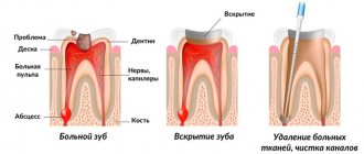
Cervical lymphadenitis is often caused by acute or aggravated periodontitis and complications of advanced caries, such as acute purulent periostitis.
If the tooth can be saved, the root canals are cleaned and filled. If it is impossible to restore a tooth, it is removed. When a purulent focus has formed, the diseased tooth is treated or removed, and the abscess is opened. If cervical lymphadenitis has developed due to a disease of the ENT organs, the source of acute inflammation should also be eliminated.
Drug therapy
- Antibacterial therapy. Broad-spectrum antibiotics are usually used, mainly with a bactericidal effect. The components of such drugs destroy the cell wall of the bacterium or disrupt its metabolic processes, which leads to the death of the microbe. If the patient's condition does not improve, biological material obtained from the lymph node is examined and the sensitivity of microorganisms to drugs is determined.
- Antiviral drugs are used for viral origin of lymphadenitis, for example, herpes.
- Anti-inflammatory drugs suppress inflammation at the cellular level, reduce pain and reduce fever.
- Antihistamines reduce capillary permeability, which prevents the development of edema and congestive processes. They also prevent leukocytes from penetrating into the lesion and inhibit the production of substances that contribute to the development of inflammation.
Physiotherapeutic treatment
- UHF (ultra-high frequency therapy) is aimed at reducing swelling, inflammation and pain.
- Ultrasound is used to speed up the resolution of the inflammatory process.
- UVR (ultraviolet irradiation) is indicated to reduce inflammation.
- Laser therapy is aimed at reducing pain, improving nutrition and blood supply to the affected area.
- Electrophoresis is a method in which a medicinal substance penetrates tissue using a direct electric current. For lymphadenitis, electrophoresis with potassium iodide and proteolytic enzymes is usually performed.
- Magnetic therapy is aimed at reducing pain, inflammation, swelling and congestion in tissues.
Physiotherapeutic methods are used in Russia to reduce the duration of drug treatment, but there is insufficient scientific evidence of their effectiveness.
Surgical intervention
Opening a purulent focus is indicated for purulent forms of lymphadenitis and adenophlegmon. Depending on the size of the lesion, the operation is performed under local or general anesthesia. During surgery, the purulent contents and tissue of the disintegrated lymph node are removed.
After surgical treatment, a drainage is placed in the wound, which ensures the drainage of pus and prevents the edges of the wound from healing. Then the wound is treated, its edges are renewed and sutured.
Detoxification therapy
Reduces the level of toxins in the body by diluting them, absorbing breakdown products and increasing diuresis. To do this, drink more fluid, and in severe cases, Hemodez and Reogluman are administered intravenously.
Diet
It is recommended to eat a balanced diet and consume enough vitamins, macro- and microelements.
Features of treatment of lymphadenitis
Treatment of cervical lymphadenitis directly depends on the stage and form of the disease.
In acute serous lymphadenitis, special attention is paid to the primary source of inflammation: inflammatory diseases of the teeth, oral cavity and ENT organs. If the primary inflammatory process is stopped in the early stages, then the symptoms of acute serous lymphadenitis also become less pronounced.
In almost 98% of cases with acute lymphadenitis, it is possible to identify the primary lesion [10]. It is eliminated and antibacterial, antiviral, anti-inflammatory or antihistamine therapy is prescribed.
When a purulent form develops, the primary focus is eliminated, the abscess is opened and the tissue of the disintegrated lymph node is removed. The patient is usually kept in the hospital under 24-hour observation. Daily dressings are performed, antibacterial, anti-inflammatory, antihistamine and detoxification therapy is prescribed.
In case of chronic hyperplastic lymphadenitis, the affected lymph node is removed, and treatment is also carried out in the hospital. Tissue fragments are sent to the laboratory, processed and examined under a microscope. This procedure allows you to exclude cancer and prevent its development.
Types of Postoperative Lymphedema
- Temporary reversible lymphedema: Occurs within a few days after surgery and usually lasts a short period of time. goes away quickly enough under the compression sleeve.
- Subacute lymphostasis of the arm: appears 4-6 weeks after surgery. It is characterized by pain and high tissue density, but goes away quite quickly under the influence of compression therapy.
- Chronic arm lymphedema: This painless swelling usually appears 18-24 months after surgery.
Stages of the disease
- Stage I: Spontaneously reversible. The swelling is quite noticeable; if you press on the skin with your finger, a hole remains. After rest (especially in the morning) it subsides, but in the evening it becomes the same. Patients rarely turn to specialists at this stage.
- Stage II: Spontaneously irreversible. The skin hardens (this is due to the growth of connective tissue), the swelling is no longer so soft and when pressing with a finger on the skin there is no hole left. The skin is very tight and sensitive. The patient may experience pain.
- Stage III: Irreversible. The skin is fibrous (the tissue scars), cysts or papillomas appear on it. The limb is deformed due to tissue damage. She moves poorly or even becomes motionless and becomes heavy. This is the extreme stage of lymphedema - elephantiasis.
Treatment of axillary lymphadenitis
Treatment tactics for axillary lymphadenitis depend on the form and cause of the disease. In most cases, lymphadenitis does not require special therapy and goes away on its own after the underlying disease is eliminated. If the process is complicated by suppuration, then the treatment tactics completely change.
General recommendations: maintain rest, immobilize the affected area (do not rub or injure the affected lymph nodes), proper nutrition, take painkillers, anti-inflammatory medications. Therapy for acute axillary lymphadenitis depends entirely on the stage of the disease. Conservative treatment is used for the initial stages: UHF therapy, sanitation of the source of infection (opening of abscesses, leaks, cellulitis, drainage of the abscess), antibiotic therapy taking into account the sensitivity of the microbial flora in the main focus.
Surgical intervention is necessary for purulent axillary lymphadenitis: adenophlegmon, abscesses are opened, pus is removed, and the wound is drained. It happens that a biopsy confirms the presence of a tumor process - benign or malignant. Treatment may include radiation and chemotherapy. In the case of lymphadenitis, as in the presence of any other diseases, it is extremely dangerous to self-medicate.
Prevention and control
Although it is impossible to predict how the disease will progress, there are steps patients can take to reduce the risk. If you already have lymphedema, it can be controlled by following some of the recommendations below:
- Perform daily stretching exercises to maintain your range of motion.
- Learn to perform lymphatic drainage self-massage.
- Lymph production is directly proportional to blood flow, so strenuous arm exercises that increase blood flow can increase the amount of lymph and therefore increase the risk of lymphedema.
- Avoid drinking alcohol.
- Avoid foods high in salt and fat. Try to eat a healthy diet that is high in fiber.
- Wear comfortable clothes and jewelry that do not constrict your hand.
- Burns can increase the risk of lymphedema, so avoid excessively hot water when bathing or washing dishes. Always use sunscreen.
- When you are sleeping or sitting, try to keep your arm elevated (for example, by placing it on a pillow).
- Take care of your skin to avoid infections. Wear gloves when doing housework or yard work. Apply moisturizer to chapped skin and use insect repellent to prevent bites.
- Watch your weight.
Your doctor may recommend a compression sleeve as a precaution. You should never buy a sleeve online or in a medical supply store and use it yourself without a doctor's advice. Choosing the right compression stocking ensures that the sleeve will perform as it should. Otherwise, the sleeve may be too tight in certain areas, which can restrict lymph flow and make the situation worse.
