Types of tooth dislocations
Depending on the degree of damage, there are three types of tooth dislocations:
- incomplete;
- full;
- hammered.
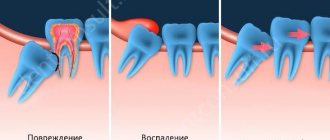
Incomplete dislocation is characterized by partial displacement of the dental crown. In this case, it can be moved forward, backward, or rotated along its axis. In this case, the tooth itself remains in the socket and its loss is not observed. A characteristic sign is that the crown wobbles with more or less force. Since the damaged tooth begins to stand out from the general dentition, this can prevent a person from closing his jaws, chewing food normally, or speaking. In some cases, the patient cannot do this at all.
Due to damage to the crowns of the teeth, pulp or nerve, soft tissues may also be damaged. Often the gums become inflamed. It swells and may bleed. All this is accompanied by severe pain.
Complete dislocation in most cases is accompanied by the tooth falling out of the socket. It can also be held on to nerves or pieces of soft tissue. A bleeding wound will form at the site of the dental crown, which may be open and require stitches. The gums may change color and become bluish. There is severe pain. Such a dislocation may be accompanied by a painful shock.
Important! Complete luxation is most often observed in children, since their baby teeth are less strong and are not held so firmly in the socket.
Impacted dislocation is characterized by complete or partial immersion of the tooth crowns into the gum tissue. At the same time, it swells and darkens, which is accompanied by severe pain. Due to deformation, this type of dislocation may be accompanied by root destruction or rupture of blood vessels.
If impacted luxation is observed in children, such immersion of a baby tooth can negatively affect the normal formation of permanent teeth.
Mechanism of injury
When a tooth is exposed to mechanical factors (an impact during a fall, a game, an accident; biting off hard food with a moving tooth; opening bottles with teeth), the tooth in the socket is displaced in any direction. At the same time, the tooth root shifts, and a partial rupture of periodontal fibers occurs; The fibers that have maintained their integrity are stretched.
The directions in which the tooth moves are different: towards the adjacent tooth, back or forward along the dentition, towards the occlusal plane; the tooth can turn around its axis. The direction of the displacement is influenced by the location of the impact, its strength and vector.
The main causes of tooth dislocation
The main reasons that can cause tooth dislocation of one type or another are often associated with mechanical damage. These can be injuries - local or general: most often blows that fall on the jaw area. However, the force of the impact does not always matter. A dislocation can also result from a fall onto a hard surface, such as from a bicycle.
Another common cause is the fact that solid food particles get on the crowns of the teeth during chewing. Often these are bone fragments from meat or berry seeds.
Dislocation can also occur if a person often opens bottles with metal caps with his teeth, chews nut shells, etc.
Causes of pathology
Tooth dislocation is understood as injury to the ligamentous apparatus, connective tissue (periodontal tissue) and bone tissue, due to which the root is displaced in the socket, i.e. changes its position. This problem is faced both at a young and adult age. According to statistics, injuries mainly occur on the front teeth; the pathology affects the incisors and canines of the upper jaw, but the lateral teeth, both the upper and lower jaws, can also be affected.
Let's consider the possible causes of dislocation:
- mechanical damage to the facial area: for example, a blow from a fall, a fight, an accident. An injury of this nature can also occur as a result of playing dangerous sports, for example, boxing,
- eating too hard foods: most often in this case, injury occurs as a result of an excessively hard particle getting on the tooth. For example, chewing meat requires a significant mechanical impact, but there may very well be a piece of bone in the meat that a person is not aware of. Therefore, with a strong closure of the jaws, this fragment can lead to damage and chips, and if the hard piece is large, then to dislocations,
- bad habits: if you like to neglect generally accepted rules of behavior and sometimes “dabble” in unconventional ways of opening bottles, for example, using your teeth for these purposes, then you can soon get such an unpleasant phenomenon as a dislocation,
- mistakes made by the dentist when removing a tooth, which lead to injury to its “neighbor”.
It is worth noting that with strong immunity, good health, in the absence of dental diseases and inflammation of periodontal and periodontal tissues, the tooth sits very firmly in its bed, and the likelihood of getting dislocated while eating, even with strong mechanical impact, is reduced to zero. But if the surrounding tissues have lost their strength and elasticity as a result of weakening of the protective functions and inflammation of the ligamentous apparatus, then the likelihood of “breakage” and dislocation increases significantly.
Consequences of dislocation
The dislocation of a tooth itself is very unpleasant, as it leads to the appearance of pain. However, its consequences can be much more dangerous. In some cases, when a dislocation occurs, not only the tooth tissue is damaged, but also cracks appear in the jaw. If they were not diagnosed on time, this may lead to subsequent inflammatory processes.
Since complete luxation causes significant damage to the gum tissue, all gaps must be repaired. If this is not done, you can get a severe infection. The same applies to the tissues of the inner surface of the cheek, which may be damaged due to improper placement of teeth after dislocation.
The cause of dislocation can also be the unprofessional actions of a dental surgeon during certain manipulations with teeth. For example, when removing a diseased tooth, the doctor can hook (press, move or partially pull out) and provoke dislocation of an adjacent tooth, which is absolutely healthy.
Important! If we are talking about the dislocation of a baby tooth in children, you should be especially careful about its consequences. It must be remembered that any deformation can lead to destruction of the dental bottom. This can cause damage to the molar or negatively affect its growth.
Diagnostic measures
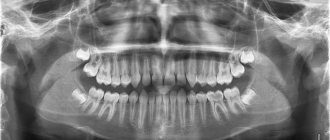
Naturally, it is not particularly difficult for a specialist to recognize the nature of the injury even with a visual examination, since the signs are quite pronounced. But for a more detailed study of the nature and form of the pathology, the dentist will prescribe an x-ray, orthopantomogram or computed tomography of the jaw. The patient may also be prescribed electrodiagnostics, the results of which will make it clear what condition the pulp, bone tissue and periodontal tissue are in. The diagnostic data obtained will determine the upcoming treatment.
Important! A dislocation requires immediate medical attention! If the pathology is neglected (especially in the case of deep trauma), dangerous complications can develop, one of which is the death (necrosis) of pulp tissue. This irreversible pathological process occurs in approximately 52% of diagnosed cases of dislocation2.
Clinical manifestations of tooth dislocation
The main clinical symptoms that accompany a dislocation are:
- bleeding and severe pain in the area of the affected tooth;
- numbness of the area of injury or the entire jaw;
- swelling or swelling of the gums and entire cheek;
- increased temperature due to inflammation;
- dizziness or even loss of consciousness, which can result from painful shock.
If one or several unpleasant symptoms appear, you should immediately consult a doctor.
Diagnostics
An experienced doctor can detect a tooth dislocation with a simple examination. At the same time, he pays attention to the location of the dental crown and the characteristics of the damaged gum. However, there are cases when damage and deformation of the teeth may not be noticeable externally and, until subsequent symptoms appear, the dislocation may go unnoticed. This, in turn, will complicate further treatment.
To avoid such situations, it is important to additionally take x-rays of the teeth and jaw during the examination. It will not only confirm or refute the presence of a dislocation, but will also identify possible damage to the jaw or internal tooth tissues. This approach will allow you to choose the most effective treatment and make it possible to save the tooth itself.
Anomalies of teeth position
In case of anomalies in the position of the teeth, the task of the orthodontist is to preliminary normalize the shape and size of the dentition and occlusion. For this purpose, various orthodontic structures are used - both removable and non-removable. In the distal position, the teeth are moved mesially if there is space in the dentition. The need for mesial tooth movement arises when the first molar is removed (for therapeutic indications), and in this case the second molar moves mesially.
Since this anomaly relates to the lateral teeth, in devices of any design the fulcrum is formed in the anterior or lateral part of the corresponding side, and the point of application of force is the tooth being moved. If a rubber rod is used to move a tooth in an inclined distal position, the point of application of force is the coronal part of the tooth; in the case of a corpus tooth, it is the coronal and root parts, for which a rod with a hook is used in the area of the transitional fold.
In plate devices and mouth guard plastic structures, the fulcrum is hooks welded into the base. In metal structures, hooks are also soldered in the front section on the corresponding structural elements. Primary and permanent teeth at the appropriate stage of formation can be moved in the mesial direction using hand-shaped springs (according to Kalvelis). Permanent teeth in the final stage of root formation are moved using a brace system both obliquely-rotational and corpusally. To move lateral teeth in the mesial direction, the use of a positioner is ineffective.
Treatment of mesial position of teeth is carried out individually. With early removal of the second primary molar or primary edentia of the second premolar of the upper jaw, mesial movement of the first molar is observed. In this regard, the closure of one pair of antagonist teeth is disrupted, namely the mesial-buccal cusp of the first molar of the upper jaw is located in front of the intercuspal fissure of the first molar of the lower jaw. In this case, it is possible to maintain the mesial position of the first molar and then it is advisable to move the second molar forward.
If the doctor decides to move the first molar in the distal direction in order to achieve good closure with the antagonist teeth, you can use a plate on the upper jaw with a sectoral cut, a Kalamkarov apparatus, or an Angle arch. The use of a facebow with cervical traction is especially effective. For the first molars, rings with facebow tubes are made. On the side of the distally moved first molar, a bend is made on the arch, which rests against the tube, and on the opposite side, the end of the arch does not have a stop and is freely located in the tube. In the anterior section, the facial bow is separated from the front teeth. When applying a cervical traction, the entire force of the facebow is directed toward the first molar, which should be moved distally. To move both first molars distally, the facebow has stops in front of the tubes on both sides and both teeth will move distally.
After moving the first molars in the distal direction, the integrity of the dentition is restored at the level of the second premolar using only prosthetics or with preliminary implantation. In the clinic, the mesial position of the lateral teeth is often encountered. This may be due to early removal of the primary canine, high position of the permanent canine bud, the presence of a supernumerary tooth bud, macrodentia of the lateral teeth, a change in the order of eruption of the canine and second premolar (the second premolar erupts first). In this case, the type of closure of the lateral teeth corresponds to Angle class II. To create space for the canine, the lateral teeth must be moved distally. For this you can use plate devices.
Devices 1 and 2 allow you to move the lateral group of teeth on both sides in the distal direction. In this case, the front teeth are moved in the labial direction. Plate apparatus 3 (a plate for the upper jaw with a sectoral cut) moves the lateral teeth in the distal direction, and apparatus 4 allows, using a vestibular arch with an M-shaped bend, to move the canine in the same direction (the end of the arch is welded into the distal part of the cut). Apparatuses 5 and 7 move molars in the distal direction, and apparatus 6 moves one molar.
The main problem encountered when moving a canine distally is its initial position. The choice of orthodontic apparatus and the direction of the acting force depend on the position of the crown and root parts of the tooth.
Treatment of lateral position of teeth . The most typical clinical sign of such an anomaly is the appearance of a gap between the central incisors - diastema .
The following types of diastema are distinguished: 1) symmetrical diastema, in which there is a lateral displacement of the central incisors; 2) diastema with preferential movement of the crowns of the central teeth in the lateral direction from the midline. The roots of the central incisors retain their position or move slightly in the lateral direction; 3) diastema, in which the crowns of the central teeth have shifted slightly in the lateral direction from the midline, and the roots of the central incisors have shifted significantly; 4) asymmetrical diastema, which occurs when one central incisor has moved significantly in the lateral direction, while the other central incisor has maintained its normal position.
It should be noted that the lateral displacement of the central incisors can be combined with their rotation along the axis of the tooth (tortoanomaly) and vertical displacement of the teeth (dental alveolar lengthening or shortening).
Treatment depends on the clinical picture and causes of the anomaly. If there is a supernumerary tooth germ between the roots of the central incisors, it should be removed. In case of microdentia of the central incisors, the diastema is eliminated only by prosthetics of the central incisors with solid cast or metal-ceramic structures. Such prosthetics are performed in adolescents after 14-15 years of age. In case of diastema caused by microdentia of the lateral incisors, the diastema should be eliminated, and then prosthetics of the lateral incisors should be performed with artificial crowns.
If the upper jaw develops excessively in the anterior region and a diastema occurs, one should try to delay the growth of the upper jaw using a plate with a loop for the treatment of diastema and a vestibular arch. At the same time, the loop and U-shaped bends of the vestibular arch are activated. The canine is removed and installed in place of the missing lateral incisor or moved distally. In the first option, this can be done when the root of the canine is located significantly ahead of its proper place in case of normal eruption. If the mesiodistal size of the canine allows filling the gap formed behind the central incisor, then the cusp of the canine crown can be ground down and given the shape of a lateral incisor. Moving the canine mesially is only possible if the antagonist teeth allow the canine to create normal occlusion with them; otherwise, contact with antagonist teeth (regardless of retention) will result in lateral movement of the canine.
When distal movement of the canine occurs, the gap formed in the area of the missing lateral incisor is eliminated by prosthetics. To do this, you can make a metal-ceramic structure with support on the canine and select the central incisor as the second point of support by making a cusp located on the palatal surface
If the diastema has developed due to the low attachment of the frenulum of the upper lip, they resort to plastic surgery of the low-attached frenulum. Surgical treatment should begin after the eruption of not only the central incisors, but also the lateral ones, i.e. at the age of 8-9 years. There are cases when, after the eruption of the lateral incisors, the diastema disappears on its own.
If there is a diastema caused by bad habits, it is necessary to wean children from them, and hypnosis therapy is also effective.
With a diastema formed as a result of the abnormal position of the primordia of the incisors and canines, the eruption of not only the incisors, but also the canines is required, after which the diastema may self-remove.
Treatment of symmetrical diastema is carried out with orthodontic appliances, taking into account the size of the gap between the incisors. If the diastema is 3 mm or less, a plate on the upper jaw with a loop for treating diastema or with arm-shaped springs can be used. Activation of the loop is carried out 2 times a week by squeezing the loop with crampon tongs or pliers. You can also use a plate on the upper jaw with two arm-shaped springs covering the incisors from the lateral side, and hooks open to the rear, between which a rubber ring is placed. To prevent rotation of the incisors as they move toward the midline, bend the wire along the palatal surface of the incisors.
When a diastema is combined with deep incisal occlusion or disocclusion, it is necessary to make a bite pad on top of the loop. When treating more pronounced diastema, devices are used that would facilitate the body movement of the incisors and would prevent their rotation during movement. To do this, orthodontic crowns (rings) are used on the incisors with rods soldered to their vestibular surface with hooks open to the rear, between which a rubber ring is placed. To prevent rotation of the incisors when moving them, you can solder a horizontal tube to the ring of one of the teeth, and a wire to the other, one end of which will be soldered horizontally to the crown on the vestibular side, and the other should fit into the tube. Thus, the problem of rotation is removed and tension is created for the teeth to move.
When treating diastema with predominant movement of the crowns of the central incisors, the main load of the orthodontic apparatus should be in the area of the coronal part of the incisors. To do this, use a plate on the upper jaw with a loop for the treatment of diastema, arm-shaped springs with hooks open to the back, with a rubber rod placed between them. You can make orthodontic crowns or rings for the central incisors, solder vertically directed rods with hooks open to the rear to them, and put a rubber rod between them.
In case of diastema, when the crowns of the central incisors have shifted slightly in the lateral direction from the midline, and their roots are more significant, it is necessary to create conditions for a more significant movement of the root part of the teeth compared to their crown part. In these cases, a rotational moment is created between the crown and root parts of the tooth for the correct vertical position of the incisors, and only then the diastema is eliminated. For this purpose, crowns or rings are made for the central incisors, and rods are soldered vertically on the vestibular side. The upper end of the rod should be extended and end with a hook, open back at the level of 2 tooth roots or K from the top of the tooth root. Then a stable Angle arch is applied to the dentition, to which a hook open to the rear is soldered in the canine area on the opposite side of the dentition. When an oblique rubber rod is applied, the tooth root experiences a load in the mesial direction, but the tooth does not rotate, since there is no second rod in the opposite direction. To do this, the lower hook from the bar is open forward, from it a rubber rod will go to the hook, open back, which is soldered to the Angle arch in the canine area on the same side of the dentition.
Instead of an arch, a plate on the upper jaw with Adams clasps on the first molars and button clasps located between the first and second premolar on both sides of the dentition can be used as a support. The ideal technique to correct this anomaly is braces.
When treating an asymmetrical diastema, which occurs when one central incisor is displaced laterally, only this tooth should be treated. The choice of orthodontic technique depends on the position of the central incisor, which can be different: parallel with an offset from the midline, when the root and crown of the tooth are displaced the same distance from the midline; the crown of the tooth is displaced more significantly than its root, the root of the tooth - more significantly than its crown. Lateral displacement of the central incisor can be combined with its tortoanomaly, as well as with dentoalveolar lengthening or shortening. With this form of diastema, the central incisor, which is normally located, can serve as a fulcrum when moving the abnormal incisor. To eliminate an asymmetrical diastema, a plate can be made for the upper jaw with a hand-shaped spring covering the moving incisor from the distal side. As a support, Adams clasps are used on the first molars, button clasps and a round clasp on the central incisor, located correctly. You can make an arm-shaped spring with hooks open back, and put a rubber rod between it and the second hook located on a round clasp and also open back.
For a more pronounced diastema, a crown or ring is made for the tooth being moved with a guide tube, as described above.
Very often, diastema is accompanied by protrusion of the upper front teeth. In this case, along with treatment of the diastema, the anterior portion of the upper dentition should be flattened. For this purpose, it is more correct to make a plate for the upper jaw with arm-shaped springs on 1|1 to correct the diastema and a vestibular blowout with U-shaped bends with a vinyl chloride coating.
In recent years, orthodontic devices - positioners - have been used to eliminate diastema in dental practice.
Treatment of vestibular position of teeth . Permanent teeth with formed roots are moved from the vestibular position with an Angle arch, and depending on the combination with anomalies in the size and shape of the dentition, both a stationary and a sliding arch are used. Since the bracket system is universal, it is intended to use its design features to normalize the position of permanent teeth in the vestibular position. At the appropriate stage of formation of the roots and periodontium of permanent teeth, it is possible to use a positioner.
Normalization of the position of the anterior teeth located vestibularly is carried out, as is the normalization of the position of the lateral teeth. However, the morphological, functional and topographical features of the anterior teeth determine the possibility of using devices of specific designs and different combinations of their structural elements. Thus, vestibular retracting arches are widely used in children with baby teeth and during their replacement period. Naturally, the design of the device is determined by a complex of clinical manifestations.
One of the features of normalization of labially located upper teeth is also the use of a face bow. It should be said that the use of positioners to eliminate the labial position of the anterior teeth is more effective than when moving other teeth.
Treatment of the vestibular (labial) position of the lower front teeth is carried out with a retracting arch with vinyl chloride coating in the presence of three and diastema between the teeth.
If there is protrusion of the lower front teeth and the absence of three and diastema between them, one should take the path of removing complete teeth (usually the first premolars). The choice of treatment method depends on the size of the teeth and the type of closure of the first molars and canines. The canine often occupies a vestibular position, which is called dystopia, and it is necessary to determine whether there is a place for it in the dentition. Canine dystopia can occur as a result of disturbances in the eruption of teeth and the sequence of their eruption. Thus, very often after the eruption of the first premolar of the upper jaw, the eruption of the second premolar, and not the canine, follows. In this regard, and taking into account the mesial position of the teeth when they erupt, the canine has no place in the dentition and it erupts either in the vestibular or oral direction.
Dystopia of the canine occurs with macrodentia of the upper front teeth, which take the place of the canine. It can also occur in the presence of supernumerary teeth, narrowing of the dentition, early removal of the primary canine (in this case, mesial displacement of the lateral teeth occurs). Clinically, the mesial shift of the lateral teeth can be determined by the closure of these teeth with the antagonist teeth. On this side of the dentition, the closure of the lateral teeth occurs according to Engle’s class II, and on the opposite side - according to class I.
In case of canine dystopia, it is necessary to find out whether there is a place for it in the dentition. If there is one, then there is only one task: to place the canine in the dentition. To do this, you can use a plate on the upper jaw with a vestibular arch and an M-shaped bend on the canine. When the M-shaped bend is activated (plastic is first cut out from under the canine on the palatal side), the canine experiences increased load and moves in the oral direction.
Teeth are moved from the vestibular position using a rubber rod and springs, arches, even screws. Moving with a screw involves placing it in activated form on a plate with a sectoral cut, which has clasps or a multi-link clasp on the moving teeth, as well as additional Adams or round support clasps on the opposite side. By activating the screw, i.e. returning it to its original position, the necessary movement of the teeth is achieved. When moving teeth using a rubber rod, a ring or crown with a hook, or a bracket is fixed on the tooth, which is the point of application of force, and the fulcrum is the hook in the base of the device.
If there is dystopia of the canine and there is no place for it in the dentition, a place should be created for it. If there is no space for a canine due to mesial displacement of the lateral teeth, they should be moved distally. Distal movement of teeth is possible in the absence of a wisdom tooth germ. For distal movement of teeth, a plate apparatus with a sectoral cut, a face bow, a Kalamkarov apparatus, and arm-shaped springs are used.
If there is a rudiment of a wisdom tooth, macrodentia of teeth, one should follow the path of removing a complete tooth in order to create a place for a canine. Most often, for orthodontic reasons, the first premolar is removed; in the presence of caries and destruction of the crown of the tooth, the second premolar and even the first molar can be removed. When removing a tooth, attention should be paid to the passage of the midline between the incisors, and the choice of the tooth to be removed should be such as not to aggravate the asymmetry of the position of the incisors of the upper and lower jaws.
Treatment of oral position of teeth should include normalization of the position of the tooth and its placement in the dentition. It is necessary to find out whether there is room for this tooth. If there is space, the tooth or group of teeth is moved using orthodontic appliances.
If the upper anterior teeth are in a palatal position, a plate is made for the upper jaw with a sectoral cut or protracting springs. A stable Angle arch can be made and by activating the ligatures or nuts the teeth will be moved in the labial direction. When the upper incisors are in a palatal position, Bynin and Schwartz mouthguards and a Reichenbach-Brückle plate with an inclined plane are used. The use of a positioner with a preliminary setup system is also shown.
In case of crowded position of the lower front teeth and their lingual position, which arose as a result of macrodentia, it is advisable to take the path of removing complete teeth. First you should pay attention to the passage of the midline. The tooth to be removed can be a central or lateral incisor, as well as a first or second premolar. It all depends on the lack of space in the dentition and the location of the lower incisors in relation to the midline. If the space deficit is greater than the size of the incisor, and the midline is not displaced, then the abnormally located tooth is removed. If the midline is displaced to one side or the other, then the tooth on the opposite side of the midline displacement is removed.
The question of removing the first or second premolars is decided depending on the lack of space, taking into account the violation of the closure of the lateral teeth.
It must be remembered that the removal of any incisor on the lower jaw leads to aggravation of the depth of the incisal overlap.
When the upper or lower teeth are in an oral position, the closure of the dentition is disrupted. Thus, with a palatal inclination of the upper anterior teeth, a deep incisal occlusion is formed. This is typical for class II of the 2nd subclass of Angle. Otherwise, it is distal occlusion of the dentition in combination with palatal inclination of the upper incisors. With a significant palatal position of the upper incisors, reverse incisal occlusion, or disocclusion, is formed.
In this case, it is necessary to take into account the separation of the dentition in order to eliminate blocking of the upper and lower incisors. For this purpose, plate devices are made with occlusal linings in the lateral areas of the dentition. To eliminate the pressure of the orbicularis oris muscle on the upper incisors, it is necessary to make a labial plastic bandage. You can separate the dentition using mouthguards or orthodontic crowns.
If the upper lateral teeth are in a palatal position, it is advisable to use a plate on the upper jaw with a sectoral cut and occlusal overlays on the opposite side of the dentition. When a palatal position of the upper incisors is combined with a mesial position of the lateral teeth, it is necessary to either move the lateral teeth distally or remove complete teeth (usually the first premolar - one or both sides). Thus, a place is created in the dentition for the frontal teeth, after which they are moved in the labial direction.
Very good results are achieved when treating crowded lower anterior teeth with a lip bumper. This device allows you to change the myodynamic balance between the orbicularis oris muscle and the tongue muscles. Treatment of anomalies in the vertical position of teeth involves reducing or increasing the dento-alveolar height in the corresponding section. Reducing the dento-alveolar height is achieved by creating vertical loads on the corresponding teeth to induce the process of bone resorption.
Dental alveolar elongation in the area of one tooth or a group of teeth may be associated with the absence of antagonist teeth or the presence of bad habits. Dentoalveolar elongation of the lateral teeth of the upper jaw is often observed, which leads to vertical incisal disocclusion. Dentoalveolar elongation of the lower anterior teeth leads to deep incisal disocclusion or occlusion. When dentoalveolar lengthening of the lateral teeth occurs, they should be introduced.
Treatment is carried out with a plate on the lower jaw with occlusal pads, and dentoalveolar lengthening of the lower anterior teeth is carried out with a plate on the upper jaw with a bite pad. An Andresen-Goipl monoblock and positioner are used.
When dentoalveolar lengthening of one tooth is carried out, it is implanted and then an apparatus is necessarily made for the opposite dentition with an artificial antagonist tooth.
When the tooth is suprapositioned, another task is to increase the dentoalveolar height in the corresponding section as a result of bone building. This is achieved by physiological irritation by applying a rubber ring and creating traction that transfers the load through the periodontium to the bone structures. The point of application of force is a hook on a ring fixed to the tooth being moved (crowns or braces are possible), the fulcrum is a hook on a mouthguard blocking antagonist teeth, or a hook in the design of an apparatus used in complex treatment. At the end of the change of teeth and after it, you can use a bracket system, as well as a stationary Angle arch. It should be noted that after eliminating such an anomaly, a long retention period is usually required.
Treatment of tortoanomalies involves creating a pair of forces directed in the directions opposite to the rotation of the tooth. This is achieved by creating two points of force application on the crown of the tooth being moved. The points of application of force can be hooks on rings, crowns or braces, and the points of support can be hooks on aligners blocking groups of teeth, or fixed in basic appliances. When elastic rings are applied, a pair of multidirectional forces is created, which leads to normalization of the tooth position. It is extremely important to maintain constant optimal traction. Tortoanomaly is also eliminated with the help of positioners.
At the end of the change of teeth and after it, the tortoanomaly can be eliminated using a brace system or an Angle arc, if there are other indications for their use. Treatment of tooth transposition. If such an anomaly is present in the area of the front teeth, the cosmetic and functional effect is often achieved by grinding (for example, when there is a canine in place of the incisor). Depending on the combination of clinical factors, it may be preferable to restore the optimal shape of the tooth using an orthopedic crown. In the area of the lateral teeth, grinding is usually sufficient.
Problems arise when there is transposition of the teeth and these teeth are abnormally positioned. For example, in the place of the canine there is a first premolar, the canine is vestibular at the level of the first premolar, and in the dentition there is a second premolar (in place of the first premolar), then the first and second molars. If there is a rudiment of a wisdom tooth, it is necessary to remove the vestibular canine. In the absence of a wisdom tooth rudiment, distal displacement of premolars and molars and movement of the canine in the dentition to its place are possible.
Distal movement of teeth is carried out using a plate with a sectoral cut, arm-shaped springs, a Kalamkarov apparatus, a face bow, and a positioner. It should be noted that dental anomalies lead to anomalies of the dentition and anomalies of occlusion.
Treatment for dislocation
Therapy for dislocation depends on the type of damage to the dental crown, root, etc. Often, treatment is aimed at preserving and restoring the tooth, relieving pain and inflammation.
Help with incomplete dislocation
If the dislocation has been diagnosed as incomplete, the doctor’s main task is to fix the crown in its natural place. To do this, the surgeon places a crown in the socket, making sure that no nerves or blood vessels are damaged. After this, the tooth must be made immovable using a special wire or plaster. The patient wears this splint for about 6 months. During this time, the tooth should completely take root, and the blood vessels should be restored.
Important! If a tooth dislocation causes severe damage to the soft tissue of the gums or the inner surface of the cheek, the wounds must be sutures. If damage to the jaw was identified during the diagnostic process, they also need to be repaired immediately.
Help with complete dislocation
This type of dislocation is in most cases accompanied by complete tooth loss. However, you should not think that this makes it impossible to restore it. A professional doctor can quickly install it in its natural place and ensure engraftment. However, to make this possible, it is very important to keep the tooth in good condition before arriving at the surgeon. This task falls on the shoulders of the victim himself.
In order for a tooth to be restored after falling out, it must be immediately placed in a biocompatible liquid. Milk is ideal for this. Under no circumstances should you use water, as after it the tooth will not be suitable for healing.
Help with impacted dislocation
After impacted damage, a tooth can often recover on its own. However, experts do not recommend waiting, wasting the time necessary for quality treatment. In such a situation, the doctor takes measures to eliminate pain and restore soft tissues if they have been damaged.
In each of these cases, the patient must systematically visit the doctor during the entire treatment period. This will avoid negative consequences and increase the chances of high-quality dental restoration.
TREATMENT OF TOOTH ROOT FRACTURE
For correct and competent treatment of a tooth root fracture, it is necessary to find out whether or not there is a connection with the oral cavity, how and where the fracture line runs, and whether the tooth is of functional value. But before answering these questions, it is necessary to understand 4 possible mechanisms of healing of a tooth root fracture line.
Mechanisms of tooth root healing
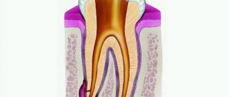
Among all the mechanisms of tooth root healing, mineralized healing is considered the most favorable, that is, dentin and cement grow into the fracture line (this healing mechanism is typical for well-immobilized teeth and without infection). The so-called “connective tissue fusion” is possible - when the connective tissue bundle ruptures and the blood supply is restored, the connective tissue grows in the fracture line (that is, the periodontal ligament) with the formation of a new apical foramen. The periodontal ligament grows into the target of the fracture, thereby forming a periodontal gap around each fragment. The third option for healing the fracture line is combined healing, that is, mineralized healing together with connective tissue. Such fusion will occur only if the alveolar processes grow and there is a large distance between the fracture fragments. And the most unfavorable thing with the possible formation of fistula tracts is healing “without fusion” with the germination of granulation tissue. This type of fusion is typical for teeth without timely endodontic treatment.
The main goal in the treatment of a tooth root fracture is to create a layer to form a mineralized connection between the fragments. Of course, vital pulp will improve the prognosis of treatment. Therefore, when treating a tooth root fracture, it is necessary to reduce the distance between the fragments as early as possible and ensure reliable immobilization of the teeth.
Treatment of a tooth root fracture that does not communicate with the oral cavity
When treating a fracture of a tooth root that does not communicate with the oral cavity, when confirming the viability of the pulp, it is necessary to reposition the fragments and immobilize the teeth, followed by monitoring on an x-ray. The duration of splinting can be up to 3 months. In this case, the patient is recommended to eat only soft foods. Every month, the splint is checked and a photo is taken to ensure proper fusion of the fragments, and pulp vitality tests are done.
Treatment of a fractured tooth root communicating with the oral cavity
Treatment of a fracture of a tooth root communicating with the oral cavity depends primarily on the passage of the fracture line, the volume of damaged tissue, and the length and anatomy of the tooth root. In case of a fracture involving more than 2/3 of the length of the teeth, the tooth is removed.
Often, when a tooth root is fractured, the pulp is already necrotic. And then the dentist must determine the indications for filling the canals with either gutta-percha or calcium hydroxide.
Indications for canal filling with gutta-percha in case of tooth root fracture
Indications for filling canals with gutta-percha in case of a tooth root fracture are as follows:
- The radiograph shows no areas of destruction in the area of the lower third of the tooth root;
- When you touch the tooth pulp with a paper pin, there is bleeding (confirmation of the vitality of the tooth pulp);
- There is no distance between the fragments, or the distance is insignificant;
- The root canal at the fracture line is not wide.
Indications for canal filling with calcium hydroxide in case of tooth root fracture
Indications for canal filling with calcium hydroxide in case of a tooth root fracture may be:
- Wide root canal (as a risk of gutta-percha removal);
- Presence of periapical changes on the radiograph;
- Internal/external resorption of root fragents;
- Viable pulp (paper point bleeding test).
When observing changes on an x-ray, if the course is favorable, a dentinal bridge may form in six months, after which the canals can be filled with gutta-percha.