Reasons for the development of micrognathia
Anomalies of jaw development can be congenital or acquired.
Congenital ones include:
- Severe acute illnesses of a woman during pregnancy,
- Anomalies of intrauterine development of the fetus,
- Congenital pathologies of the development of the dental system,
- Genetic predisposition.
An acquired anomaly can result from:
- Maxillofacial trauma,
- Artificial feeding and improper sucking in infancy,
- Diseases of the endocrine system,
- Past infectious diseases and inflammatory processes,
- Early loss of baby teeth,
- Problems with nasal breathing.
Possible consequences
In addition to the aesthetic aspect, underdevelopment of the lower jaw can cause serious harm to a person’s health. The following problems may occur:
- malocclusion;
- disruption of the auditory and respiratory organs;
- persistent problems with chewing and swallowing, which ultimately leads to problems in the gastrointestinal tract;
- the absence of some teeth and deformation of existing ones;
- weakening of the immune system in general, and, as a result, frequent illnesses.
For adults, the treatment process may take longer. If microgenia is congenital, then it is necessary to contact a professional as soon as possible.
Diagnosis and treatment of narrow jaw
Deformed dentition, crowded teeth, malocclusion, unerupted teeth are consequences of an underdeveloped dental system. The earlier these abnormalities are diagnosed, the more effective the treatment will be. To expand the lower and upper jaws, therapeutic methods, mechanical expanders and plates are used. In rare cases, only surgery helps.
In children under 10-11 years old, jaw expansion gives the best results. This is a time of active growth and formation of bone tissue, so the defect can be corrected with the help of orthodontic structures.
Description of the term
In orthodontics, there is such a thing as micrognathia, which is literally translated from Greek as “small jaw” and is underdevelopment of the upper or lower jaw. The opposite concept is macrognathia, in which the jaw is large in size compared to normal.
Microgenia. With macrognathia, the chin, on the contrary, is strongly pushed forward
Abnormal development of the lower jaw is called microgenia. Such disturbances in the development of the masticatory apparatus contribute to uneven tooth growth and the formation of malocclusion. In some cases, crowns may protrude beyond the teeth’s growth line or be completely absent. The upper jaw has normal development.
Expansion of the lower jaw
A narrow lower jaw, or microgenia, can be corrected in childhood with special devices - distractors. This is a device that gradually stretches the bones of the lower jaw. New bone and soft tissue grows at the site of the sprain. When installing a distractor, first increase the distance between the canines of the lower jaw. Every day the device allows you to increase the lower jaw by 1 mm. When the jaw is wide enough to accommodate tooth growth, the distractor is placed in place for at least two weeks to stabilize the sprain.
If you have a problem similar to that described in this article, be sure to contact our specialists. Don't diagnose yourself!
Why you should call us now:
- We will answer all your questions in 3 minutes
- Free consultation
- The average work experience of doctors is 12 years
- Convenient location of clinics
Single contact phone number: +7
Make an appointment
In adult patients, the lower jaw bones are fused and cannot be stretched, as in children. Therefore, to enlarge the lower jaw, the surgeon cuts the bone in several places with an ultrasonic knife. On the fourth day after surgery, an expansion device is installed. The first adjustment of the stretching is carried out by an orthodontist, then the patient himself controls the stretching process, which can take up to six months.
After expanding the lower jaw, braces are installed on the lower front teeth to correct the bite. Crowded teeth gradually occupy the resulting space.
The photo below shows a narrow jaw before and after expansion.
Symptoms and signs
Signs of micrognathia include:
- A malocclusion that can be easily detected by a dentist.
- Distortion of facial features, violation of proportions and symmetry, this is especially noticeable in the photo.
- With an underdeveloped lower jaw, the lower lip “sinks” and the chin appears too small.
- With insufficient development of the upper jaw, the upper lip “sinks”, the lower jaw seems to protrude forward.
- A cleft in the palate may appear.
- Difficulty breathing, speaking and swallowing, and the tongue may not develop well or retract into the throat when lying on your back.
- Teeth wear down greatly due to constant friction; they can also grow unevenly, falling out of an even row.
It is important to know: signs of micrognathia are often found in other pathologies, and therefore treatment should be prescribed by a dentist after a full examination.
Hypoplasia
Hypoplasia of baby teeth, which form during the prenatal period, is caused by disturbances in the body of a pregnant woman, and hypoplasia of permanent teeth, which begin to form in the 5-6th month of a child’s life, is caused by disturbances in metabolic processes in the child’s body. Diseases in a child are observed much more often than in a fetus, so hypoplasia of permanent teeth is more common than primary teeth.
Currently, hypoplasia of primary teeth is observed more often than before, which is explained by a decrease in perinatal mortality. Hypoplasia of primary teeth occurs when a child becomes ill in the first weeks and months of his life, which affects the formation of incisors, canines and large molars.
It is noted in the literature that the higher the incidence in childhood, the greater the incidence of dental hypoplasia. Thus, dental hypoplasia is observed in 50% of children with chronic somatic diseases accompanied by metabolic disorders that arose before or shortly after birth.
Hypoplasia of primary incisors is observed in children whose mothers suffered from diseases such as rubella, toxoplasmosis and toxicosis during pregnancy. Hypoplasia is observed in premature infants, children with congenital allergies, who have suffered a birth injury or hemolytic jaundice resulting from incompatibility of the blood of mother and fetus according to the Rh factor, and those born with asphyxia. With hemolytic disease of the newborn, enamel hypoplasia in most children develops in utero (at 25-32 weeks of pregnancy), and sometimes during the 1st month of life.
Hypoplasia of permanent teeth develops under the influence of various diseases that occur in children during the formation and mineralization of these teeth. Hypoplasia is detected in children who have suffered from rickets, tetany, acute infectious diseases, diseases of the gastrointestinal tract, toxic dyspepsia, nutritional dystrophy, diseases of the endocrine system, brain disorders, and congenital syphilis. About 60% of hypoplastic defects in permanent teeth occur in the first 9 months of a child’s life, when the adaptive and compensatory capabilities of his body are poorly expressed.
Hypoplasia of the tooth crown, as well as the group affiliation of the affected teeth, largely depends on the age at which the child suffered the disease. Thus, when the disease occurs in the first months of life, hypoplasia develops in the area of the cutting edge of the central incisors and the cusps of the sixth teeth, since their formation begins in the 5-6th month after birth. At the 8-9th month of life, the second incisors and canines form. If a child becomes ill during this period, areas of hypoplasia will be localized at the cutting edge of the lateral incisors and canines, while in the central incisors and sixth tooth, areas of underdeveloped enamel will appear approximately at the level of the equator (since half of the crown has already formed).
In cases where the disease in a child continues for a long time, changes in the enamel occupy significant areas on the surface of the tooth crown. Some children have an uneven enamel structure throughout the crown of a certain group of teeth.
The severity of hypoplasia depends on the severity of the disease: with mild metabolic disorders, only chalky spots are formed, and with severe diseases, underdevelopment of the enamel is observed, up to its absence (enamel aplasia).
Hypoplasia of the hard tissues of teeth that form in the same period of time is called systemic, and hypoplasia of a single tooth is called local.
Systemic hypoplasia. Clinically, three forms of systemic enamel hypoplasia are distinguished:
- color change;
- underdevelopment;
- absence.
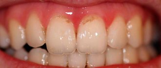
A weak degree of enamel hypoplasia can appear in the form of white, less often yellowish spots, with clear boundaries and the same size on the teeth of the same name. The spots are usually localized on the vestibular surface and are not accompanied by any unpleasant sensations. A characteristic feature of the stain is that the outer layer of enamel is not painted with dyes. During life, the size, shape and color of the spot usually do not change. The thickness of the enamel in the area of the stain is the same as in the area of intact enamel next to it. This form of hypoplasia is usually not detected on x-ray.
A more severe form of enamel hypoplasia is its underdevelopment, which manifests itself in different ways (wavy, dotted, grooved enamel).
Wavy enamel is revealed when the surface of the crown is dried: upon examination, small ridges can be distinguished, between which there are depressions covered with unchanged enamel.
The most common form of hypoplasia is in the form of pinpoint depressions in the enamel, located on the vestibular and lingual surfaces at different levels in teeth of different groups. Over time, the enamel in the depressions gradually becomes pigmented, but remains dense and smooth. Sometimes hypoplasia appears as a single transverse groove on the crown (interception). This form of hypoplasia is called sulcal by some. There may be several such grooves; they alternate with unchanged tooth tissues. Rarely, grooves are present along the entire height of the crown of the teeth of some groups. This form is called scalene hypoplasia. It is characteristic that even with severe manifestations of hypoplasia (sulcal and scalene), the integrity of the enamel is not compromised.
The most rare occurrence is the absence of enamel (aplasia) in a certain area. With this form of hypoplasia, pain may be experienced when exposed to irritants, after which the pain disappears. Clinically, this form is manifested by the absence of enamel on part of the crown, but more often - at the bottom of the cup-shaped depression or in the groove covering the crown of the tooth.
Histological examination reveals enlarged interprismatic spaces and extended lines of Retzius; the boundaries of the prisms lose clarity. The degree of change depends on the severity of the process. Thus, with a point form, changes in dentin are more noticeable: the zone of interglobular spaces increases, and intensive deposition of replacement dentin is observed. The number of cellular elements in the pulp decreases. Degenerative changes are detected in the nerve elements of the pulp.
Electron microscopic examination of the enamel reveals changes in the width of the prisms, the orientation of hydroxyapatite crystals, and the structure of the dentinal tubules.
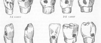
One of the varieties of systemic hypoplasia are the teeth of Hutchinson, Pfluger and Fournier, the crowns of which have a peculiar shape.
Hutchinson's teeth - upper central incisors with a screwdriver and a barrel-shaped crown (the size at the neck is larger than at the cutting edge); the semilunar recess can be covered with enamel, but sometimes enamel is observed only at the corners of the tooth, and in the middle part the dentin is not covered with enamel.
Fournier's teeth are central incisors with a screwdriver-shaped crown (the same as Hutchinson's teeth), but without a semilunar notch along the cutting edge.
It was previously believed that Hutchinson's and Fournier's teeth were part of the triad of symptoms of congenital syphilis: parenchymal keratitis, congenital deafness and Hutchinson's teeth. It was later found that this anomaly can be observed not only in syphilis.
Pflueger's teeth are the first large molars, the crown size of which near the neck is larger than that of the chewing surface, and the tubercles are underdeveloped and, converging, give the tooth the appearance of a cone. The development of Pflueger's teeth is explained by the action of a syphilitic infection.
Enamel hypoplasia is differentiated from initial and superficial caries.
With initial caries, the white spot is usually single, located at the neck of the tooth, and with hypoplasia, the white spots are multiple and localized on any part of the crown. In addition, with hypoplasia, the spot is not stained with a 2% solution of methylene blue, but with caries it is stained.
With hypoplasia, the enamel surface is smooth, and with superficial caries, it is rough (detected by probing), its integrity is compromised.
“Getran wedge” teeth. Separately, we should consider this type of systemic hypoplasia, such as “tetracycline” teeth. These are teeth with discoloration as a result of taking tetracycline during the formation and mineralization of tooth tissue. Tetracycline is deposited in the enamel and dentin of developing teeth, as well as in the bones of the fetus or child when tetracycline is administered to a pregnant woman or child as a therapeutic agent for various diseases. Tetracycline can cause not only tooth staining, but also enamel hypoplasia. The nature of the change depends on the dose and type of drug. When small doses are administered, the color changes, and with very large doses, along with a change in color, underdevelopment of the enamel is observed. When taking dimethylchlortetracycline, the color change is more significant; when using oxytetracycline, the color is less intense.
Treatment of a pregnant woman with tetracycline leads to a change in the color of the child's front teeth, namely the crowns of the incisors, starting from the cutting edge, and the chewing surfaces of large molars. It is believed that tetracycline penetrates the placental barrier. Taking tetracycline by a child, starting from 6 months of age, causes staining not only of primary molars, but also of permanent teeth, as a rule, part of the tooth crown, which is formed during this period.
The intensity of tooth coloring from light yellow to dark yellow also depends on the type of tetracycline and its amount. Teeth stained yellow with tetracycline have the ability to fluoresce under the influence of UV rays. This property can be used to differentiate from tooth staining caused by other factors, such as bilirubin in hemolytic disease of the newborn.
Staining of tooth enamel with tetracycline is persistent and further tooth tissue cannot be whitened, so tetracycline should be prescribed to children and pregnant women only for health reasons.
Local hypoplasia. Local hypoplasia is a violation of the formation of enamel on permanent teeth as a result of involvement of tooth buds in the inflammatory process or mechanical trauma of the developing follicle. This pathology manifests itself in the form of spots - from white to yellowish-brown or, more often, pinpoint depressions located on all surfaces of the tooth. In severe cases, aplasia (absence) of enamel may occur. Sometimes the enamel on the crown of a tooth is partially or completely missing. These teeth are called “Turner teeth.”
More common is local hypoplasia of permanent small molars, the rudiments of which are located between the roots of primary teeth. The disease can be prevented by extensive measures to prevent caries of primary teeth or by treating them at an early stage of the lesion in order to prevent the occurrence of periodontal inflammation.
In case of enamel defect, preference is given to composite filling materials, and in case of significant deformation of the tooth crown, orthopedic treatment is indicated.
Enamel hyperplasia is the excessive formation of tooth tissue during its development.
“Enamel drops” are observed in 1.5% of patients. The diameter of these formations ranges from 1 to 2-4 mm. They are located in the area of the tooth neck, at the border of enamel and cement, sometimes in the bifurcation (trifurcation) zone of the roots. “Enamel drops” consist of dentin covered with enamel, often containing a cavity filled with pulp.
Hyperplasia usually does not manifest itself clinically and does not cause functional impairment.
Essentially, these formations are closer to another form of anomaly - the fusion of the crowns or roots of well-formed teeth. It is believed that this is due to the close location of the tooth germs in the dental plate. More often, fusion of central incisors with lateral incisors is observed, less often - normal and supernumerary teeth.