Enamel is the hardest tissue in the human body and a natural protective barrier for teeth. However, some people are diagnosed with its deficiency. Hypoplasia of dental enamel - as this condition is professionally called - can have different causes, but appropriate treatment methods must be used each time.
The main function of enamel in the oral cavity is to protect teeth from harmful factors: the action of bacteria, thermal and chemical irritants, as well as abrasion during chewing. Although there are proven ways to strengthen enamel, its gradual erosion is associated with various reasons. Some patients, even children, are also diagnosed with underdevelopment of this tissue. Dental hypoplasia can have many causes and each time requires appropriate treatment.
Causes
The disease can be genetically determined or acquired in the early stages of development of the organism. At the same time, depending on the timing of the manifestation of the pathology, the list of its causes may vary.
Attention! Genetic hypoplasia is transmitted through a damaged gene on the sex X chromosome and is inherited as a dominant trait. In the case of the father's illness, it affects only daughters (100% of girls are born with hypoplasia, boys are healthy), and in the case of the mother's illness, it affects 50% of all children of both sexes.
In addition to heredity and spontaneous mutations, the main pathogenic factors for hypoplasia of primary teeth are disorders in the mother’s body and neonatal complications. Other factors:
- hormonal imbalances;
- viral infections (rubella, syphilis, toxoplasmosis, etc.);
- discrepancy between the Rh factor of the mother and the fetus - hemolytic disease of the newborn develops;
- severe toxicosis;
- alcohol abuse;
- irradiation - one-time or cumulative;
- lack of vitamins, micro- and macroelements;
- prematurity, asphyxia and birth injuries - due to the high risk of developing encephalopathies with disruption of the regulatory functions of the central nervous system.
The impact of these causes disrupts the formation of tooth germs or disrupts metabolism, which immediately affects the future of various dental structures, including enamel.
Attention! Pregnancy infections disrupt the formation of hard tissues of the fetus, including tooth enamel. Research confirms that severe cases of aplasia are more common in children whose mothers suffered from rubella, toxoplasmosis, “banal” ARVI or toxicosis during pregnancy. Moreover, statistics show that various manifestations of enamel hypoplasia occur today in every third child of preschool age.
Hypoplasia of the enamel of permanent teeth begins in childhood, starting from 5–6 months, and is often associated with metabolic disorders:
- Failures in the central nervous system disrupt the metabolism of essential minerals. The skeleton, including teeth, does not receive enough of the necessary elements to build its structure.
- Hormonal diseases - hypothyroidism and parathyroid insufficiency reduce the calcium and phosphorus content in bones, nails, and teeth.
- Disorders of the gastrointestinal tract - various types of dyspepsia and enzyme problems reduce the ability to absorb essential microelements.
- Hypovitaminosis, especially with a deficiency of vitamins D, C, E, affects the normal formation of the skeleton as a whole (up to rickets).
- Acute infectious diseases in the first years of life slow down mineral metabolism and inhibit the natural development of the body.
The condition of baby teeth also affects the condition of permanent teeth developing in their immediate vicinity. Chronic periodontitis or trauma due to unsuccessful removal with damage to the tooth germ can cause disruption of enamel formation.
What are the anomalies in childhood?
As can be seen from all of the above, in clinical practice there are a variety of anomalies associated with the shape, number and structure of teeth. Below is a simplified classification, which includes the most common forms of such pathologies:
- the size of the crowns – too small or large,
- quantity – hyperdontia and adentia,
- shape – spiky, barrel-shaped, etc.,
- deformation of the structure of enamel and dentin – hypo-, hyper- and aplasia,
- position – curvature of elements, inclined position, rotation around its axis,
- color – fluorosis, tetracycline teeth.
All these deviations must be treated, preferably on time. Otherwise, the child risks developing serious psychological complexes due to external defects, as well as encountering no less serious dental diseases caused by such anomalies.
Symptoms
Signs of pathological development of the surface layer of the dental crown:
- violation of the integrity of the enamel in the form of grooves, ridges, lines, spots, up to the complete absence of tissue;
- pigmentation of white, yellow, yellow-brown shade;
- deformation - shortening of roots, curvature of the crown;
- increased sensitivity to irritants.
At the same time, the enamel can retain its smoothness and shine, and the defects present during cutting are not prone to growth. However, by the appearance of the defect one can also judge some characteristics of the damaging factor:
- Localization on certain teeth depends on at what stage of development the body suffered a negative impact. If the child was ill at the age of 5–6 months, the edges of the front incisors and the cusps of the “sixes” will be affected on the permanent teeth; at 8–9 months, the marginal incisors and the cutting surface of the canines will be affected.
- The size of the defect determines the duration of exposure. If the disease lasts throughout the preschool period, hypoplasia evenly covers the entire surface of the teeth.
- The number of pathogenic structures within one tooth depends on the frequency of the pathogenic influence. For example, if hypoplastic changes are uneven and have a wave-like structure, this indicates a history of infections or chronic diseases with periods of remissions and exacerbations.
Symptoms
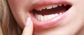
In most cases, enamel hypoplasia is diagnosed by a dentist; clinical symptoms of the disease are difficult for the patient to detect. The disease may appear as white or brown spots on the surface of the enamel, small pits on the teeth, grooves on the crowns or curled edges of the incisors of the teeth.
Only microscopic examination can give a reliable picture of enamel hypoplasia. Then it is discovered that the layer is much thinner than in healthy people. In addition, the disease is sometimes accompanied by disturbances in the dentin structure.
Classification of the disease
The main current taxonomy of hypoplasia is based on the etiology of the pathological process and its size. The following types are distinguished:
- Systemic hypoplasia affects all teeth in the oral cavity or individual symmetrical groups with similar periods of formation. The main defects are located parallel to the gum, closer to the chewing/cutting part, mainly on the vestibular surface and are noticeable immediately after eruption.
- Local hypoplasia damages individual teeth in the permanent dentition. It looks like a whitish or yellow-brown clouding of the enamel with areas of erosive process. The main reason is acute periapical inflammation of baby teeth. Most often it affects premolars, less often – molars.
- Focal - usually involves several consecutive teeth. Hypoplasia affects not only the enamel, but also the dentin of the tooth with a serious violation of its functionality. Such structures erupt late and on x-ray show low tissue density, large pulp chambers and wide short root canals.
On a note! Local hypoplasia is externally similar to fluorous lesions and the first signs of caries, therefore, to make a diagnosis, differential diagnosis with staining of hard tissues is required (areas of hypoplasia, unlike fluorosis, cannot be stained).
A common type of childhood hypoplasia is molar-incisal hypomineralization, when the pathology covers the incisors and 1–4 molars. It appears as blurry spots of white, yellow or yellow-brown color on various parts of the crown. Such enamel is characterized by increased sensitivity and often chips, exposing dentin and contributing to the development of caries.
In addition, there is a whole set of forms of external manifestation of pathology:
- Spotted - stripes or spots of white, yellow, yellowish-brown color. The damage itself can be located in the subsurface layer of the enamel, and then the enamel itself remains smooth and shiny, or directly on the surface - the structure becomes dull and rough.
- Erosive - the main damage is concentrated in the surface layer of the enamel and in the form of spotty areas of erosion.
- Grooved - stripes of erosion form grooves parallel to the cutting edge of the teeth.
- Wavy - thin areas of hypoplasia parallel to the cutting edge almost merge with each other, forming a wavy surface structure.
- Aplastic – complete absence of enamel with pronounced brown pigmentation of the tooth surface.
Changing tooth shape
In this case, there are no visible changes in the enamel, but the tooth takes on pathological forms during abnormal development. The most common options:
- spike-shaped teeth - the crown takes the form of a triangle and resembles the teeth of a shark;
- Hutchinson's teeth are incisors with a curved, capsule-shaped edge;
- Fournier's teeth - the wide base of the incisors tapers to a spade-shaped cutting edge;
- Pflueger teeth - a change similar to the previous two, but typical for the first premolars (the chewing surface is narrower than the neck of the tooth);
- teeth with disordered distortion of shape (ugly teeth).
The main reason for such changes is late congenital infections, mainly syphilis. However, treponema penetrates into the fetal tissue during the transition to placental circulation in the 4th–5th month of pregnancy, therefore, if the infection is detected in a timely manner and specific treatment is carried out, the child may well be born completely healthy.
Systemic enamel damage
Systemic hypoplasia is a more serious problem, which can be expressed in changes in the color of the enamel, its underdevelopment or even complete absence. In dental practice, grooved, dotted and wavy forms are encountered; in other more serious clinical cases, obvious deformation of the crown shape occurs.
Hypoplasia does not have painful symptoms, but often leads to concomitant dental diseases. In this case, the lesion covers all teeth and can progress quickly, so in such a situation, urgent help from a specialist is required.
Diagnosis and treatment
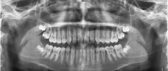
Hypoplasia is easily detected by visual examination at a dentist's appointment. To make an accurate diagnosis, radiography is additionally prescribed and a differential analysis is carried out by staining hard tissues.
The choice of treatment methods depends on the type of defect, its depth, area and degree of mineralization of surrounding tissues. Complete restoration is not always possible, so the final aesthetics largely depends on the technical capabilities of the clinic and dentistry in general, as well as the capabilities and desires of the patient’s parents.
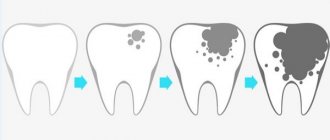
Important! In case of hypoplasia, special attention should be paid to the prevention of caries. Thinning or partial/complete absence of enamel opens access to microbes, which, at the first favorable opportunity, begin their pathogenic activity.
Conservative treatment
The approach is relevant for damage to the surface layer of enamel and general weak mineralization of dental tissues. Includes:
- Endogenous intake of vitamins , minerals and drugs that increase their absorption by the body.
- Local remineralization of hard dental tissues with fluoride, calcium, and phosphorus preparations.
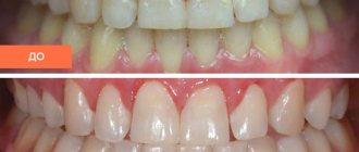
- Microabrasion or bleaching is light etching and grinding of enamel with a minimally abrasive bur. After polishing, the tooth takes on a natural, healthy appearance. The procedure allows you to eliminate clouding of the enamel and slight erosion of its upper layer - in all other cases, such a measure of influence is inappropriate.
Treatment with tooth preparation
As a rule, it is carried out after preliminary mineralization of tissues, to restore more extensive and deep aesthetic defects. To do this, the affected area of enamel is treated for the installation of fillings and/or veneers. In severe cases of hypoplasia, as well as complete aplasia of the enamel, more radical methods are used - depending on the condition of the dentin, either the installation of crowns or complete removal followed by prosthetics are prescribed.
How to adjust sizes
Treatment of such anomalies is directly related to its specific type and severity. If the dimensions slightly deviate from the norm and the bite is not disturbed, if there is no serious aesthetic defect, you can leave everything as is. In other cases, the size of the crowns can be restored using photopolymer composites, prosthetic crowns or the installation of veneers. Sometimes the doctor has to remove one to several large teeth and then replace them with fixed dentures on implants.
Prevention of hypoplasia
The greatest attention should be paid to pregnant women and preschool children. The prenatal and infant periods are especially important for the formation and growth of healthy teeth.
Basic preventive measures:
- When planning a pregnancy, pay attention to your health (including teeth) and diet.
- During all trimesters, avoid infectious diseases and acute toxicosis.
- Provide the expectant mother with a balanced diet, avoid vitamin deficiencies and micronutrient deficiencies.
- After birth, provide your baby with high-quality nutrition with sufficient levels of vitamins A, B, C, D, calcium minerals, phosphorus, and fluorine. The ideal option for the first months of life is breastfeeding (provided the mother is well-nourished). This will not only provide the child’s body with everything it needs, but will also provide a reliable level of immune protection.
- Eliminate somatic diseases in preschool children in a timely manner.
- From the first days of life, take care of the hygiene of baby teeth. For this you can use special silicone fingertips or napkins.
- Treat baby teeth efficiently and in a timely manner - prevent the development of complicated caries. If this cannot be avoided, atraumatic removal of the damaged tooth will be required to avoid periapical tissue inflammation.
- Try to protect your child from possible dental injuries.
Hypoplasia is an irreversible change in tissue, so failure to comply with these simple preventive standards will subsequently become a “toothache” for a person for the rest of his life. Remember: treatment provides only temporary protection and an aesthetic solution to the problem.
Reviews
It is important to take care of baby teeth in the same way as permanent teeth - everyone should remember this. At the slightest suspicion of caries in a child, it is important to immediately contact a dental clinic for medical help. After all, destructive processes in baby teeth can have serious consequences.
Have you or your relatives ever had to deal with Turner hypoplasia? If yes, then share with our readers the features of the disease and effective methods of treatment in the comments below.
If you find an error, please select a piece of text and press Ctrl+Enter.
Tags: tooth enamel
Did you like the article? stay tuned
No comments yet