Dental phlegmon is a complex infectious inflammation of the tissues of the oral cavity and face, provoked by the development of pathogenic microorganisms (streptococcus, staphylococcus, etc.). The basis is an acute purulent process in the subcutaneous fatty tissue, which spreads to the surrounding tissues and causes their necrosis.
Lack of timely diagnosis or improper treatment can lead to a number of serious complications that pose a threat not only to the aesthetics/functionality of the dentition or facial tissues, but also to human life. If you find characteristic symptoms of dental phlegmon in yourself or a loved one, contact your dentist immediately. He will perform an initial examination, determine the cause of the problem and prescribe an adequate treatment regimen that will eliminate the pathology with minimal consequences for health.
What is phlegmon
This is purulent necrotization of soft tissues, which has no clear boundaries. Unlike an abscess, it tends to affect adjacent intercellular spaces. It can occur anywhere in the human body. But the head and neck area is the most dangerous, due to the presence of a large number of blood vessels through which the infection can spread to the brain.
Causes
In 99% of cases, this disease develops due to dental problems. As practice shows, it is always a consequence of untreated caries. However, purulent melting does not develop immediately. This must be preceded by other pathologies, such as inflammation of the pulp, periodontium, development of a radicular cyst, or osteomyelitis. An obligatory element leading to infection must always be a source with microbial contamination.
Not always purulent-inflammatory processes can lead to the spread of infection. The human body’s immunity plays a huge role. When it decreases, pathogenic microbes in the purulent focus can lead to necrosis of nearby tissues. The head and neck area is rich in cellular spaces that smoothly merge into each other. Therefore, pus can freely move from the lower jaw area down the neck.
The following may be subject to melting:
- Subcutaneous fat tissue;
- Fiber of the intermuscular space;
- Interfascial tissue;
- The lymph nodes;
But there is another factor - an infected wound, which could be the result of injuries. The development of a purulent process is very rarely observed in this case, but with reduced immunity it occurs. Therefore, every wound must be subjected to antiseptic treatment.
DIAGNOSTIC MEASURES
Often, to identify phlegmon, the dentist only needs an external examination of the patient and collection of complaints.
However, to determine the exact location of the source of inflammation and the extent of its spread throughout the tissues of the oral cavity, additional diagnostic methods are required - computed tomography and ultrasound.
To clarify the severity of the inflammatory process, a general blood test is prescribed, and to determine the causative agent of the disease, culture of purulent contents is performed.
Using this analysis, the sensitivity of pathogenic microflora to various antibiotics is subsequently studied.
Odontogenic phlegmon
The cause of the development of this kind of disease is always a dental problem. Any pathology of the masticatory organs can lead to this outcome.
Problems may arise at the following stages:
- Before tooth eruption;
- At the moment when he is already in the row;
- After removing it.
The most common cause of this disease is untreated caries. The pathological process in hard tissues, having gone through certain stages of disease development (pulpitis -> periodontitis), can lead to the development of a radicular cyst. When immunity decreases, it can fester, and then the contents surrounded by the capsule have the opportunity to go beyond its limits.
Wisdom teeth can also cause problems as they are the most advanced teeth and may not have enough space in the row. Having appeared at least half, they become vulnerable to the carious process. Under the gingival hood, which is located above the tubercles, food begins to accumulate. Then cariogenic microorganisms join this substrate and begin to ferment leftover food into organic acids. As a result, a cavity forms in the hard tissues. The wisdom tooth is anatomically designed in such a way that it is very difficult to treat, and if preservation is not practical, then it is better to remove it.
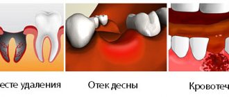
A very rare phenomenon occurs when the disease develops after tooth extraction. The following reasons may contribute to this:
- Part of the organ was left in the socket;
- Food getting into an unhealed hole;
- Permanent injury to the socket with a blood clot, followed by infection.
If any element of the tooth remains inside, then in most cases it will lead to inflammation, since it will be perceived by its own tissues as a foreign body. In addition, local immunity will be activated, which will actively produce lymphocytes. When pathogenic microflora attaches, this phenomenon can spread to nearby tissues. Therefore, it is necessary to remove everything that remains in the hole after tooth extraction.
At first, a person complains of swelling in the area of the extracted tooth and painful opening of the mouth. Throughout the course of the disease, the puffiness of the face becomes more and more. Then, every day the person’s general condition begins to deteriorate. The temperature rises, and blood tests change, which begin to indicate an inflammatory process occurring in the body.
Characteristic symptoms of the pathology
At the beginning of its development, phlegmon may not manifest itself in any way. Symptoms of the pathology become obvious when it enters the active stage:
- it becomes difficult to open your mouth,
- swelling of the affected area, face, neck, cheekbones appears: it all depends on the area in which the infectious process is localized. This is usually the most noticeable symptom - part of the face suddenly swells,
- pain syndrome occurs
- bad breath bothers you,
- problems with speech, chewing and swallowing cause discomfort,
- body temperature rises to 39–41˚С and above,
- A local increase in temperature occurs: there may be a feeling that the skin in the area of localization of inflammation is warmer than usual or even “burns” or “bakes”.
As the disease progresses, breathing problems arise, a grayish coating appears on the tongue, and the mucous membrane at the site of inflammation becomes red and swollen. If phlegmon is localized in the zygomatic region, discomfort occurs when blinking, swelling and cyanosis of the eyelid, pain in the eyes, and blurred vision.
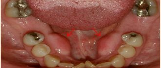
Cellulitis of the floor of the mouth
Oral phlegmon is a diffuse purulent melting of the subcutaneous tissue located between the muscles that make up the floor of the oral cavity. Either one side or both can be affected. This disease is expressed in severe clinical symptoms.
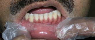
Externally, the patient has a disturbance in the configuration of the face and its asymmetry due to edema. The nearby submandibular and submental areas also have inflammatory swelling. The skin over the purulent infiltrate is hyperemic and does not fold. Nearby lymph nodes are enlarged.
Patients complain of increased salivation, difficulty moving the tongue and lower jaw due to pain. Due to swelling, the tongue does not fit in the oral cavity, so it sticks out slightly. There are teeth marks on its surface. From the outside it seems that the person is thirsty.
The tongue and teeth are coated with plaque, since the patient’s condition does not allow for proper oral hygiene. The composition of saliva also changes, it becomes cloudy and viscous. Body temperature ranges from febrile to pyretic. Depressed or, on the contrary, excited behavior is observed. The general condition of the body is characterized as serious.
The centers of microbial contamination are the lower teeth, and especially the wisdom tooth. The teeth of the upper jaw are not anatomically connected to the area of the diaphragm of the mouth, so they cannot cause this disease. Infection from the upper dentition will only spread to nearby tissues.
Will traditional medicine methods help?
The use of traditional methods to treat the disease is possible only in agreement with a doctor and simultaneously with taking prescribed medications, as well as after surgery to remove the source of inflammation. Under no circumstances should you self-medicate at home - it is important to remember the negative consequences of phlegmon.
Traditional medicine is mainly used in the postoperative period. The oral cavity can be rinsed with decoctions and water infusions of chamomile, eucalyptus, yarrow, sage, and honey solution. Such rinses eliminate inflammation, pain, swelling, increase local immunity, and have a wound-healing and antiseptic effect.
Category Uncategorized Posted by Mister stomatolog
Putrefactive-necrotic phlegmon of the mouth, or Ludwig's angina
This pathology is similar in appearance to the previous disease, but its main difference is that with soft tissue necrosis there is no accumulation of pus. Symptoms of Ludwig's angina are very specific. This disease carries a huge danger, since sepsis can develop if you do not consult a doctor in a timely manner. Fortunately, the incidence is very low.
The specificity of Ludwig's angina is that basically only the muscles are the main point of damage. The perimuscular tissues may be in a state of inflammation, but it is the muscles that take the main “blow”. Their consistency becomes very dense and woody.
With this disease, patients complain of headaches and high fever. Moreover, in the first two days it may not reach high levels. However, every day the patient gets worse. Externally, a person’s face looks asymmetrical, the chin and neck are swollen. It is impossible to remove the tongue inside as the swelling prevents this from happening. There is a putrid odor and abundant gas formation from the source of infection.
Diagnosis of oral phlegmon
Pathology can be diagnosed through external examination and examination of the oral cavity. The doctor collects complaints and detailed information about what preceded the disease. Visually identifies the source of infection. If the purulent process is located in the superficial layers, then diagnosis is usually not difficult. But if the pus is localized in deep places, then additional clinical and laboratory tests will need to be done. And also carry out microbiological seeding in order to establish the microbial flora. This is necessary in order to prescribe appropriate antibacterial drugs in the future.
Differential diagnosis
When making a diagnosis, doctors differentiate the disease from furunculosis, erysipelas, cysts, and inflammation of the salivary glands. As a rule, an experienced dentist can easily recognize phlegmon by visual signs and complaints. However, to clarify the localization of pus, its volume, and the degree of tissue damage, a computed tomography or ultrasound examination is required. In addition, the contents of the phlegmon are taken for bacterial culture. This makes it possible to determine the type of infectious agent and prescribe the optimal treatment, i.e. choose the right and most effective antibiotic.
Treatment methods for phlegmon of the floor of the mouth
The first place a person comes with this disease is dentistry. Although this is a dental problem, the doctor should send him to the hospital. Since such patients are treated only by maxillofacial surgery.
Treatment of phlegmon of the floor of the mouth combines conservative and surgical methods. If there is pus, it is always necessary to open the source of infection, otherwise it will not resolve on its own. Access for drainage of infiltrate can be made through the mouth or through the skin, here the surgeon starts from where the infection is closest.
The most rational access to the purulent contents is an external incision along the midline going down from the lower jaw. It is safe and cosmetic. The scar remains practically invisible. Then a drain is inserted into the wound for several days. And the causative tooth is removed.
Afterwards, during the recovery period, the doctor must prescribe antiseptic care and antibiotics to eliminate the development and proliferation of microorganisms. In order for the wound to heal faster, vitamins and physical therapy can also be prescribed.
In Moscow, any maxillofacial hospitals, as well as specialized clinics, provide such treatment, each of which has its own price list for such an operation.
Treatment
Despite the severity of the disease, in general, with timely treatment, perimandibular phlegmon has a favorable prognosis. Treatment is carried out only in a hospital setting, but at the initial stage everything is often limited to the use of conservative methods - antibiotic therapy, the prescription of restoratives, vitamins, and a special diet. But if the process has already started and the spread of infection is observed, complex surgical treatment is indicated, which includes the following points:
- opening the inflammation site under general anesthesia;
- cleansing the wound from pus;
- installation of drains for drainage of wound contents;
- daily treatment with bactericidal solutions;
- powerful antibiotic therapy - often a complex of several drugs is prescribed;
- detoxification of the body;
- use of antipyretic and painkillers;
- prescription of immunomodulating, restorative agents, vitamins;
- physiotherapy – ultraviolet irradiation, UHF, magnetic therapy, etc.
If treatment is successful and there are no complications, the patient’s recovery occurs on average in 14-21 days.
What are the consequences
Oral phlegmon can lead to various complications. The most severe consequence will be the development of sepsis. This condition occurs when germs travel through the blood throughout the body, colonizing internal organs. Also, the purulent process through the lymphatic and blood vessels can penetrate through their walls, and then the development of thrombophlebitis of the facial veins is possible. The surrounding tissues also do not remain unchanged. The location of a purulent focus close to the bone can provoke the development of osteomyelitis.
A threatening situation is the development of bleeding due to damage to the walls of blood vessels. Such complications arise when the process is advanced and are much more difficult to treat. Therefore, to avoid such consequences, you must immediately seek help.
Classification
Depending on the mechanism of occurrence, phlegmon can be:
- independent form;
- complication – the result of surgical diseases or surgery;
- resulting from damage to any part of the body.
Classification by type of pathogen: fungal, streptococcal, staphylococcal, pneumococcal, clostridial, mixed.
Classification according to the localization of the purulent process:
- phlegmon of the neck;
- phlegmon of the hand (symptoms of phlegmon of the hand will be discussed below);
- phlegmon of the century;
- Fournier's phlegmon;
- phlegmon of the soft tissues of the thigh.