Giuseppe Cantatore
University of Verona (Italy) Department of Endodontics. Assistant professor.
In a classic study published in 1985, Bystrom et al. compared the sterilizing effectiveness of three different methods of endodontic treatment of infected canals and found that mechanical treatment in combination with irrigation with saline solution ensures canal sterility in 20% of cases, while replacing NaCl with a 5% sodium hypochlorite solution leads to canal sterility in 50% of cases. cases, and the addition of the latter scheme with a single temporary filling of the canal with calcium hydroxide increases the percentage of canal sterilization to 97%. Does this mean that when treating infected root canals, temporary filling with medicated paste is required in all cases? More than 15 years have passed since the Bystrom et Al study; Today we know much more about the properties of microorganisms associated with pulp-periodontal pathology: from virulence to mobility, from the ability to penetrate dentinal tubules to sensitivity to various antiseptics. 15 years of research experience have shown that many irrigation solutions have pronounced bactericidal activity against microorganisms such as enterococcus faecalis or candida, which are resistant to calcium hydroxide or chlorophenol. In this article, we will discuss in 7 points how to improve the root canal irrigation procedure, using the right products in the right sequence, in order to reduce the need for additional medicinal treatment of the canals between visits.
Techniques
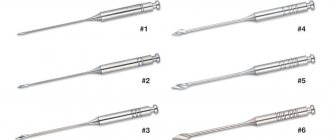
Today there are 5 main washing methods used:
- Manual.
- Ultrasonic.
- Sound.
- Laser.
- Hydrodynamic.
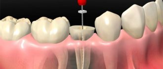
In classical irrigation, a special syringe and an endodontic needle are used, which perfectly cleans the coronal and middle third of the canal.
However, these instruments do not reach the apex area. For optimal results, the irrigator must treat the entire tooth cavity.
At the moment, the ultrasonization technique is considered the most effective. When exposed to ultrasonic waves, a cleaning solution is supplied to the cavity.
The antibacterial effect increases when using sodium hypochlorite, but it is very important to control the temperature (overheating of the irrigant can cause undesirable reactions).
Another method is sonic irrigation. In this case, cleaning takes place under the influence of sound vibrations. This method is much more effective than manual cleaning. In addition, disposable tips are used for irrigation, which increases the antiseptic effect.
The video provides additional information on the topic of the article.
Key Factors for Effective Cleaning and Irrigation of the Root Canal System
- Thorough diagnosis of existing pulp-periodontal pathology
- Taking into account the condition of dental tissues and the complexity of the anatomy of the root canal system
- Removal of the endodontic smear layer
- Compliance with indications when choosing irrigation products
- Optimization of active components of irrigation solution
- Correct sequence of application of irrigant solution during root canal treatment
- Mandatory costs of at least 5 minutes for irrigation before filling
Effectiveness factors
In order to get the maximum result during the procedure, you must consider the following:
- The dentist must conduct a thorough examination of the pulp and periodontal tissues.
- Take into account the tissue structure of the problem element and the structure of the root canals.
- Remove the top layer of dentin.
- Follow all safety rules when working with aggressive irrigator forks.
- Follow a consistent algorithm of actions during root disinfection.
- Allow at least 5 minutes for irrigation before filling.
Only by fulfilling all these conditions can the doctor achieve effective cleaning and disinfection of cavities. The slightest mistake increases the risk of developing associated complications.
Pros and cons of the method of vertical condensation of gutta-percha, technique.
Come here to learn more about the purpose of profiles in dentistry.
At this address https://www.vash-dentist.ru/lechenie/zubyi/plombyi/kak-rabotaet-atatsamit.html we offer detailed information about cement for filling Atatsamit canals.
Diagnosis of pulp and periodontal pathology
The primary concern that arises from the very beginning of endodontic treatment is the risk of bacterial contamination, even in teeth with viable pulp. Already 40 years ago, in 1960, when cultural methods of microbiological research were not as advanced as they are now, Engstrom showed that viable inflamed pulp in a closed tooth cavity is characterized by microbial contamination in more than 7% of cases. This indicator is characterized by an exponential increase to 80% in chronic pulpitis, acute apical periodontitis and pulp necrosis. Root canal infection is usually associated with the presence of radiological signs of periapical pathology and clinical symptoms of acute or chronic periodontitis. According to the Abourass and Bogen classification, periapical lesions are divided into open and closed, regardless of the associated symptoms. Open lesions most often contain opportunistic microflora of the oral cavity, penetrating directly through the pulp or through the extrapulpal portal of entry. This group includes foci of infection associated with poor-quality endodontic treatment and violation of the marginal seal of restorations after endodontic treatment (Fig. 1, 2).
Rice. 1
Rice. 2
Closed lesions are characteristic of teeth with pathology of the pulp or apical periodontium, which do not have signs of direct penetration of the microflora of the oral cavity. This category includes lesions in the area of teeth with symptoms of severe obliteration of canals or with post-traumatic necrosis of the pulp (Fig. 3), as well as persistent periapical lesions after adequate endodontic treatment. The challenges in treating open and closed lesions differ significantly. Open lesions are associated with contamination by microflora of the oral cavity, therefore the prognosis for the treatment of such lesions is favorable provided that the root canal system is completely cleaned and all entrance gates of infection are hermetically sealed in order to prevent further infection.
Rice. 3
The origin and location of the microflora associated with closed lesions often remains a mystery and is one of the most controversial issues in scientific circles: are bacteria present in a periapical granuloma, and if so, in what quantity? And, in the case of bacterial contamination, are the microorganisms localized to the root area or is there a primary periradicular infection while the apex remains sterile?
Abou-Rass et al conducted a study of a series of closed lesions selected according to strict criteria. During the study, bacterial infection was detected in all cases, with 63% being obligate anaerobic microflora, and 36% being facultative anaerobic pathogens.
Among the identified microorganisms, the authors indicate strains
- Actinomyces (22.7%)
- Propionibacterius (18.2%)
- Streptococcus (13.6%)
- Staphylococcus (4.6%)
- Porphiromonas gingvalis (4.6%)
- and Gram-negative enterobacteriaceae
All studied samples taken from the root apex area contained microorganisms, while no infection could be isolated from the surgical access area or from the peripheral zone of the apical lesions. Data obtained by Abou-Rass suggest that periapical infection is localized mainly in the root apex area, and only in some cases do microorganisms spread deep into the periapical tissues. Contrasting data obtained by a number of authors, who identified infection of endodontic origin in periapical lesions, may be explained by the high likelihood of contamination during sample collection due to the difficulty of strictly adhering to the sampling protocol. This conflicting opinion corresponds to opposing therapeutic approaches. If we assume that a granuloma can be primarily infected when the root apex is sterile, we can prescribe the patient a course of antibiotic therapy for at least a few weeks with full confidence that this will lead to the elimination of the granuloma. On the contrary, if we consider the root canal system to be the primary source of infection, we are left with only two options: clean and disinfect the canals or, if this is not possible (obliterated or impassable canals, complex canal topography), apply periapical surgery.
Diagnostic measures
Most problems associated with endotherapy are caused by the presence of bacterial microflora. Infection is accompanied by signs of periodontitis.
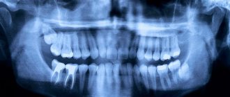
Therefore, before irrigation and disinfection, it is necessary to undergo diagnostic measures , which include x-ray diagnostics.
In this case, the image will clearly show signs of pathology in the periapical area.
The most common types of microorganisms that cause disorders are streptococci, staphylococci, actinomycetes, propionic acid bacteria and enterococci.
It is very important to accurately identify the pathogen in order to select the optimal solution for irrigation. Otherwise, you should not count on high disinfection efficiency.
Characteristics of the root canal system and hard dental tissues
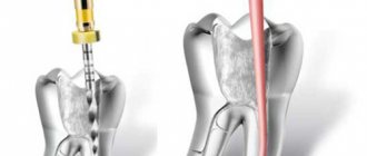
The root canal system can have a very complex morphology, which is often characterized by the presence of lateral canals and anastomoses, and a loose structure in the apical part. Complete cleaning, shaping and sterilization of root canals is not possible in all cases (Fig. 4).
Rice. 4
The histological structure of the root canal is even more complex: as you can see, from the center of the canal to the periphery it is represented by the following tissues: pulp tissue, odontoblast layer, predentin, i.e. a dentin zone corresponding in mineral composition to the concept of “dentin mineralization boundary” (Fig. 5) and dentin with a complex tubular structure system.
Rice. 5
The number and orientation of dentinal tubules may vary depending on physiological characteristics or pathological changes. According to Garberoglio and Brannstrom, the concentration of dentinal tubules can range from 20,000 to 40,000/mm2, and the average diameter is in the range of 2-4 µm. Dentinal tubules create a complex three-dimensional communication system between the oral cavity and the root canal system: in teeth with a viable pulp, the lumens of the dentinal tubules are filled with processes of odontoblasts and dental cerebrospinal fluid, which provide protection to the pulp by forming a barrier to the penetration of bacteria and their toxins. (Fig. 6).
Rice. 6
In the event of pulp death, dehydration of the dentinal tubules occurs, in the lumen of which only tissue decay of the processes of odontoblasts remains; along the lumen of the tubules, the migration of microorganisms, toxins, and medications that are capable of penetrating dentin easily occurs (Fig. 7).
Rice. 7
Infection of dentinal tubules by various types of microorganisms has been demonstrated in many studies: Buck reports the ability to penetrate dentinal tubules, characteristic of Micrococcus Luteus and Bacillus megaterium, Berkiten - for Streptococcus sanguis, Prevotella intermedia, Actinomyces naeslundii, Siqueira - for Porphyromonas endodontalis, Fusobacterium nucleatum, Actinomyces israelii, Porphyromonas gengivalis, Propionibacterium acnes, Enterococcus faecalis, in the work of Waltimo - for Candida Albicans. In general, dentinal tubules can harbor bacteria from both the oral cavity and the root canal system. Since these bacteria can lead to failure of endodontic treatment, they must be eliminated.
Irrigator models
Today, the oral irrigator market is divided into main types:
- family;
- portable;
- stationary.
The family irrigator has a large capacity that can be attached to the wall in the bathroom. The device has a power switch for changing the pressure of the water jet and several nozzles.
The travel device runs on a battery and is indispensable on trips and hikes where there is no opportunity to brush your teeth.
Stationary irrigators include devices connected to a tap. Their advantage is that they do not require electricity to operate. This device is equipped with a special valve to reduce water pressure (if necessary). I am attracted by its noiseless operation, safety and compactness. Like a portable irrigator, you can take it with you on a business trip or vacation. You don't have to worry about the battery running out.
All irrigators include a set of attachments:
- for cleaning the tongue;
- for washing and massaging gums;
- washing away food residues in hard-to-reach places.
Removal of the endodontic smear layer
During the preparation of hard tooth tissues with hand or machine instruments (high-speed or low-speed burs, curettes, endodontic instruments), a microscopic layer of sawdust is formed on the surface of the dentin (Fig. 8).
Rice. 8
The thickness and composition of this layer vary depending on the properties of the hard tissues being processed and the characteristics of the cutting tools, but a mandatory feature is the presence of organic and inorganic components in it. The smear layer formed during endodontic treatment is characterized by a high content of organic components in the form of pulp fragments, odontoblasts, and weakly mineralized predentin. At the same time, there are also inorganic components, the source of which is dentin. In this regard, to remove the smear layer from the walls of the root canal, it is necessary to use solutions that are effective against both organic and mineral components. The smear layer of the root canal, first described by MacComb and Mader, is tightly connected to the root canal wall through "smear plugs" embedded in the dentinal tubules. The thickness of the superficial smear layer ranges from 1 to 6 µm, while the depth of its penetration into the dentinal tubules can be higher, reaching 50 µm (Fig. 9).
Rice. 9
According to several authors, the smear layer of the root canal should not be removed, since it reduces the permeability of the dentinal tubules to microorganisms and their toxins by sealing the dentinal tubules. But in a CEM microphotograph of the root canal dentin surface covered with a smear layer, it is easy to notice that this layer does not have a uniform, homogeneous, dense consistency, but, on the contrary, is interrupted in many areas, that is, it does not form an airtight barrier. Moreover, the smear layer of the root canal can contain microorganisms and serve as a breeding ground for them, as well as disrupt the adhesion of filling materials to the walls of the root canals (Fig. 10).
Rice. 10
Due to the above, the smear layer of the root canal must be completely removed. After removing the smear layer, the lumens of well-cleaned dentinal tubules with gaping and moderately widened orifices are clearly visible on the walls of the root canal. Peritubular dentin, highly sensitive to EDTA or acid solutions, is completely dissolved. While intertubular dentin may exhibit moderate demineralization phenomena. Filling materials, such as root cements or gutta-percha, when applied correctly, easily penetrate and obstruct open dentinal tubules, promoting the tightness of the root filling and preventing the possibility of further penetration of infection into the root canal system (Fig. 11, 12).
Rice. eleven
Rice. 12
Irrigation solutions
a) Water
Water is characterized by high biocompatibility and low cost. This makes it quite suitable for mechanical jet washing of bacteria due to the return flow of liquid. Unfortunately, routine canal rinsing with water does not completely sterilize the root canal system or eliminate the smear layer formed during instrumentation. However, we consider jetting the canal with water to be an important component of the process of cleaning and shaping root canals, which ensures the removal of large tissue fragments, washing out temporary filling materials from the canals, and can also be used for emergency rinsing of the periapical area in case of accidental removal of solution beyond the root apex sodium hypochlorite. To reap the full benefits of jetting, we highly recommend using a Stropko Surgical Irrigator, preferably with a fine plastic needle to reduce water and air pressure.
b) Chlorine-containing solutions
■ Sodium hypochlorite
Sodium hypochlorite (NaOCl) has both oxidizing and hydrolyzing properties: it has bactericidal and proteolytic effects. The solution was proposed for use as a wound irrigant no later than 1915, and began to be used as an irrigant solution for endodontics around 1920. Sodium hypochlorite is traditionally prepared by passing chlorine gas through a solution of sodium hydroxide, according to the following reaction:
Cl2 + 2NaOH -> NaOCl + NaCl + H2O (Clarkson)
Ready-to-use sodium hypochlorite solutions for endodontics have a pronounced alkaline reaction, hypertonic properties and a nominal concentration of 1-5% active chlorine. It is important to remember that many NaOCl solutions used for root canal irrigation are household bleaches and contain other components such as stabilizers (sodium chlorate and sodium hydroxide), cleaning additives (surfactants, fatty acids), and fragrances. In this regard, it is preferable to use solutions specially designed for endodontic use and with an indication of expiration date and concentration on the packaging. The popularity of NaOCl as an irrigation solution for endodontics is determined by the general availability and low cost of the solution. Many studies have demonstrated its antiseptic and solvent properties. In particular, NaOCl has a rapid bactericidal effect against vegetative forms, spore-forming bacteria, fungi, protozoa and viruses (including HIV, rotavirus, HSV-1 and -2, hepatitis A and B viruses). The exact mechanism of the antimicrobial activity of NaOCl is not entirely clear, but it may be determined by the formation of hypochlorous acid and the release of active chlorine, which leads to the oxidation of sulfhydryl groups of important bacterial enzymes. However, there is no consensus on the optimal concentration of sodium hypochlorite for use in endodontics. Spandberg demonstrated that a 5.25% sodium hypochlorite solution was too toxic and that the bacterial strains most common in infected root canals could be eliminated with a much lower concentration of the solution. On the other hand, Th and Wesilsoy showed that 5.25% NaOCl did not differ in toxicity from a hypochlorite solution of lower concentrations, as well as from a solution of chlorhexidine bigluconate. Even data on antimicrobial activity are contradictory. Some studies have found no significant difference in antibacterial effectiveness between 0.5 and 5% NaOCl, while other studies have reported that the antiseptic effect is significantly reduced after dilution. Heling emphasizes the importance of exposure time, which should not be less than 10 minutes, in order to allow sodium hypochlorite to have a bactericidal effect even against resistant species of microorganisms. Sodium hypochlorite has pronounced dissolving properties against pulp residues, even those located in the lateral and additional canals (Fig. 13, 14), and is also partly effective against the collagen matrix of predentin; however, its effect on calcospherites and on the inorganic smear layer matrix is negligible (Fig. 15).
Rice. 13
Rice. 14
Rice. 15
The dissolving effect is determined by the concentration of sodium hypochlorite: the maximum severity of the effect is manifested in a 5% NaOCl solution. To increase the effectiveness of sodium hypochlorite as a solvent for tissue decay, it is recommended:
- use a heated solution with a temperature of about 40°C;
- activate and heat the solution using ultrasonic files;
- use temporary root canal filling with calcium hydroxide to take advantage of the synergistic effect of these two substances;
- Use in combination with surfactants to enhance the penetration of sodium hypochlorite into dentinal tubules, thereby reducing the number of intratubular bacteria.
■ Sodium dichloroisocyanurate
Sodium dichloroisocyanurate is a chlorine-producing disinfectant and is usually used to disinfect contaminated surfaces and solutions. Coates reports that sodium dichloroisocyanurate solution contains 4,000 ppm active chlorine, exhibiting a bactericidal effect similar to a NaOC solution containing 17,000 ppm active chlorine. In the presence of organic material, the antiseptic and solvent properties of sodium dichloroisocyanurate are significantly higher than those of NaOCI. HeIing in a recent study evaluated and compared the bactericidal effect of sodium dichloroisocyanurate and NaOCI against the following pathogens: Streptococcus sobrinus, Streptococcus saIvarius, Enterococcus faecaIis, Streptococcus mutans. The data obtained indicate that the minimum inhibitory and bactericidal concentrations relative to the studied bacteria were similar for both solutions studied. Overall, sodium dichloroisocyanurate may be an effective alternative to NaOCI, even if further research and clinical experience will be required to confirm the preliminary data.
c) Hydrogen peroxide
Hydrogen peroxide is widely used as a solution for washing root canals alternately with NaOCl due to the short-term but pronounced foaming effect when mixing these substances, which contributes to the mechanical leaching of tissue debris and microorganisms from the canal. According to Grossman, the benefits of using NaOCl and H2O2 in combination are:
- Foaming
- Solvent effect of NaOCl
- Disinfecting effect of both solutions
- Whitening effect of both solutions
The interaction between NaOCl + H2O2 is classically described by the following reaction:
NaOCl + H2O2 -> NaCl + H2O + 2•O•2
(where O• is atomic oxygen converted to O2). Shiozawa insists that the reaction is much more complex and results in the formation of two reactive oxygen species (ROS): the superoxide anion radical (O2-) and the hydroxyl radical (•OH). Both ROS are active and demonstrate a more pronounced antibacterial effect against Streptococcus strains and Staphylococcus aureus than pure hydrogen peroxide. However, ROS reactive radicals can cause irritation to periapical tissues; In this regard, it is recommended to rinse the root canal with clean distilled water before filling. An acceptable alternative to H2O2 is Glyde (Dentsply-Maillefer®reg;). Glyde is a clear gel containing 15% EDTA and carbamide peroxide in a water-soluble methylcellulose base. Alternating Glyde and sodium hypochlorite solution provides a pronounced and long-lasting foaming effect as well as a bactericidal effect, while 15% EDTA provides a dissolving effect on mineralized sawdust. In addition, Glyde has beneficial lubricating and whitening properties. The only limitation to the use of Glyde is the slight tendency to underclean the apical third of the root canal, as demonstrated in our recent study. To completely remove these contaminants (it is not entirely clear whether they are gel residue or a mixture of gel and smear layer), it is necessary to use a liquid form of EDTA activated by ultrasonic files (Fig. 16).
Rice. 16
d) EDTA components
EDTA is commonly used in endodontics as a liquid or gel as a chelating agent that extracts calcium ions from the hydroxyapatite, thereby dissolving the mineral fraction of the root canal smear layer. The effectiveness of EDTA solutions depends mainly on their pH. The optimal pH value should be between 6 and 10. Most formulations are based on disodium EDTA, which has a neutral pH and chelates isomolar calcium until the EDTA is completely consumed. This explains the importance of constantly updating the solution in the root canal to use active EDTA. O'Connell, in a recent study, indicates that the isolated use of EDTA without sodium hypochlorite did not completely remove the smear layer of the root canal in any case. This explains the advisability of alternating EDTA-containing agents and hypochlorite during endodontic treatment; their combined effect provides an excellent degree of cleaning of the dentinal walls in the apical third of the root canal, provided that both substances are brought to the apex and activated using ultrasound or hand files (Fig. 17).
Rice. 17
Surfactants such as Centrimide are often added to preformulated EDTA solutions (Largal Ultra® Septodont or R-EDTA®) to enhance their penetration into accessory canals and dentinal tubules. EDTA-containing gels include RC-Prep® from Premier Dental and Glyde® from Dentsply Maillefer.
e) Acidic components
The acids used in endodontics for washing canals are phosphoric and citric in concentrations from 6% to 30%. Acid solutions are highly effective in removing the mineral component of the smear layer of the root canal and in the treatment of obliterated canals. However, since their effectiveness as antiseptics and organic solvents is limited, combined use with sodium hypochlorite is recommended. In a recent study, we examined the effectiveness of two different irrigation solution combinations (NaOCl + EDTA and NaOCl + phosphoric and citric acid) for smear layer removal. Our results indicate that both concentrations were effective, although the use of EDTA was characterized by a more gentle effect on peritubular and intertubular dentin, while phosphoric acid demonstrated a more aggressive effect with signs of dentin demineralization on the canal walls (Fig. 18).
Rice. 18
Since acid solutions are quickly inactivated in the presence of mineralized sawdust, it is necessary to periodically refresh the solution, especially if it becomes cloudy or begins to dry out. After using acids, it is recommended to rinse the canal with distilled water, since there is a tendency for crystallization and precipitate to form on the canal walls.
f) Chlorhexidine
The antibacterial effect of chlorhexidine gluconate at a concentration of 0.2 to 2% has been shown in many studies. Leonardo in an in vivo study showed the elimination of 100% of streptococcus mutans and 78% of anaerobic microorganisms using a 2% solution of chlorhexidine gluconate. Vahdaty confirmed the disinfectant effect of chlorhexidine against bacteria located in the dentinal tubules, Hakan-Sen revealed an antifungal effect against Candida albicans. Since the dissolving effectiveness of chlorhexidine relative to organic and mineralized tissues is not pronounced, it is recommended to alternate it with NaOCl solutions. Moreover, when used in combination, these two solutions are characterized by a summative effect, possibly due to the formation of chlorhexidine chloride components, which increase the ionizing properties of the chlorhexidine molecule. Ready-made forms based on chlorhexidine include “Cetrexidin®” manufactured by VEBAS. The drug contains 0.2% chlorhexidine in combination with 0.2% cetrimide, a surfactant ammine, which reduces the surface tension of the product, increasing its penetration and antiseptic properties. Chlorhexidine can finally be used for short-term temporary filling of root canals. In fact, this drug inhibits microbial activity “in vivo” within 48 hours after use.
g) Alcohol
The use of alcohol as an irrigation solution is not recommended. Yesilsoy found that 11.6% alcohol was extremely ineffective against most microorganisms; Ayhan obtained more favorable results only when the concentration was increased (21%). On the other hand, final rinsing of the root canal with alcohol is recommended to ensure maximum dehydration of the canal walls, which facilitates deeper penetration of root cement into the dentinal tubules and improves the tightness of the obturation.
Optimization of irrigation solutions
In 1917, Walter Hess published images of the root canal system after perfusion with Indian ink dye, which convincingly proved the high degree of diversity and complexity of the morphology of the root canals, which in some cases makes their complete cleaning and obturation impossible. Kim, based on Hess's illustrations, created three-dimensional images of root canals using computer modeling. Computer analysis of these models showed that up to 93% of the lateral canals and 96% of the branches of the apical delta are located in the terminal 3 mm of the root. Therefore, it is extremely important to provide sufficient volume of irrigation solution to the apical third of the root to optimize canal cleaning.
Possible ways to increase the cleaning efficiency of irrigation solutions are:
- Increasing the diameter of the apical part of the root canal
- Increasing the total volume of irrigation solution
- Excretion of irrigation solution directly into the apical region
- Prolonging the exposure of the irrigation solution to microorganisms and tissue decay
- Mechanical activation of irrigation solutions.
The first three methods are interrelated: when the lumen of the apical part of the canal expands, it becomes possible to immerse the needle of the irrigation syringe deeper and directly deliver the irrigation solution to the apical part of the canal. The use of extra-taper nickel-titanium instruments easily and quickly provides “deep shaping”, allowing a 27- or 30-gauge endodontic needle to be inserted directly into the apical third of even curved root canals (Fig. 19-21).
Rice. 19
Rice. 20
Rice. 21
Regarding the preferred type of needles, we recommend those with a rounded sealed tip and a lateral opening to prevent irrigant leakage into the periapical tissue. Syringes with irrigation solution should ideally be stored at a temperature of 37 ° C in a special electric thermostat: “Siringe warmer” (Vista-Dental®) or “Appli-Vac System” (Vista-Dental®), which allows the use of up to 6 different irrigation solutions, simply by switching from one to the other using the buttons on the irrigation gun. For curved root canals, the use of flexible nickel-titanium needles (Vista-Dental®) may be recommended. Insulin needles (too short), anesthesia needles (too sharp), or syringe needles (too thick) are not recommended. When the solution is in the apical third of the canal, it must be activated with an ultrasonic file. You can use ultrasonic devices that provide direct injection of NaOCl or EDTA into the canal through the tip, or you can simply fill the canal with an irrigant solution, immerse the ultrasonic file in it and keep it running for a while until the solution becomes cloudy, which indicates the need for it. refreshment. The mechanism of activation of solutions by ultrasound is not entirely clear: the important components are considered to be the development of acoustic vortex flows, the process of cavitation (formation of microbubbles), mechanical stirring and heating of the solution. Using 5% and 12% sodium hypochlorite and ultrasound, Huque demonstrated complete elimination of the smear layer and disinfection of the canal surface, outer and deep layers of the root canal wall on extracted teeth. Similar results were obtained by Turkun, Ahmad, Cameron. It is recommended to use the endodontic file only after completing the root canal preparation, so that when deeply immersed in the canal, the instrument does not come into contact with the walls. The free oscillating vibration of the tip of the endodontic ultrasonic file improves acoustic vortex flows and, at the same time, reduces the risk of damage to the canal wall (Fig. 22).
Rice. 22
Irrigators in everyday life
If you conduct a survey among patients in dental clinics, you will find that few have heard of household irrigators.
The number of patients using irrigators at home is only a few. The irrigator is indispensable for high-quality cleaning of the oral cavity and massage of the gums. A thin stream of liquid under pressure washes away food debris from places where a toothbrush cannot reach, and effectively removes plaque. This daily procedure serves as a reliable prevention of diseases of teeth and gums. In addition, the irrigator can be filled with a special solution for rinsing teeth, which enhances the therapeutic effect.
Considering that in our fast-paced times, few people spend enough time brushing their teeth for five minutes, it’s worth thinking about purchasing a device. Irrigation can serve as a reliable way to clean the oral cavity during travel, business trips, and at work after lunch. It is especially recommended to wash away food debris using an irrigator for people who have dentures. Many people know how difficult it is to clean the cavity under a fixed bridge with a toothbrush. A jet of water under pressure will do a great job of solving this problem.
Irrigation sequence during root canal preparation
- A. Remove the roof of the pulp chamber and use a jet wash with water or sodium hypochlorite to remove residual pulp and identify root canal orifices;
- b. Begin instrumental treatment of the canal lumen, alternating it only with sodium hypochlorite;
- c. When starting to excise dentin, fill the canal lumen with Glyde material;
- d. Continue instrumenting until Glyde becomes cloudy and viscous;
- e. Rinse the channel with sodium hypochlorite until foaming stops;
- f. Complete instrumentation by ensuring that the canal is always filled with Glyde and flushing the canal with sodium hypochlorite after every 3-4 instruments;
- h. In infected canals, alternate rinsing the canal with NaOCl and chlorhexidine.
Irrigation before filling
>Before filling, we have one last chance to remove the smear layer and disinfect the root canal system; Usually for this purpose the combined use of a solution of sodium hypochlorite with EDTA or citric acid is recommended. The effectiveness of final irrigation depends on the chemical properties and concentration of the solution, as well as on the total volume of solutions and the duration of exposure in the canal (Fig. 23-26).
Rice. 23
Rice. 24
Rice. 25
Rice. 26