Tooth canal treatment: when is it necessary and how is it carried out?
Tooth canal treatment is a procedure that is carried out for certain indications and allows you to stop active inflammation in the internal tissues of the tooth and avoid the removal of a dental unit. Treatment of tooth canals has its own specifics and is complex; only an experienced and competent specialist can carry out the procedure efficiently. In the article we will look in detail at the process of root canal treatment: we will find out under what circumstances the procedure is carried out, how it goes, what methods are used for treating tooth canals.
Where is the tooth canal located, how is it structured
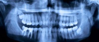
Human teeth have almost the same anatomical structure, including three main parts: crown, neck and root. Inside the tooth there is a pulp chamber, from which canals originate and extend all the way to the root. The pulp chamber contains the pulp or nerve of the tooth, which is a bundle of nerve fibers and blood vessels. Nerve fibers and vessels also pass through the entire internal space of the dental canal. The shape of the tooth canals can be either straight or curved, and their number varies depending on the type of tooth. Thus, the canines and incisors of the upper jaw have one canal, the canines and incisors of the lower jaw have two. There may be two or three canals in molars, and the maximum number of canals is observed in wisdom teeth - from three to five. The exact number of dental canals is determined using radiographic examination.
As mentioned above, the shape of the canals can have bends and turns, which complicates the process of their high-quality cleaning and treatment. Meanwhile, when treating canals, it is important to thoroughly clean their internal space - otherwise it will not be possible to stop the inflammatory process and save the diseased tooth.
Indications for dental canal treatment
Tooth canal treatment is a specific dental procedure that is advisable to perform in the following clinical cases:
When an active inflammatory process is detected inside the root of a dental unit. The inflammatory process in this part of the tooth can lead to gradual tissue necrosis. If tissue necrosis begins, the diseased tooth will have to be removed.
The condition is diagnosed by radiography;
Treatment of tooth canals is required for pulpitis - inflammation affecting the pulp bundle;
Endodontic treatment is carried out for periodontitis, a disease affecting the apical part of the tooth root;
In some cases, cleaning and treatment of dental canals is required for advanced forms of caries.
Dental canal treatment is also indicated for abscesses, the onset of an inflammatory process under an old filling, or a fractured tooth root. The need for measures to treat tooth canals may be indicated by such symptoms as: severe, excruciating pain in the tooth, occurring mainly at night, swelling of soft tissues, discoloration of the gums and tooth enamel.
In some cases, tooth canal treatment is carried out before prosthetics. The nerve is removed from the tooth, and the canal cavities are cleaned, treated with antiseptic drugs and filled.
When is the procedure applied?
Before performing root canal treatment, the doctor conducts a thorough examination of the tooth, including clinical and x-ray examinations. Endodontic therapy, that is, work with root canals, is carried out if:
- Pulpitis is inflammation of the pulp. Symptoms of the condition: painful sensations, increased pain at night.
- Necrosis is the death of the pulp; it can occur asymptomatically and is detected by x-ray or complete tooth destruction.
- Swelling – increased sensitivity, swelling of the gums, change in color.
- Neoplasms on the gums - can be periodic or constant in the form of lumps, fistulas, different in size.
- Trauma – after physical impact after an accident, fight, etc. The integrity of the tooth may be damaged, including the root. Some of the pathologies are asymptomatic, but it is worth paying attention to the color of the tooth - if it darkens, an inflammatory process occurs or increases.
Main stages of root canal treatment
Treatment of tooth canals is a complex, specific process that includes several successive stages. Below we will consider all stages of the treatment process in detail.
Diagnostics
During the initial visit to the dentist, the doctor will conduct a thorough examination of the patient’s oral cavity and will also prescribe x-rays. An image of the tooth will allow you to assess the condition of the dental canals, see their number, determine the stage of development of the inflammatory process, and also select adequate treatment for a specific clinical case.
Anesthesia
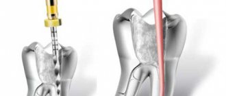
To gain access to the canals of a tooth, the dentist will drill out the coronal part of the tooth to reach the pulp chamber, behind which the canals are located. Both the pulp and the canals are penetrated by nerve fibers and therefore the specialist’s actions can cause some discomfort to the patient. To relieve a person of discomfort, local anesthesia is performed before starting root canal treatment. The drug is selected individually for each patient.
Moisture insulation of the area of the diseased tooth
To prevent pathogenic microorganisms that may be contained in saliva from entering the internal space of the tooth, the area of the diseased tooth is first protected from moisture by covering with a special latex bandage - a rubber dam.
Reaming a tooth
To gain access to the dental canals, the doctor will drill the tooth, removing all tissue affected by caries. The access hole is made either on the chewing surface of the tooth (when treating molars) or on the inside (when treating incisors and canines). After drilling, the doctor will remove either the entire pulp bundle or only part of the tissue affected by the inflammatory process.
Treatment of dental canal cavities
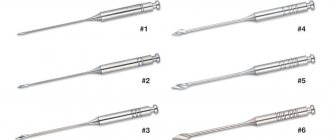
For high-quality treatment of dental canals, a specialized tool is used - files. Before the cleaning process, it is important to obtain detailed information on the structure of the dental canals, as well as their length. To do this, a photograph of the diseased tooth is taken, and a special type of device is used - an apex locator. The dentist will use the files to clean the canal cavities, and then rinse them with an antiseptic solution and treat them with antibacterial agents. During cleaning and antiseptic treatment, the dentist will expand the cavity of the canals, clean and smooth their internal surfaces. Then the cavities of the dental canals are thoroughly dried with paper points.
Next, the doctor puts medicine into the canal cavity, places a temporary filling on the tooth and sends the patient home. After a few days, you should come back to the dentist to evaluate the results of the treatment. If the image shows that the inflammation has been stopped, the canals are filled.
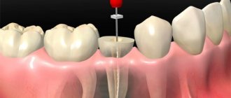
Sealing
Gutta-percha pins are used to fill dental canals. It is important that the dental canal is sealed correctly and efficiently, otherwise a recurrence of inflammation is possible and the tooth cannot be saved. After filling the canals, the crown part of the tooth is restored with a photopolymer filling or orthopedic structures. It will be useful to know that mild pain in the tooth within 14 days after the procedure is considered normal by dentists. Taking painkillers prescribed by your doctor will help eliminate discomfort. However, if the pain does not go away, its character is sharp, and does not subside even after taking anesthetics, you should immediately contact the dentist!
Material requirements
To install a temporary filling, a specialized material is used that can be easily removed by the dentist if necessary. The composition for temporary filling must meet the following requirements:
- X-ray resistant. To check the correctness of the filling, the dentist prescribes an x-ray examination. The image allows you to timely identify the presence of cracks in the filling material and eliminate them.
- Good grip on the surface. It is necessary to exclude the formation of microcracks or incomplete adhesion of the material to the tooth tissue. Violation of this rule may result in medication entering the oral cavity.
- Elasticity. The material should easily fill the pores formed in the tooth tissue.
- Strength. The material is installed by the dentist for a certain period of time. When eating, the filling is subjected to high stress. In this regard, the seal must be resistant to mechanical damage.
- Harmlessness. The filling should not cause allergic reactions or negatively affect human health.
- Easy to remove. At your follow-up appointment, the doctor quickly removes the temporary filling.
A mandatory requirement for temporary material is a high curing speed. The filling completely hardens in 2 – 3 hours.
How long does root canal treatment take?
The duration of tooth canal treatment depends on the complexity and characteristics of the clinical case, as well as the number of canals that the specialist will have to treat. On average, the duration of the procedure can range from 20 minutes to 1.5 hours.
It is also worth considering that for high-quality root canal treatment, you will have to visit the dentist’s office several times.
Is it painful to treat root canals?
Thanks to the use of modern anesthetics, root canal treatment has become a painless procedure. The patient may feel mild discomfort only during the injection of the anesthetic.
Therefore, there is no need to be afraid of root canal treatment; moreover, you must remember that you should never postpone the procedure due to fear of the dentist! Inflammation in a tooth that has progressed to a certain stage of development cannot be cured, which means that the diseased tooth will have to be removed and a prosthesis put in its place. In addition, more serious complications may occur.
How long can you walk with a temporary filling?
The material used for temporary filling is highly durable. The filling does not break down for a long time. The specialist determines the period of wearing temporary filling material based on the nature and degree of neglect of the disease. The duration of wearing the filling is individual for each patient.
At the end of the established period, the person must immediately contact a specialist. An increase in the time period can lead to the development of diseases. In some cases, the filling may crumble at the end of its period, and the drug will negatively affect the patient’s body.
Modern method of treating tooth canals: treatment of the canal cavity under a microscope
The quality of dental canal treatment depends on the correctness and accuracy of cleaning and processing of their internal space. This is a very painstaking work, since the channels themselves are quite narrow (no more than one millimeter), and in addition, they can have bends and turns. If a specialist makes a mistake when treating the canals and does not remove particles of the affected dentin, a relapse of the inflammatory process is guaranteed. The use of a special tool in treatment - a dental microscope - will help eliminate possible errors and inaccuracies in the treatment of dental canals.
Using a microscope, the dentist will perform a high-quality removal of all infected tissues, while preventing injury to healthy tooth tissues. The use of the device simplifies the process of cleaning and processing channels with significant lengths, anomalous structure,
with curvatures and branches. In addition, the use of a dental microscope during the treatment of tooth canals improves the quality of tooth restoration as a whole, and also contributes to the timely detection of hidden caries.
At our dental clinic “Uni Dent” in St. Petersburg, you can receive services for dental canal treatment under a microscope. The procedure is carried out by highly qualified doctors using modern instruments and materials, and therefore when you contact us, you can rest assured that the treatment result will be of high quality! Dentistry "Uni Dent" - come to us for a beautiful smile and painless dental treatment!
Smear layer: to remove or not
When the endodontic instrument comes into contact with the root canal wall, a smear layer is formed. The smear layer consists of the remains of dentinal filings, microorganisms and their toxins. The organic part consists of proteins, microbes, cells, and pulp residues. The inorganic part consists of dentin components.
It has been studied that the smear layer is able to penetrate to varying depths into the lateral tubules, spreading the infection throughout the root canal system. At the same time, it is a physical barrier to microorganisms. It has been scientifically proven that the presence of a smear layer disrupts the adhesion of the sealer in the canal, promotes microleakage, which triggers even greater bacterial growth. Being an obstacle to the penetration of sealer and irrigant into the tubules, it significantly reduces the quality of permanent root canal filling.
The organic component is a breeding ground for bacteria and is easily removed by sodium hypochlorite. The inorganic component of the smear layer is unstable to EDTA and citric acid. The mechanism is associated with demineralization of peritubular dentin and opening of dentinal tubules.