Tooth extraction is a surgical procedure that can lead to complications. Especially if the operation is performed incorrectly when the root configuration is complex, the enamel is fragile, or when the tooth has crumbled but the root remains. Today we will look at all the reasons why fragments of teeth remain in the gums, and we will tell you whether it is worth removing fragments of crowns and roots.
Reasons why tooth fragments remain in the gums
Pieces of teeth remain in the following cases:
- Removal of wisdom teeth or multi-rooted teeth of complex configuration. If the tooth is severely damaged or the enamel is thinned, the walls may crack when it is removed. A small piece goes unnoticed by the doctor, and fragments measuring 1-3 mm are often not visible even on a control x-ray. In such cases, everything depends on the professionalism of the dentist - the more experience, the less likely it is that after tooth extraction there are fragments left in the gums.
- Crushing of an adjacent tooth during extraction. Sometimes, during the operation, a nearby tooth may be damaged due to fragile enamel or incorrect actions of the dentist. A small fragment gets into the fresh socket of the extracted tooth, and if it is not removed in time, it becomes a cause of discomfort, pain or suppuration.
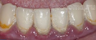
- The crown crumbled, and the remains of the tooth were overgrown with gum. This applies to pulpless teeth - a living nerve would not allow the patient to delay a visit to the dentist. Most often this happens when the old filling is destroyed, and caries gradually eats up the crown part, reaching the roots. Remains of a tooth root overgrown with gums become a breeding ground for infection for neighboring units, negatively affecting the gums.
Important!
If you see a dentist in time, you can save on prosthetics or implantation. Today, doctors restore a tooth, even when a minimal amount of tissue from the crown of the tooth remains and the root is untouched by caries. The remainder of the tooth root is more vulnerable to pathogenic bacteria - it lacks integrity, and the microflora penetrates deep into the tooth very quickly. A weak, fragile and carious root cannot serve as a support for an artificial crown and must be removed.
Why does wisdom tooth hurt?
Wisdom teeth almost always grow crooked. Problems with teething are dictated by the following anatomical features:
- “Eights” appear later than all other teeth. As a rule, the jaw bones are fully formed, the dentition is stable, and the “newbie” simply does not have enough space;
- The third molars do not have milk “predecessors”, and the rudiments of wisdom teeth developed for too long and not in very favorable conditions. Because of this, they erupt chaotically, damaging the adjacent tooth or causing inflammation of the gums.
When talking about the causes of pain associated with one or more molars, it is worth distinguishing between some concepts. Toothache can occur against the background of abnormal eruption, due to deformations of soft tissues of neighboring teeth. This clinical picture is typical only for “eights”; other teeth are cut much more smoothly.
Causes
However, the cause of toothache may also lie in fairly classic causes associated with common dental pathologies: caries, periodontal disease or periodontitis. If you systematize all the reasons that cause tooth pain, you will get the following list:
- The wisdom tooth rests too much on the tissues of the oral cavity, squeezing and injuring them;
- The tooth is rotated around its axis. The greater the angle of rotation, the more tense the dental nerve is, which provokes pain;
- Carious cavities have formed on the surface of the wisdom teeth; if they reach the dental tissues, the pain will intensify;
- Tartar deposits have formed, which rub the gums and cause inflammation;
- Due to the fact that the rapid growth of the “eight” and crooked eruption damaged the root system of the neighboring molar, it is this neighboring tooth that hurts;
- If the wisdom tooth, due to its incorrect position, begins to pinch the jaw nerve, pain occurs in the lower jaw. In this case, neuritis manifests itself in this way;
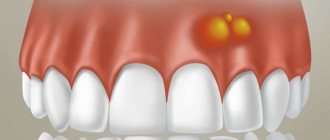
- A follicular cyst has formed at the neck of the tooth. It often occurs against the background of permanent damage to the gum tissue and the tooth itself.
It is worth noting the fact that a follicular cyst near wisdom teeth is often accompanied by all sorts of complications. For example, an abscess may occur that can even deform the jaw in a neglected state. The periodontal tissues may also become inflamed and pericoronitis may develop.
Inflammatory processes of the tissue covering the wisdom tooth
Inflammation of the hood is a common phenomenon. If a wisdom tooth erupts through several crown cusps at once, phenomena may occur when the chewing surface of the crown is partially covered by tissue. The part of the gum and mucous membrane that overhangs the molar and is called the hood. He is constantly susceptible to injury and prone to inflammation.
Under the surface of the hood, particularly favorable conditions arise for the accumulation of various biological “garbage” and the proliferation of pathogenic bacteria. Often even the slightest damage causes irritation. The gums around the “eight” may not hurt much, so they don’t always turn to people with such a problem.
However, if the inflammatory process is severe, the tissues not only swell, but are also irritated. The patient complains of painful swallowing, sometimes it is difficult even to simply open his mouth. There may be pus and severe hyperemia on the gum near the wisdom tooth. Only a doctor can save you from such an obsessive and severe dental inflammation by excision of the hood.
Lack of space during teething
If there is not enough space for the wisdom tooth in the dentition, neighboring tissues are injured during eruption. The pain is total. It hurts not only the place where the molar erupts, but also the neighboring teeth. Swelling, hyperemia, and severe pain that are permanent may also be observed. It is difficult for the patient to open his mouth, especially if the “eights” grow in the direction opposite to the jaw. Such teeth must be removed. This is due to the fact that a crooked tooth can damage the entire dentition, up to its noticeable curvature.
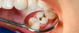
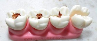
Caries
If a wisdom tooth is affected by caries, the first symptom indicating the disease will be pathological increased sensitivity of the teeth. They will react painfully to any irritants - sour, spicy, salty foods, sweets, cold irritants.
Situations often occur when the curvature of a wisdom tooth injures the neighboring tooth, which causes caries in it. The most dangerous situation is when the “eight” damages the root of the neighboring “seven”. Caries develops just below the gum, and the doctor is not always able to visually identify the carious lesion. At the same time, a severely advanced form of “root” caries is difficult to treat and the tooth can almost never be saved.
Pulpitis
In acute pulpitis that affects the wisdom tooth, patients complain of acute pain that occurs in attacks. The disease and its main manifestations are characterized by a spontaneous nature. Unpleasant sensations can occur spontaneously and last up to 15 - 30 minutes. At night the pain intensifies.
In the chronic form of wisdom tooth pulpitis, the pain is not so pronounced. Unpleasant, aching sensations occur against the background of exposure to cold or heat. If the irritant is removed, the pain continues for some time.
Periodontitis
Periodontitis is characterized by aching pain that constantly worries the patient. At the same time, the diseased tooth will react to any irritant with acute pulsating, shooting pain. Slight mobility of the affected tooth is also noted, and swelling and soreness of the gums are often observed.
It is interesting that with chronic periodontitis the pain is almost not disturbing. Very minor unpleasant sensations can only appear when the “figure eight” is tapped on the patient’s tissues.
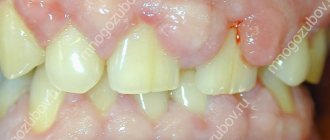
By what symptoms can you tell that there is a tooth fragment left in the gum?
An accurate diagnosis can only be made by a doctor during a face-to-face appointment. But there are some symptoms by which the patient can independently determine that after extraction the tooth remains in the gum:
- aching or throbbing pain in the socket area;
- swollen, reddened gums;
- pain when biting or pressing on the jaw;
- temperature increase;
- sensation of a foreign body in the thickness of the gums.
Sometimes, after removal, the fragments painlessly come out on their own and patients notice them only when the sharp edge of a dental fragment protruding from the gums cuts the tongue. But even in such painless cases, consultation with a specialist is required.
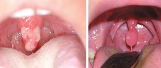
Signs and symptoms of hyperplasia
Those who have this problem rarely experience pain right away. But almost all patients notice a visual violation of the aesthetics of a smile, because the gums become voluminous and loose, which, as a rule, is the reason for visiting a doctor.
Discomfort and pain appear after the tissues have grown significantly, and they involuntarily have to be constantly injured during hygiene procedures and eating food. Another sign is the appearance of bad breath, which does not go away after careful hygiene or quickly returns again.
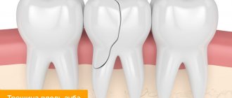
What to do if there are tooth remains
- After the blow. If, due to an injury, half a tooth or only the root remains, in most cases, a crown can be built up. It all depends on the condition of the teeth, the complexity of the fracture, and a number of other factors.
- After removal. Fragments of tooth fragments may remain in the socket and come out a couple of days, a week or even a month later. And they can lead to inflammation, fever, and complications. When the hole is fresh, there is a risk of confusing the remainder of the tooth after extraction with the interroot septum and, when trying to remove the fragments, damage the unhealed wound, which will lead to alveolitis. Therefore, if you have any suspicions, you should consult a doctor as soon as possible for advice.
- After complete destruction of the crown due to caries. The best solution is to pull out the remains of the tooth, then perform prosthetics or insert an implant. The longer rotten stumps remain in the gums, the more negative the consequences and the more expensive the treatment.
Which doctor should I contact if a tooth falls out but the root remains?
If you have a dental therapist whom you trust, contact him first. He will examine the gums and give a direction for CLT or X-ray. If there really are remains of a tooth left and the edge of the fragments is visible, the attending physician can get them out himself without sending his patient to the surgeon who performed the operation.
In cases where the removal took place in a random clinic, make a second appointment with the same doctor or get a second opinion on removing the fragment at another dental center.
Important!
When a toothache takes you by surprise at night, on a trip or out of town, do not wait until you visit your doctor - contact the first dentist you come across: it is important that you remove the fragment, relieve swelling of the gums, relieve pain, and stop the spread of infection due to fragments. You can visit your healthcare professional later for a follow-up examination.
When is tooth root removal required?
It is not always necessary to remove the remaining fragments after extraction: small fragments of 1-3 mm can painlessly come out on their own after a while. But if you feel sharp edges with your tongue, be sure to visit a doctor for a consultation, even if there is no pain or swelling of the soft tissues. The doctor will quickly remove the tooth fragment from the gum after removal and use an x-ray to check whether there is still a piece.
The remainder of the rotting tooth root in the gum must be removed when the crown has already crumbled due to carious lesions. Such fragments have a porous structure, on which bacterial plaque and tartar quickly form. In most cases, cysts and granulomas appear at the top of a rotten stump, leading to even greater problems with the gums.
Important!
Complex tooth extraction is often accompanied by fracture of the root apex. The splinter provokes increased bleeding, which obscures the dentist’s view. In such cases, the procedure develops according to two possible scenarios. The doctor can continue the extraction, relying on his many years of experience, or schedule a second appointment, telling the patient in detail what to do at home before the next removal. When the dentist suggests waiting until the tooth pieces come out on their own, this indicates inexperience. If a fragment remains after a tooth has been pulled out, it can cause various complications: from alveolitis to purulent inflammation.
Lump after crown installation
Improper denture fitting is a very common cause of lumps on the gums. Most often, errors occur at the tooth preparation stage, when the depulpation procedure is carried out. Poor canal filling provokes the development of infection, which, in turn, leads to periodontitis. Another common reason is a poorly made prosthesis or defects during its installation. In this case, the prosthesis injures the mucous membrane and causes swelling, abscesses and growths. Treatment almost always involves removal of the prosthesis and subsequent replacement of the prosthesis (if possible). The sooner a complication is detected, the higher the chance that it can be successfully treated. A well-installed prosthesis should not cause pain, discomfort or injure the mucous membrane. The tooth under the crown should also not hurt, since the dental nerve is killed before permanent prosthetics.
How to remove tooth debris
Most often, patients ignore visiting the dentist because of the common fear of pain during extraction: the memories are still too fresh of how in the 90s, in many clinics, teeth were mercilessly pulled out without pain relief. Modern dentistry has good anesthetics that make the procedure painless. The most unpleasant sensations are an injection into the gums when frozen and a possible aching pain after the drug wears off.
There are several methods for removing residue. The choice of technology depends on where the tooth is located and the reason why the fragment appeared:
- Forceps. The doctor separates the circular ligament and soft tissue before applying forceps, tightly grasping the tooth pieces with his cheeks and removing them from the gums. Instruments may differ in shape and structure, depending on which teeth and on which jaw need to be removed. For example, straight forceps are used for incisors and canines, bayonet-shaped forceps are used for molars, and premolars are removed with S-shaped forceps. The teeth of the lower jaw are the easiest to remove: teeth with short roots and thin socket walls most often do not cause complications after extraction.
- Elevator. If the root is deep in the hole, then the forceps give way to elevators. With their help, the dentist expands the space between the root part of the tooth and the socket, while simultaneously turning the instrument along the longitudinal axis. The concave part of the cheek is used to remove the fragment that remains after tooth extraction.
- Surgical removal. When the remains cannot be removed with instruments, the surgeon performs an operation. After an injection of anesthesia, the mucous membrane is incised, the flap is peeled off and trephination or sawing out of part of the tooth with a small bur is carried out. When all parts are removed, the flap is sutured into place.
Is it possible to grow a tooth if only the root remains?
If the crown is broken and the root remains in the gum, the tooth can be restored. But this is not always realistic, but only under certain circumstances:
- The root is not rotten, not affected by caries.
- The length of the root is twice as long as the crown being restored.
- There are no cysts or granulomas at the root tips.
How to restore a tooth:
- Artificial crowns and inlays. For this method, it is important that the fragment of the tooth wall protrudes above the gum by at least 1 mm. First, an inlay is made from the impression, then a crown. The procedure can take up to two weeks, and the price depends on the materials chosen.
- Fiberglass post with metal-free crown. Fiberglass posts are transparent and look aesthetically pleasing, especially when it comes to teeth in the smile area. It takes from a week to ten days to make a crown, depending on the clinic’s capabilities: if the center has its own technical laboratory, it will take less time.
- Restoration with composites. The advantages of this method include comparative cheapness and short recovery time, but you need to understand that this is more of a temporary solution due to the unreliability and fragility of the materials. On average, a composite restoration lasts 5 years.
What happens if the remaining tooth fragment is not removed?
Leaving a tooth fragment in the gum is a lottery. Maybe it will come out on its own, or maybe it will smoothly flow into phlegmon, which is extremely life-threatening. In addition, pushing out a fragment from soft tissue is an unpleasant process. The gums swell, painful sensations appear when chewing and communicating, and the sharp edge of the fragment cuts the tongue. Therefore, after removal, our dentists recommend calling or coming to an appointment immediately if the patient suspects the presence of dental splinters.
If we are not talking about extraction, but about tooth destruction due to deep caries, then the consequences can be even more serious. While the remains of a dead tooth gradually rot in the thickness of the gums, the infection penetrates deeper and pathogenic organisms spread throughout the body. Entire organ systems are affected: cardiac, gastrointestinal, respiratory. A small root fragment can incur a huge medical bill and irrevocably waste the body's health.
Prevention
To prevent the mucous membrane from growing into the tooth hole, you need to take some preventive measures to avoid the formation of carious lesions:
- visit the dentist promptly when the first signs of caries appear;
- monitor oral hygiene;
- use a toothbrush of medium or low hardness;
- pay special attention to the selection of toothpaste;
- lead a healthy lifestyle: do not abuse smoking and alcohol;
- eat a balanced diet.
The growth of mucous membrane in a tooth hole is often provoked by carious processes, which lead to the development of various pathologies in the oral cavity. Surgery, as well as the use of drug therapy, will help get rid of the unpleasant symptoms of mucous membrane ingrowth into a tooth hole.
Bibliography
- Barer G.M. – Periodontal diseases. Textbook, M.: GEOTAR-Media, 2008.
- Maksimovsky Yu.M., Mitronin A.V. - Therapeutic dentistry - M.: Geotar-Media, 2012.
- Grigoryan A.S., Grudyanov A.I., Rabukhina N.A., Frolova O.A. — Periodontal diseases, M., 2004.
- O. O. Yanushevich, V. M. Grinin [and others] - Periodontal diseases. Modern view on clinical, diagnostic and therapeutic aspects, M.: GEOTAR-Media, 2010.
- Ijndhe J. - Textbook of Clinical Periodontology. 1995. Copenhagen.
- Ivanov V.S., Urbanovich L.I., Berezhnoy V.P. — Inflammation of the dental pulp, M., Medicine, 1990.
- Stephen Cohen - Pathways of the Pulp - Mosby - 1980
- Ivanov V.S., Urbanovich L.I., Berezhnoy V.P. — Inflammation of the dental pulp, M., Medicine, 1990.
Find a clinic