In this article
- Who is susceptible to developing interdental caries?
- How to detect caries in interdental spaces?
- Features of the treatment of interdental caries
- Interdental caries of the anterior teeth: what should be done?
- Specifics of treatment of interdental caries of chewing teeth
- How is caries between teeth treated when molars and premolars are affected?
- Prevention of interdental caries
Interdental caries is one of the most “inconvenient” and difficult to treat. In this article we will tell you why it appears, how caries between two teeth is treated, and whether its occurrence can be prevented using preventive measures.
What is interdental caries called and why does it occur? Caries between the lower or upper teeth located next to each other, joint to joint, is called interdental. The denser the arrangement of adjacent teeth in relation to each other, the greater the likelihood of developing carious lesions of the tooth enamel in the area of the joints. When the teeth are tightly packed together, food particles become stuck in the spaces between the teeth, which are difficult to remove with a brush, even if a person regularly brushes their teeth twice a day. To clean the interdental spaces, you need to constantly use dental floss, floss or a special device - an irrigator.
Unfortunately, most people limit themselves to using a toothbrush and toothpaste, as a result of which high-quality cleansing of the interdental spaces does not occur. Accumulated food debris is a favorable environment for the development of microbes and the formation of plaque, which leads to the development of caries between teeth. Due to the location and anatomical features, interdental caries of the anterior teeth most often occurs. Its early detection and timely treatment play an important role from the point of view of not only health, but also aesthetics, because caries between the front teeth, which is located in the most visible place, spoils a person’s smile and appearance. Its specificity is also that the enamel of the front incisors is thinner than the enamel of other teeth, so carious lesions spread deeper and deeper, and treatment presents certain difficulties.
Description of the pathological process
Its occurrence is provoked by cariogenic microorganisms, streptococci and lactobacilli (some types of them), which appear due to insufficient oral hygiene. Typically, bacteria begin to multiply in plaque, which is difficult to clean with a brush (most often on the back surface of the incisors). The carious area is difficult to notice on your own, especially in the early stages, since darkening begins to appear only when the area is already very widespread. That's why regular checkups with a dentist are so necessary - only he can carefully examine the entire oral cavity using a small mirror or take an x-ray.
Sometimes people wonder: “can children have caries on their front teeth?” Unfortunately, yes, it is also called “bottle” or “carob”.
It occurs in a small child if he often eats sweets (baby formula, cotton candy, candy, etc.). Such products contain a large amount of carbohydrates, which quickly destroy fragile tissues.
You also need to be careful with breast and cow's milk. It often sticks to the teeth in the upper jaw when children accidentally fall asleep with a bottle in their mouth. This product also contains sugar (albeit natural), and it quickly oxidizes and destroys the enamel. During sleep, this is especially dangerous due to the fact that the intensity of salivation, which is created for self-cleaning of the oral cavity, decreases. You can see the result of the defeat in the photo.
Who is susceptible to developing interdental caries?
Carious lesions on the lateral surfaces of adjacent teeth can occur in anyone, including children. But the following categories of people are especially susceptible to the formation of interdental caries:
- those who eat poorly: consume a lot of sweet foods and drinks, few solid vegetables, fruits and foods rich in fluoride, calcium, phosphorus;
- those who have problems with oral hygiene (do not brush or brush their teeth poorly, do not use dental floss or irrigator to clean the interdental spaces);
- those whose normal salivation process is disrupted;
- when there is a gap between the teeth that is too narrow or closed at the bottom and widened at the top, into which food easily gets clogged.
Caries on the front teeth: what to do and why it occurs
In fact, there can be many reasons for its appearance:
- Hereditary predisposition. Our genes determine the thickness of the enamel. The thinner this tissue is from birth, the higher the likelihood of disease. But often the protection is weakened due to a person’s fault when he abuses hard, spicy, sour foods, carbonated drinks and sweets.
- Lack of hygiene procedures. Sometimes we are too lazy to carry them out thoroughly, so we either brush superficially, forgetting about hard-to-reach places, or we completely neglect this process. Because of this, plaque accumulates, and this is “fertile soil” for microbes.
- Poor nutrition. Vitamins and microelements are important for healthy teeth; without them, the enamel becomes thinner and changes its structure.
- Deformed jaw. It can be this way from birth or change during life from injuries or improper growth of the incisors. Most often, caries affects curved units: they are more difficult to clean, so plaque often accumulates on them. People with a narrow interdental gap or too wide fissures (grooves on the chewing surface) are also at risk.
- If a person works in a hazardous enterprise, he often unknowingly inhales air through his mouth containing chemicals that corrode enamel.
- Artificial injury. This applies most of all to seed lovers and seamstresses who bite threads instead of using scissors.
- Bad habits. Alcohol and nicotine not only thin the dental tissue, but also change its color, which makes their appearance unpleasant.
Causes and stages of development of caries between teeth
The causes of interdental caries are exactly the same as those of classic caries. This is the activity of pathogenic microorganisms that destroy the protective shell of the enamel and then penetrate into deeper tissues. The spaces between teeth are the main places for food debris to accumulate. Without aids, they are quite difficult to clean. Complicating the situation is the fact that in this area the enamel is thinner, especially in the front teeth, where the protective layer of enamel itself is less dense compared to molars. Hidden caries between teeth is difficult to detect. The disease can be deceptive: a cavity that is small in diameter is often very deep and has already affected the dentin and pulp. This means that in addition to installing a filling, the patient must undergo endodontic treatment of two teeth at once, which significantly increases the cost of treatment.
Four stages of development
- Initial.
Appears as a chalky stain and does not disturb the enamel structure. - Superficial.
Affects the top layer of enamel. - Average.
It affects not only enamel, but also dentin. - Deep.
The inflammatory process covers the tooth pulp.
Symptoms
You need to understand that the enamel on the incisors is very thin, so the disease develops on them much faster. Many people do not know what caries on the front teeth looks like initially. It begins with the appearance of a small white or yellowish spot. Further, the enamel becomes thinner and sensitivity increases: it becomes difficult to drink cold drinks or eat ice cream. Later, periodic pains of aching or throbbing nature appear. In the final stages, pulpitis (inflammation of a bundle of nerves and blood vessels) or periodontitis (damage to tissue around the root) develops.
It is best to go to the doctor at the first stage, so the treatment process will be easier, faster and more comfortable. But usually they do not pay attention to a minor formation - then the lesion begins to spread at high speed.
Symptoms of contact caries
The disease develops gradually - just like other types of caries. First, yellow or grayish spots appear on the contact surfaces of adjacent teeth. If left untreated, small cavities form at the site of the spots, affecting only the thickness of the enamel. Moving to the next stage, the disease affects dentin. And the most difficult form of interdental caries is deep, when the infection reaches the root canals.
If the crown defect is not yet visible, the following symptoms may indicate the presence of caries:
- increased sensitivity of teeth: they begin to react sharply to hot, cold, sweet, salty and sour, and pain during eating goes away quite quickly;
- unpleasant smell and taste in the mouth: with contact caries, they come from a certain place where food debris accumulates and decomposes;
- change in the color of the tooth in the area of the lateral surfaces: by shining a light on the tooth, you can notice that it is unevenly colored, and darker areas are visible under the enamel.
Is it possible to determine the onset of the disease yourself?
In most cases, it is not possible to notice the problem at an early stage of its development. Most often, caries occurs in hard-to-reach places (between the teeth or on their back surface).
But sometimes they show specific signals that there is trouble with the incisors:
- when there is a sharp change in temperature, short-term pain occurs;
- sometimes the uneven edges of a carious cavity can be felt tactilely, using the tongue;
- a specific odor appears from the mouth, especially in the morning before meals;
- a dark spot becomes noticeable.
It is important to understand that such signs are not always present, and only a doctor can determine the onset of the disease. It has all the necessary tools and devices for high-quality diagnostics. Therefore, you should not neglect a visit to the doctor and check in time whether everything is in order.
Treatment of the disease
Caries between teeth is treated in the same way as regular one, with the only difference being that two units have to be “fixed” at once. In some special cases, in order to get to the affected part, the dentist is obliged to get rid of healthy tooth tissue by drilling. First, the specialist must prepare the cavity and then treat it.
It is important to be careful about the process so as not to touch or even damage the nerve. After which the doctor installs a plate that separates the teeth and inserts a filling. At the end of the process, it is put in order by grinding and polishing.
Types of damage
Before you find out how caries on the front teeth is treated, you need to understand to what extent it has developed. There are several types of the disease based on the scale of spread:
- Superficial (shallow damage to tooth enamel). Usually at this stage the patient does not yet experience discomfort.
- Average. Slightly affecting contacts and causing aching pain when sugar enters.
- Deep. At this stage, the dentin (the main part of the incisor) is damaged.
- Radical. Here, the carious formation is already difficult to eliminate without complete removal.
- Atypical. It first affects the cutting edge as a result of heredity, smoking or poor nutrition.
Before understanding how to treat cervical caries on the front teeth and whether it hurts, the doctor needs to determine at what stage the disease is.
If the dentist does not do this and only carries out superficial work without completely cleaning the canals, then in the future a cyst may form in the jaw. And it will already be necessary to remove it with the help of surgical intervention, until the pathology has grown to the brain. The sooner treatment begins, the less pain the patient will feel, so it is better not to put off going to the doctor until tomorrow.
Causes of cervical caries
Cervical caries appears more often than other carious lesions. The enamel in the area at the gingival margin is thin and is destroyed more quickly. Cleaning of the cervical area is often not of sufficient quality, and plaque and tartar accumulate more quickly on the cervical surface.
Cervical caries will appear more often if:
- The technique of brushing your teeth is incorrect - the movements of the toothbrush are not vertical, but horizontal;
- the patient takes antibiotics, antihistamines and other drugs that affect the condition of tooth enamel, making it porous;
- the diet is not balanced: with frequent consumption of acidic foods, consumption of fruit juices, carbonated or alcoholic drinks, sweet foods, high-carbohydrate foods;
- the patient has endocrine diseases;
- There is a deficiency of vitamins and minerals in the diet.
You have questions?
We will call you back within 30 seconds
+7
Possible complications
There are those who want to learn how to get rid of caries on their front teeth or hide it themselves. But we must understand that a simple disguise will not please you for long. Soon the person will feel severe pain, which will mean that the problem has reached a severe stage. If the patient is not provided with timely medical assistance, pulpitis may form, which develops in just a few hours. In such cases, the patient is called an ambulance. He feels an increasing throbbing pain, which intensifies in the lying position.
When a carious formation reaches the root, it is useless to treat it; the unit must be removed completely. If this is not done, inflammation may begin, which will soon affect the soft tissues of the face and cause osteomyelitis, abscess (damage to the root zone) or phlegmon (swelling of the jaw and impaired mobility).
Why is cervical caries dangerous?
Develops faster. Due to the small thickness of the enamel, the affected area quickly darkens and begins to collapse. If plaque and tartar accumulate in the root part of the crowns, it is better to contact the dentist as soon as possible - caries can form in just a few weeks.
Leads to tooth loss. Without treatment, the tooth is destroyed at the base, becomes brittle, and can break even under light load. Such fractures may extend below the gum line, in which case the tooth will have to be removed.
Source of infection in the mouth. It can provoke caries on other teeth, increase the risk of problems with the gastrointestinal tract, cardiovascular, respiratory, immune systems, etc.
Gum diseases. With cervical caries, the affected area is located at the edge of the gum or goes under it. This provokes the proliferation of bacteria in the periodontal pocket, inflammation, the appearance of gingivitis and other gum diseases.
Front teeth - how to remove caries if it has spread deeply
In this case, very careful treatment must be carried out. It is necessary to take an x-ray to determine the exact depth of the damage. If you make a mistake during processing, you can hit the pulp (neurovascular bundle), and it will begin to die. Typically, the dentist uses a drill and then fills the drilled area. Sometimes special tabs are used that are more resistant to external influences and are easier to chew. In this case, you do not need to drill much into the fabric to insert them, and preliminary heat treatment makes the material as durable as possible.
Features of the treatment of interdental caries
Treatment tactics depend on several factors:
- stages of the carious process;
- localization of caries.
In terms of the depth of damage, interdental caries can vary from the spot stage, when there is no carious cavity yet, but only a section of demineralized enamel is found, to medium and deep caries, when a large carious cavity forms in the dentin and there may be a risk of pulpitis and other complications.
At the stain stage, it is possible to treat teeth using conservative methods, without preparation. As a rule, this is remineralization by simple or deep fluoridation, infiltration method or ozone therapy. These methods strengthen tooth enamel, increase its protective functions and resistance to external influences.
At the stages of superficial, medium and deep caries, the disease will have to be treated surgically - through tooth preparation, removal of affected tissue and further filling of the affected areas.
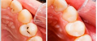
At the same time, the main difference between interdental caries and other types is that it is necessary to treat not one, but two teeth at once in the area of contact surfaces.
Features of restoration
It is important not only to correct the appearance of the tooth, but also to cure the disease. At an early stage, when white or brownish spots just appear on the surface, sanding or bleaching can be done. But you don’t need to do this at home, the doctor will make the procedure much more effective and protect against complications. He can use professional grinding discs and polishers. Severe cases require the use of a drill.
Air abrasive is a more gentle processing method. It involves spraying a special substance that affects dental tissue. But this technique is not effective in all cases and is very expensive. Some clinics use laser; this is a simpler method, but requires more investment. Moreover, its effectiveness has not been proven one hundred percent; there is still debate among experts about it.
Types of caries diagnostics in dentistry
It is very difficult to detect a carious lesion on your own, so it is important to pay attention to the signs described above that indicate its appearance. The earlier caries is diagnosed, the higher the likelihood of successful treatment and prevention of its development on neighboring teeth.
In modern dentistry, there are different diagnostic methods that will help identify the disease and determine its stage.
Types of diagnostics:
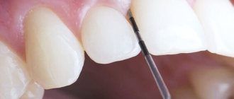
- Visual examination of the dentist using a mirror and probe;
- X-ray diagnostics. The image shows affected areas that are invisible from the outside;
- Use of coloring compounds;
- The use of a special apparatus with pulsed light waves reflected on the surface of the enamel. When the machine sees a carious lesion, it beeps;
- CT scan. Makes it possible to see the structural features of the jaw and the condition of all teeth in a 3D image.
Elimination of the disease
There are several options for treating caries; the choice of one or the other depends on the stage of its development and the financial capabilities of the patient.
Installation of veneers
Recently, this method has become very popular, especially among show business stars. It is good because with the help of special ceramic or polymer crowns you can hide all defects (shape, shade, etc.). But to install the plates, you need to grind off the dental tissue by about half and drill out all the affected areas. The custom-made veneer is then bonded using a non-toxic substance.
With high-quality installation, it can remain unchanged for up to 10-15 years.
The price of such a procedure at Dentic is about 30,000 rubles per tooth.
Crowns
They are used when the disease progresses to a deep stage and covers both sides of the incisor. They are usually made of metal and ceramics: these materials are very durable and are relatively cheap: 20 thousand rubles. But they also have a drawback: it is very difficult to choose a shade that will not stand out in the teeth. To avoid big differences, it is necessary to choose crowns made entirely of ceramic or zirconium.
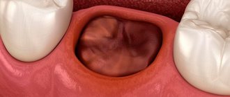
Implantation
When caries destroys tissue down to the root, more serious measures must be taken. The diseased area is removed, and in its place an implant is implanted into the jaw. It is a strong rod that will hold the crown. Nowadays they make it very natural and aesthetic. The cost at Dentic starts from 30,000 rubles.
Prosthetics
There are cases when installing an implant becomes impossible, for example, when there is a risk that it will not take root, or the gums are not thick enough. Then special zirconium or ceramic prostheses are created, which are attached to adjacent units.
Treatment of caries between the front teeth with pinning
If only the root of the affected incisor remains intact, then a pin is inserted into it, having been carefully processed beforehand. It is then masked with a temporary crown for about a week while it takes root. Next they set a constant.
Black classification
American dentist Green Black developed his own classification of caries, dividing it into classes:
- 1st class: carious lesions are localized in fissures;
- 2nd class: the pathological focus is localized between the molars;
- 3rd class: caries affects the areas between the incisors and canines;
- 4th class: the disease develops along the edges of the front teeth;
- 5th class: the affected area includes the cervical area;
- 6th grade: caries affects the cutting edges of the front teeth.
During the diagnosis, the doctor determines the degree and type of caries of the front teeth, based on which he selects treatment.
Diagnostics
Only a dentist can correctly determine the nature and stage of development of the disease.
Therefore, you should not neglect going to the doctor at least once a year. Now clinics use various examination methods:
- Vital coloring. A solution of 1% methylene blue is mixed with 0.5% red and applied to the tooth surface, after which the lesion acquires a different color. This is a very economical option, but not the most effective: the depth of distribution cannot be determined.
- Fluorescent radiation testing. If a carious process begins in any area, it will not glow.
- Electroodontometry. When exposed to electric current, the condition of the pulp is determined. This method is absolutely safe for all ages.
Treatment for severe crown lesions
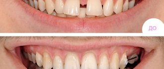
If caries affects more than 50% of the incisor or canine tooth, it will not be possible to install a filling. After removing the carious areas, the patient is sent to an orthopedist for installation of an orthodontic structure: a bridge, crown, veneer or lumineer. They allow you to hide a cosmetic defect that appears after an illness.
If the tooth is completely destroyed by caries, but the root remains, the doctor installs a pin and a crown. If a tooth is completely lost, an implant will be required. This is one of the most expensive dental procedures.
How to remove caries on the front teeth in adults
If the disease overtakes children, then nothing terrible will happen, although it must be treated in the early stages so that the infection does not penetrate the jaw. Over time, the milky affected incisor will simply fall out, and a new strong one will begin to grow. For a mature person, the situation is more complicated: it is necessary to cure and preserve it, otherwise the element will be lost forever. We have given the main treatment methods above, but it is worth adding a few more important nuances:
- When the doctor completes the procedures, he must prescribe preventive measures. If this is not done, the problem will reappear.
- After the filling is installed, it must be sanded and polished, otherwise it may damage the lips, cheeks or tongue.
- The material for it should be selected so that the patient’s smile looks natural.
How to treat caries: stages
To get rid of caries, you need to make an effort, because although modern drills do not vibrate like hammer drills, they still make us wait for the sudden appearance of acute pain - while drilling out carious tissues. Fortunately, modern anesthetics allow the dentist to properly numb the teeth during treatment - unlike the ineffective novocaine and lidocaine that were widely used previously.
Proper treatment of dental caries in dentistry consists of a number of successive stages, each of which has a clear goal. But nevertheless, the most important thing is the complete removal of caries, because... if the removal of tissue affected by caries is incomplete, it will immediately develop under the filling and will certainly lead to the development of pulpitis and the need to remove the nerve from the tooth. Watch the video below to see how hard tooth tissues affected by caries are removed.
Treatment of dental caries: video 1-2
In detail about the stages of treatment of average caries -
But before we move on to drilling out the carious tissue, which you could see in the video above, you still need to perform a number of procedures to prepare the tooth for treatment, as well as anesthetize it with an injection of a local anesthetic. For those who like stronger anesthesia, there are methods of sedation and general anesthesia.
- Cleaning a tooth from plaque (Fig.4) –
Before starting treatment, it is necessary to hygienically clean the tooth, as well as neighboring teeth, from plaque and tartar. For this purpose, ultrasonic attachments are used to remove massive dental plaque, as well as special brushes and abrasive pastes to remove soft microbial and pigmented plaque. - Determination of tooth color using a special scale (Fig.5) –
Hygienic treatment of the tooth also helps the doctor to accurately select the color of the filling material. In this case, the filling will match the color of the tooth, and will not stand out against the background of the tooth’s own tissues. This is especially important for teeth that are visible when you smile. - Anesthesia (Fig. 6) – is it painful to treat caries: for painless drilling of carious tissues if the tooth is alive, local anesthesia is necessary. Modern painkillers in dentistry, for example, ultracaine or ubistezin, make the intervention absolutely painless. Depending on the amount of anesthetic administered and the method of anesthesia, the anesthesia time can last from 40 minutes to several hours.
The only pain that the patient can feel is the moment the needle is inserted into the gum, as well as the process of removing the anesthetic into the tissue. This process can sometimes be painful, which largely depends on the patient’s level of pain sensitivity, as well as on the speed at which the anesthetic is injected into the soft gum tissue. The faster the solution is administered, the more painful the injection. - Drilling out carious tissues – As can be seen in Fig. 7, enamel is always destroyed with average caries to a lesser extent than the underlying tissue (dentin). This is due to the fact that enamel is much, much stronger and harder than dentin. Therefore, the carious cavity usually expands in depth, and the entrance hole in the enamel can even be quite small.
The dentist must drill out the edges of the enamel overhanging the carious cavity, and also remove all carious dentin. If you leave even a small amount of dentin affected by caries and put a filling on top of it, then very soon you can expect complications - the rapid development of caries under the filling and destruction of the tooth crown, with the subsequent development of pulpitis and periodontitis (24stoma.ru).
In Fig. 8, the dotted line shows the approximate boundaries of tooth tissue removal. In this way, the cavity is given a relatively correct shape and the next stages of treatment can begin. It should be noted here that recently new methods of tooth preparation have appeared, which help to do without traditional drilling. Recently, it has become possible to remove caries with a laser.
- Isolation of tooth from saliva – this is a very important stage! After the carious tissue is drilled out, and before filling the tooth, the doctor must carefully isolate the tooth from saliva and even the patient’s wet breath. These factors will greatly affect how long the filling will last. Previously, cotton balls and gauze balls were used for insulation, which were used to cover the tooth on all sides. It should be noted that this is a very unreliable and ineffective protection.
For the last 10 years, “cofferdam” has been used for these purposes. The latter is a thin latex “scarf” in which holes are made for the teeth. This scarf is pulled over the teeth (Fig. 9-10), after which 1-2 special metal clasps are installed on the necks of the teeth, which hold the rubber dam against the gums. The edges of such a latex scarf are attached to a special frame (Fig. 11), and we see the result - a group of teeth is completely isolated from the oral cavity.Installing a rubber dam is quite labor-intensive. Some doctors refuse to use it on principle to save their time. The doctor’s use of a rubber dam in the treatment of caries indicates that the doctor is very attentive to the quality of his work, because the quality of the filling will be affected not only by the accidental contact of saliva on the tooth being filled, but also simply by the moist breath of the patient himself.
- Medical treatment of a carious cavity - a cavity formed in the tooth during the removal of carious tissues - is treated with antiseptics.
- Restoring the contact point between teeth – If caries is treated on the contact (interdental) surface of the tooth, then it is also necessary to restore the side wall of the tooth.
This is a rather labor-intensive and complex task than simply treating average caries, for example, on the chewing surface of a tooth. In this case, another stage is added - the installation of special devices to restore the side wall of the tooth. Such devices include wedges (a) and matrix (b) in Fig. 12. Read more about the treatment of interdental caries in the article: → “Treatment of caries between teeth” - Etching the enamel with acid (Fig. 13) is necessary so that the adhesive (something like glue), which will be applied to the surface of dentin and enamel at the next stage, can penetrate deeply into the tooth tissue. For this purpose, a gel based on phosphoric acid is used. After etching, all the gel should be thoroughly washed off, and the tooth surface should be slightly dried.
- Treatment of dentin and enamel with adhesive - for better fixation of a permanent photopolymer filling, enamel and dentin are treated with a special adhesive, which (after absorption) is illuminated with a photopolymerization lamp.
- Applying a gasket under the filling (Fig. 14 b,c) - an insulating gasket, usually made of glass ionomer cement, is applied to the bottom of the cavity. The need for lining material under the filling is explained by the complex mechanisms of polymerization shrinkage of the filling material and other factors (we will not dwell on them).
- Filling – dental filling is necessary to restore the shape of the tooth, its aesthetics, as well as to restore chewing efficiency. For this purpose, photopolymer composite materials are usually used. They are applied in layers and each layer is illuminated with a special lamp, which allows the material to harden.
- Grinding and polishing of the tooth - after the shape of the tooth has been restored using filling material, it is necessary to grind and polish the filling, because it is rough and uneven. Final polishing gives the filling a shine and aesthetics comparable to that of tooth enamel. This completes the treatment of average caries.
→ Cost of treatment of dental caries
Filling a carious defect: video 3-4
Please note that dentists use special metal strips (matrices) and wedges to restore the side walls of teeth. In addition, tooth filling in both cases is carried out using a rubber dam.
How to avoid getting sick
You need to regularly monitor the condition of your teeth, not only from the outside, but also from the inside. It is no coincidence that doctors advise cleaning them 2 times a day - this is required to prevent infections. But don’t forget about following your diet; you need to eat enough:
- hard vegetables and fruits (apples, carrots, cucumbers, beets, etc.);
- greens (parsley, lettuce, dill, celery);
- berries (currants, grapes, strawberries, cranberries);
- nuts (almonds, cedar, cashews);
- dairy products (cheese, cottage cheese, natural yogurt without additives);
- citrus fruits (grapefruit, lime, orange);
- seafood (fish, shrimp);
- eggs (chicken, quail);
- honey;
- water and tea without dyes (green and black).
Do not forget about the correct cleaning technique: you need to brush each unit from the gum to the cutting edge so that the dirt does not remain in the crevices and does not oxidize. It is convenient to use dental floss for this.
About the cost of treatment
Most often, a paradoxical situation occurs: a person wants to save money on the dentist and waits until the tooth is destroyed. But you need to understand that the later you see a doctor, the more expensive his services will cost. If the problem has already appeared, it will need to be solved, and the sooner, the cheaper. Conventional preparation of a carious cavity in the early stages costs only about 4,000 rubles. If you wish to install ceramic fillings, the amount will increase to 10 thousand rubles. But some patients wait until the destruction is complete, and then they have to pay 30-50 thousand for one incisor.
We described in detail how caries of the front teeth in adults is treated. For the process to go well, it is better to choose a good private clinic. You can contact: there the work with clients is at a high level, they feel comfortable physically and psychologically. In addition, the clinic received the title of one of the best in Russia in the StartSmile rating. And on the site there are often many special offers and promotions that will significantly reduce the cost of services. Hurry up to sign up for a consultation!
Weak and strong caries of anterior teeth
Spot
A chalky stain is a clear sign of a lack of minerals and the beginning of the destruction of the enamel structure. They are white in color and respond well to treatment at the initial stage. There may be a reaction to sweets.
Surface
At this stage, the enamel loses its integrity (which is especially noticeable upon probing), and the stain acquires a brownish tint. A painful reaction to temperature and chemical stimuli appears.
Average
The stage at which dentin is affected. A noticeable carious cavity appears on the surface of the tooth; over time, the crown part begins to change color. Severe pain occurs when eating cold and hot food and a reaction to sweets.
Deep
The most severe caries of the front teeth occurs when the deep layers of dentin are damaged. Severe pain from temperature stimuli and a pronounced aesthetic defect are observed.