With the birth of a child, parents are deprived of peace for a long time, if not forever. After the worries caused by childhood illnesses comes the fear of bullying at school, and then the anxiety associated with going to college... One of the parental worries can be the condition of the child’s teeth, especially in those rare cases when it comes to something more serious than correcting the bite with braces. Hutchinson's teeth is a pediatric dental condition you may never have heard of. According to Big medical dictionary, Hutchinson's teeth are called "modified upper central incisors with a screwdriver-shaped crown and a lunate notch on the cutting edge."
In this article, you will find important information about this abnormality, including its causes, symptoms and how to treat the disease.
Damage to primary occlusion
Diagram
Pathology is formed during the prenatal period. The main factor here is the serious illness of the pregnant woman. Baby teeth suffer much less often than permanent teeth. However, nowadays such cases have become more frequent. This is associated with a significant reduction in perinatal mortality.
Even in the first weeks of life, the occurrence of systemic diseases in a child can provoke the occurrence of hypoplasia. Abnormalities are more common in the incisors and canines of the upper jaw. This is why such anomalies as Hutchinson, Fournier and Turner teeth are so common.
Deciduous incisors undergo changes most often if a pregnant woman has suffered from diseases such as toxoplasmosis, rubella, or suffered from toxicosis. Hypoplasia is also observed in premature babies who have a history of birth trauma, congenital allergies or hemolytic jaundice.
Anomalies develop in utero at approximately 25-30 weeks of pregnancy. Sometimes this process occurs within 1 month of life.
Reasons for the development of pathology
As mentioned above, the etiology of the development of hypoplasia is usually associated with changes that occur in the child’s body at the stage of intrauterine development. The “trigger” in such a situation may be one of the following factors:
- infection of the mother's body during pregnancy,
- severe toxicosis,
- birth injuries,
- premature birth,
- rickets in a child,
- dystrophy,
- systemic disorders in the body,
- gastrointestinal pathologies,
- somatic diseases,
- brain disorders
- infections in the baby suffered in the womb or in the first months of life,
- serious jaw injuries,
- acute deficiency of vitamins and nutrients.
Most often, the problem arises at the stage of intrauterine development
. Often during differential. For diagnosis, you have to turn to such specialized specialists as an endocrinologist, otolaryngologist, pediatrician and geneticist. That is, to find out the reason that led to the development of the anomaly, a comprehensive examination of the entire body is required.
Damage to permanent dentition
The higher the incidence rate in childhood, the greater the possibility of dental anomalies. Half of the children who had chronic diseases, especially of the endocrine system, subsequently experienced anatomical changes in the crowns.
In addition, a special role is played by:
- gastrointestinal problems;
- toxic dyspepsia;
- congenital syphilis;
- acute infectious diseases;
- rickets;
- brain disorders.
It is noted that 40-60% of hypoplastic changes occur already in the first 9-10 months of life. During this period, the protective properties of the body are still weakly expressed. The child’s adaptive and compensatory capabilities are not able to withstand aggressive factors of the internal and external environment.
Hypoplasia in permanent dentition
In permanent teeth, the first molars are the first to suffer. The lesion is observed primarily on tubercles and convex surfaces. By 8-9 months, canines and incisors are formed.
If before this period the child suffered from some serious illnesses, then the risk of hypoplasia in the form of Hutchinson’s and Fournier’s teeth is quite high.
Treatment process
If the disease manifests itself in the form of minor depressions and grooves, then the specialist can fill the tooth according to the classic scenario.
After such treatment, the restored elements must be treated with care. Only competent and high-quality care will prolong their functionality and aesthetic appearance.
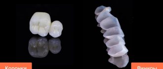
The damaged front element can be covered with a veneer on the front side. This plate has an impeccable appearance and is an excellent solution for the treatment of Turner hypoplasia.
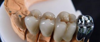
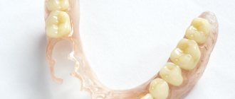
Dentists resort to the use of optopedic crowns in cases of pronounced changes in the shape of the tooth or in connection with extensive lesions. Installation of such products requires meticulous work.
The most popular materials for crowns in the treatment of Turner hypoplasia:
- one-piece crown;
- metal-free ceramics based on zirconium or aluminum oxide;
- pressed lithium disilicate ceramics.
For a spotted form of hypoplasia, expressed as spots without defects, specialists can use bleaching therapy followed by mineralization.
Pathology cannot be ignored. Lack of treatment can result in tooth loss.
Systemic damage
Local changes do not pose a serious threat to the health of the dentition. Systemic hypoplasia poses a great danger. It has three clinical forms: discoloration, underdevelopment and absence of the enamel layer.
Underdevelopment of hard tissues manifests itself in different ways. Furrowed, wavy and dotted forms may occur. There are also more severe changes in the anatomical shape of the crown. Microscopic examination reveals a violation of the structure of dentinal tubules, the width of prisms and hydroxyapatite crystals.
Hypoplasia always occurs without general symptoms. If there are additional clinical manifestations, one can judge the manifestation of a concomitant disease. Many parents are interested in how to distinguish it from a virus if fever appears on the teeth.
It should be understood that in this case, manifestations from the ENT organs are possible, which are expressed in pain, nasal congestion, lacrimation, coughing and voice changes. With a virus, nasal discharge usually has a transparent structure, and if bacterial microflora is attached, the mucus becomes viscous and cloudy with a greenish tint.
The incidence of hypoplasia is presented in chart form.
Hutchinson's teeth concept
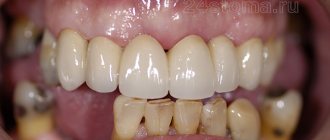
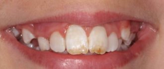
This anomaly affects the central incisors on the upper jaw. They have a specific shape, sharply different from the same units on the lower jaw.
This is a barrel-shaped crown, the size of which increases towards the cervical region. A semilunar notch is found on the cutting edge. Sometimes the characteristic is given as a screwdriver-shaped crown.
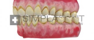
The enamel on the cutting edge is preserved only at the corners. In the middle of the formation it is completely absent, and the exposed dentin is clearly visible. Over time, it may change in color and become dark brown.
Hard tissues lose their strength. If too much pressure is applied, they can chip off in large sections. The sensitivity of such teeth is somewhat increased, but there is no sharp reaction to irritants. The exception is significant enamel separation due to mechanical stress.
For a long time it was believed that Hutchenson's teeth were exclusively a consequence of congenital syphilis. Recent research and accumulated practical knowledge suggest that such changes may also occur for other reasons.
Fournier's teeth
Such units have a similar barrel shape, but lack the lunate notch. The crown is resistant to mechanical stress.
Barrel-shaped maxillary incisors
Enamel, as a rule, does not change color and is fully functional. A distinctive feature is the absence of caries. Microscopic examination reveals underdevelopment of enamel and dentin.
Pflueger's teeth
In this case, permanent molars are involved in the pathological process. They are large in size, especially in the neck area. The chewing surface is underdeveloped, especially the tubercular areas. However, this does not interfere with the act of chewing food.
The tubercles have the appearance of an unopened flower bud or tree bud. The crown converges to the chewing surface with a cone. This phenomenon is observed in severe infections, including syphilis.
Turner or Turner teeth
Local hypoplasia is caused by factors such as:
- mechanical injury to the follicle;
- inflammatory process of the primordia;
- frequent infections;
- diseases in the milk occlusion.
As a result of such impacts, the enamel may be completely absent. For this anomaly, the concept of amelogenesis imperfecta is used.
Tetracycline teeth
It is known that taking certain medications negatively affects the development of the entire maxillofacial region of the child. Thus, tetracycline can stain enamel if it is prescribed to a woman during pregnancy. Particles of the substance are deposited as a result of mineralization of hard tissues.
Attention!!! Taking tetracycline drugs during pregnancy is strictly prohibited. In any condition, this product can be replaced with an analogue.
Changing the color of crowns
The most dangerous is dimethylchlortetracycline. It can cause not only staining, but also affect histological structures.
Discoloration is observed in all groups of dental units. However, the incisors are stained much more intensely. In the direction of the masticatory groups, the lesion is observed only in 1/3 of the crown, starting from the tubercles.
Proper nutrition as an important part of prevention
A balanced diet plays a key role in this matter. You need to eat properly and nutritiously at every stage of immensity and even while planning a child. Also, after birth, the baby should eat well, receiving all the important vitamins and microelements with food. After the stage of breastfeeding or artificial feeding, the following products should be gradually introduced into the child’s diet:
- milk and cottage cheese, as well as other products rich in fluoride and calcium,
- foods high in vitamin C - spinach, broccoli, citrus fruits in later life,
- foods with vitamins A and B - legumes, seafood, mushrooms and poultry.
Proper nutrition is an important condition for oral health.
The child should also get enough vitamin D, and for this, experts recommend spending more time in the fresh air in good sunny weather. During pregnancy, you need to monitor your health, regularly visit doctors for preventive measures and try to be less nervous.
1Belyakov Yu. A., Elizarova V. M., Krotov V. A., Blinnikova O. E. Hereditary pathology of enamel and dentin. Review of Molecular Genetic Research, 2000.
Elimination of hypoplasia, reliable methods and immediate measures
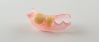
It is advisable to preserve the teeth of Hutchinson, Pfluger, and Fournier with the placement of an artificial crown. Minor changes can be corrected by using modern filling agents. Moreover, tissue preparation should be minimal. Help with hypoplasia has not only physiological significance, but also psychological significance. This is especially true for children and adolescents.
If there are stains on the enamel, remineralizing therapy is indicated. So if the size of pigmentation is 4-5 mm, then the procedures must be carried out within 1 year on average. In this regard, it is necessary to prepare the child for a long period of treatment.
In fact, complex remotherapy gives positive results for everyone. In addition, it is shown even in the absence of color change.
To strengthen hard tissues, the following drugs are prescribed:
- calcium gluconate solution for applications;
- sodium fluoride in the form of varnish;
- vitamins;
- antioxidants;
- natural biologically active substances.
The doctor prescribes all medications in courses every 3 months. Applications can be carried out both at home and in the clinic. Treatment is prescribed for the whole year. Systematic monitoring and conscientious implementation of all therapeutic recommendations are necessary.
Great importance is paid to personal oral hygiene. The use of fluoride toothpastes is required.
Dentists often recommend the following brands:
- President Classic;
- Silka;
- SPLAT Arktikum;
- Elmex - protection against caries;
- New pearls;
- Arbat;
- Children's pearls.
Toothpastes can be used as an application. They can be applied for 15 minutes. 2 times a day.
In case of severe changes in Pflueger’s teeth, it is possible to eliminate the anomaly by preparing and applying composite filling materials. They must have not only aesthetic qualities, but also have good adhesion, and also withstand mechanical stress.
Hypoplasia is much less common than carious lesions. However, it requires no less attention, both from the person himself and from dentists. With timely assistance, it is possible to preserve the dentition for a long time. When there is no treatment, this leads to complete destruction of the crown part and eventual loss of the dental unit.
Diagnostic measures
Diagnosing anomalies based on the main signs of hypoplasia is quite difficult. An accurate diagnosis can be made if the doctor visually observes the singularity and asymmetry of the defects.
To put it another way, asymmetrically located lesions on one or two teeth will be a clear sign of thinning of the enamel layer from a local cause.
Main signs of the disease:
- change in color of the protective shell to yellow or even brown;
- complete destruction of enamel on the coronal part (pronounced form);
- exposure of dentin over large areas of the crown.
For a detailed dental examination, three methods are used:
- Visual inspection of enamel . With hypoplasia, the enamel surface is smooth. Roughness is a sign of carious lesions.
- Counting the number of spots and features of their location.
- Staining the affected areas with a solution of methylene blue . This dye can only stain carious lesions.
Remarkable! In aesthetic dentistry, artistic restoration of Turner incisors is a serious problem.
Causes of enamel hypoplasia in primary teeth and methods of its treatment.
This publication is all about endodontic treatment of primary teeth.
Here https://zubovv.ru/lechenie/zubyi/plombyi/retrogradnoe-kornevyih-kanalov.html we will consider modern materials for retrograde canal filling.
Treatment
If the hypoplasia is weak and there are stains on the teeth that are invisible to the naked eye, then treatment may not be necessary. When stains are noticeable or the process of tooth decay has begun, it is necessary to urgently consult a doctor, who will immediately take appropriate measures. No matter how sad it may sound, the disease cannot be cured completely. Dentists can correct cosmetic defects, but there is a chance that after a while you will have to contact them again.
The main treatment is teeth whitening. This helps remove stains from the enamel. However, this method is not used in severe stages of the disease. Sometimes doctors grind teeth, which helps get rid of bumps and uneven cutting edges.
Signs
Clinically, MGE manifest themselves differently depending on the severity of the process. A minor degree is manifested by spots with a color ranging from white to yellow-brown (due to post-traumatic bleeding).
The most common form has the form of pinpoint depressions, grooves, and bandages, which can be located on any area of the crown.
The most severe case is a significant or complete absence of enamel and a violation of the shape and size of the crown. This is the condition that is actually called “Turner teeth.” The death of the follicle is also possible.
There are usually no general symptoms with MGE. Patients or their parents complain of loss of aesthetics, in some cases of hyperesthesia - increased sensitivity to chemical and temperature stimuli.
You need to know that enamel underdevelopment in all forms of GE is irreversible. That is, despite the measures taken, the defective element will remain without enamel until the end of its days. But, of course, it will be protected by a filling or crown.
Like other hypoplasias, “Turner teeth” can be combined with various defects and anomalies of the jaw system. In particular, caries can develop in their recesses, a combination of MGE with malocclusions, etc. is possible.
Reviews
If a child is found to have Turner teeth, his parents have no reason to panic. This is a relatively mild form of hypoplasia that can be corrected with appropriate treatment.
If your child has been diagnosed with MGE, please share your personal experience with us. How severe was the destruction of the enamel, what treatment method was used, what was the result? The feedback form is at the bottom of this page.
If you find an error, please select a piece of text and press Ctrl+Enter.
Tags: enamel destruction
Did you like the article? stay tuned
Previous article
How effective are braces for diastema and are they always necessary?
Next article
What filling materials are used in pediatric dentistry?
Tips for parents
Many parents do not even suspect that dental hypoplasia is a very common disease among children. In order to alleviate the child’s condition, the following actions must be taken:
- Eliminate all sour and sweet foods from your diet.
- Use special toothpastes.
- For small children, purchase silicone finger brushes for oral hygiene.
- Carry out the procedure of silvering your teeth regularly.
- Monitor their condition and promptly fill teeth if necessary.