5.1. Innervation of teeth and jaws
12
ANATOMICAL FEATURES OF THE STRUCTURE AND INNERVATION OF THE MAXILLOFACIAL AREA. GENERAL AND LOCAL ANESTHESIA IN DENTISTRY. Innervation of teeth and jaws. General anesthesia. Local anesthesia. Anesthetics used for local anesthesia. Instruments for anesthesia.
Most dental procedures are accompanied by a painful reaction. It is no coincidence that pain relief was first used in dentistry. Modern dentistry involves the use of anesthesia when performing any dental intervention.
There are general (anesthesia, neuroleptanalgesia) and local anesthesia. A combination of them is possible. To properly perform pain relief, it is first necessary to know the features of the anatomical structure and innervation of the maxillofacial area.
The teeth and jaws are innervated by motor and sensory nerves.
Sensory nerves: trigeminal, glossopharyngeal, vagus and nerves coming from the cervical plexus (greater auricular and lesser occipital) - innervate the skin of the face, soft tissues and organs of the oral cavity, jaw. (SL Sineln T3, P.143, fig. 819) In the face area along the branches of the trigeminal nerve there are five vegetative nodes: 1) ciliary (gangl. ciliare); 2) pterygopalatine (gangl. pterigopalatinum); 3) ear (gangl. oticum); 4) submandibular (gangl. submandibulare); 5) sublingual (gangl. sublinguale). The ciliary ganglion is connected to the first branch of the trigeminal nerve, the pterygopalatine ganglion is connected to the second branch, and the auricular, submandibular and sublingual vegetative ganglia are connected to the third branch.
Sympathetic nerves to the tissues and organs of the face come from the superior cervical sympathetic ganglion.
(SL Sineln. T3, P.135, Fig. 812) Trigeminal nerve (p. trigeminus) mixed. It contains motor, sensory and parasympathetic secretory nerve fibers. The branches of this nerve provide mainly sensory innervation of the organs and tissues of the oral cavity. Three branches depart from the trigeminal ganglion: 1) the optic nerve (n. ophthalmicus), sensitive; does not take part in the innervation of the jaws and oral tissues; 2) maxillary (p. maxillaris); 3) mandibular (p. mandibularis).
The maxillary nerve is sensitive, leaves the cavity of the skull through the round opening (foramen rotundum) and goes to the pterygopalatine fossa (fossa pterigopalatinum), where it gives off several branches: the infraorbital nerve (p. infraorbitalis), the posterior superior alveolar branches (rr. alveolares, superiores posteriores) , middle alveolar branch (r. alveolaris superior medius), anterior superior alveolar branches (rr. alveolares). In addition, the zygomatic nerve (n. zygomaticus), pterygopalatine nerves (nn. pterigopalatini) and palatine nerves (nn. palatine) depart from the maxillary nerve. Let us consider the anatomical and topographical features of each of them in more detail.
The infraorbital nerve (n. infraorbitalis) is a continuation of the maxillary nerve. From the pterygopalatine fossa through the inferior orbital fissure it enters the orbit, where it lies in the infraorbital groove (sulcus infraorbitalis), and through the infraorbital foramen (foramen infraorbitalis) it exits the orbit. It then divides into terminal branches that form the minor crow's foot (pes anserinus minor), which branches in the skin and mucous membrane of the upper lip, lower eyelid, infraorbital region, wing of the nose and the skin of the nasal septum. In the pterygopalatine fossa, the posterior superior alveolar branches (rr. alveolares superiores posteriores) extend from 4 to 8 from the infraorbital nerve. A minority of them are not included in the thickness of the bone tissue, but spread down the outer surface of the tubercle of the upper jaw towards the alveolar process . The branches end in the periosteum of the upper jaw, adjacent to the alveolar process, in the mucous membrane of the cheek and gums on the vestibular side at the level of the molars and premolars of the upper jaw. Most of the posterior superior alveolar branches enter through the openings on the surface of the maxillary tubercle into the bone canals of the upper jaw, taking part in the formation of the posterior section of the superior dental plexus. These nerves innervate the tubercle of the maxilla, the mucous membrane of the maxillary sinus, the molars of the maxilla, the mucous membrane and periosteum of the alveolar process in the area of these teeth on the vestibular side. The posterior superior alveolar branches take part in the formation of the posterior section of the superior dental plexus.
In the pterygopalatine fossa, less often in the posterior part of the infraorbital groove, the middle superior alveolar branch (m. alveolaris superior medius) departs from the infraorbital nerve. It passes through the thickness of the anterior wall of the upper jaw and branches in the alveolar process. This branch takes part in the formation of the middle section of the superior dental plexus and has anastomoses with the anterior and posterior superior alveolar branches. The middle upper dental plexus innervates the bone tissue of the anterior wall of the upper jaw, the alveolar process, the premolars of the upper jaw, the mucous membrane of the alveolar process and the gums on the vestibular side in the area of these teeth.
In the anterior section of the infraorbital canal, the anterior superior alveolar branches (alveolares superiores anteriores) - 1-3 trunks - depart from the infraorbital nerve. These branches form the anterior section of the superior dental plexus. They innervate the incisors and canines, the mucous membrane and periosteum of the alveolar process, and the mucous membrane of the gums on the vestibular side in the area of these teeth. The nasal branch departs from the anterior alveolar branches to the mucous membrane of the anterior floor of the nose, which anastomoses with the nasopalatine nerve. The posterior, middle and anterior superior alveolar branches, passing through the thickness of the walls of the upper jaw, anastomose with each other, form the superior dental plexus (plexus dentalis superior). It anastomoses with the same plexus on the other side. The plexus is located in the thickness of the alveolar process of the upper jaw along its entire length above the apices of the roots of the teeth and near the mucous membrane of the maxillary sinus.
A number of branches depart from the upper dental plexus: a) dental (rr. dentales) to the dental pulp; b) periodontal and gingival (gg. periodontales et gingivales), innervating the periodontium and gum tissue; c) interalveolar, going to the interalveolar septa, from which branches extend to the periodontium of the teeth and the periosteum of the jaw; d) to the mucous membrane and bone walls of the maxillary sinus. From the infraorbital nerve, as it exits the infraorbital foramen, the lower branches of the eyelids (palpebrales inferiores) depart, which innervate the skin of the lower eyelid; external nasal branches (rr. nasales externi); internal nasal branches (rr. nasales interni), innervating the mucous membrane of the vestibule of the mouth; superior labial branches (rr. labiales superiores), innervating the skin and mucous membrane of the upper lip to the corner of the mouth. These branches have connections with the branches of the facial nerve.
In the pterygopalatine fossa, the zygomatic nerve (n. zygomaticus) departs from the maxillary nerve, which penetrates the orbit through the lower orbital fissure, where it divides into 2 branches - the zygomaticofacial (r. zigomaticofacialis) and the zygomaticotemporal (r. zigomaticotemporalis). These branches enter the thickness of the zygomatic bone through the zygomaticotemporal foramen (foramen zigomaticoorbitale), and then exit it, branching in the skin of the zygomatic region, the upper part of the cheek and the outer corner of the palpebral fissure, the anterior part of the temporal and posterior part of the frontal areas. The zygomatic nerve has connections with the facial and lacrimal nerves.
The pterygopalatine nerves (nn. pterigopalatini) depart from the lower surface of the maxillary nerve in the pterygopalatine fossa. They go to the pterygopalatine ganglion, giving sensory fibers to the nerves starting from it. The pterygopalatine ganglion (gang. pterigopalatinum) is a formation of the autonomic nervous system. It receives parasympathetic fibers from the ganglion (gang, geniculi) of the facial nerve in the form of the greater petrosal nerve (n. petrosus major). The node receives sympathetic fibers from the sympathetic plexus of the internal carotid artery in the form of the deep petrosal nerve (p. petrosus profundus). Passing along the pterygopalatine canal, the large and deep petrosal nerves unite and form the nerve of the pterygoid canal. Branches depart from the node, including secretory (parasympathetic, sympathetic) and sensory fibers: orbital (rr. orbitales), upper and lower posterior nasal branches (rr. nasales posteriores superiores et inferiores), palatine nerves. The orbital branches branch in the mucous membrane of the posterior cells of the ethmoidal labyrinth and the sphenoid sinus.
The superior posterior nasal branches (rr. nasales posteriores superiores) enter the nasal cavity from the pterygopalatine fossa through the foramen sphenopalatinum and are divided into 2 groups - lateral and medial. The lateral branches branch in the mucous membrane of the posterior sections of the superior and middle nasal concha and nasal passages, the posterior cells of the ethmoid sinus, the upper surface of the choanae and the pharyngeal opening of the auditory tube. The medial branches branch in the mucous membrane of the nasal septum. The largest of them - the nasopalatine nerve (p. nasopalatinus) - runs between the periosteum and the mucous membrane of the nasal septum down and forward to the incisive canal, where it anastomoses with the nerve of the same name on the other side and through the incisive opening enters the hard palate. Passing along the incisive canal, sometimes before entering it, the nerve gives a series of anastomoses to the anterior part of the dental plexus. The nasopalatine nerve innervates the anterior mucous membrane of the triangular-shaped hard palate from canine to canine.
The lower posterior lateral nasal branches (rr. nasales poteriores inferiores laterales) enter the greater palatine canal (canalis palatinus major) and exit from it through small openings. They penetrate the nasal cavity, innervating the mucous membrane of the inferior turbinate, lower and middle nasal passages and the maxillary sinus. Motor fibers travel from the facial nerve through the greater petrosal nerve.
The palatine nerves (nn. palatini) go from the pterygopalatine ganglion through the greater palatine canal. These include the greater palatine nerve and the lesser palatine nerves.
The greater palatine nerve (n. palatinus major) is the largest branch, enters the hard palate through the greater palatine foramen, where it innervates the posterior and middle sections of the mucous membrane of the hard palate (up to the canine), minor salivary glands, the mucous membrane of the gums on the palatine side, partially mucous membrane of the soft palate.
The lesser palatine nerves (nn. palatini minores) exit through the lesser palatine foramina. They branch in the mucous membrane of the soft palate and palatine tonsil. In addition, they innervate the muscle that lifts the soft palate and the muscle of the uvula (m. levator veli palatini, m. uvulae).
The mandibular nerve (p. mandibularis) is the third branch of the trigeminal nerve - mixed. (SL Sineln. T3, P.141, Fig. 816) It contains sensory and motor fibers, exits the cranial cavity through the foramen ovale and branches into a number of branches in the infratemporal fossa. Nodes of the autonomic nervous system are associated with some of its branches: with the internal pterygoid nerve and auriculotemporal - the auricular node (gangl. oticum), with the lingual nerve - the submandibular (gangl. submandibulare); The hypoglossal node (gangl. sublingualis) is connected to the hypoglossal nerve (gangl. sublingualis) by a branch of the lingual nerve. From these nodes postganglionic parasympathetic secretory fibers go to the salivary glands and gustatory fibers to the taste buds of the tongue. Sensory nerves make up the majority of the mandibular nerve. Motor fibers from the third branch of the trigeminal nerve go to the muscles that lift the mandible (muscles of mastication).
The masticatory nerve (p. massetericus) is predominantly motor; it often has a common origin with other nerves of the masticatory muscles. Having separated from the main trunk, the masticatory nerve goes outward under the upper head of the lateral pterygoid muscle, then along its outer surface and through the notch of the mandible it enters the masticatory muscle. Branches extend from the main trunk to muscle bundles. Before entering the muscle, the masseteric nerve gives off a thin sensory branch to the temporomandibular joint.
Motor nerves of the same name extend from the main trunk to other groups of masticatory muscles. The temporal muscle is innervated by the deep temporal nerves (nn. temporales profundi), the lateral and medial pterygoid muscles are innervated by the nerves of the same name (nn. pterigoidei lateralis et medialis). The mylohyoid muscle and the anterior belly of the digastric muscle are innervated by the mylohyoid nerve (n. mylochyoideus).
The following sensory nerves arise from the mandibular nerve. The buccal nerve (p. buccalis) is directed downward, forward and outward. Separating below the foramen ovale from the main trunk, it passes between the two heads of the lateral pterygoid muscle to the inner surface of the temporal muscle, then, passing at the anterior edge of the coronoid process, at the level of its base it branches along the outer surface of the buccal muscle to the corner of the mouth, in the skin and mucous membrane of the cheek , in the skin of the corner of the mouth. The nerve gives off branches to the area of the mucous membrane of the gums of the lower jaw (between the second small and second large molars). Has anastomoses with the facial nerve and ear ganglion.
The auriculotemporal nerve (p. auriculotemporalis) contains sensory and presympathetic secretory fibers. Separating under the foramen ovale, it runs backward along the inner surface of the lateral pterygoid muscle, then goes outward, bending around the neck of the condylar process of the mandible from behind. Then it goes upward, penetrating through the parotid salivary gland, approaches the skin of the temporal region, branching into terminal branches.
The lingual nerve (n. lingualis) begins near the foramen ovale at the same level as the inferior alveolar nerve. Located between the pterygoid muscles. At the upper edge of the medial pterygoid muscle, the tympanic chord (chorda tympani), which contains secretory and taste fibers, joins the lingual nerve. Next, the lingual nerve passes between the inner surface of the lower jaw and the medial pterygoid muscle, then over the submandibular salivary gland, bending around the outside and below the excretory duct of this gland, and is woven into the lateral surface of the tongue. In the oral cavity, the lingual nerve gives off a number of branches: branches of the isthmus of the pharynx, hypoglossal nerve, lingual branches. The lingual nerve innervates the mucous membranes of the pharynx, sublingual region, lower jaw on the lingual side, anterior 2/3 of the tongue, sublingual salivary gland and papillae of the tongue.
The inferior alveolar nerve (p. alveolaris inferior) is mixed and is the largest branch of the mandibular nerve. Its trunk lies on the inner surface of the external pterygoid muscle behind and lateral to the lingual nerve. It passes through the pterygomaxillary cellular space formed by the lateral and medial pterygoid muscles. Through the mandibular foramen (foramen mandibulare), the nerve enters the mandibular canal (canalis mandibularis) and gives off branches, which, anastomosing among themselves, form the lower dental plexus (plexus dentalis inferior), located slightly above the main trunk. The lower dental and gingival branches extend from it to the teeth, the mucous membrane of the alveolar part and the gums of the lower jaw on the vestibular side. At the level of small molars, a large branch departs from the lower alveolar nerve - the mental nerve (n. mentalis), which exits through the mental foramen and innervates the skin and mucous membrane of the lower lip, the skin of the chin. The section of the inferior alveolar nerve, located in the thickness of the bone in the area of the canine and incisors, after the departure of the mental nerve, is called the incisive branch of the inferior alveolar nerve (g. incisivus n. alveolaris inferioris). The incisive branch innervates the canine and incisors, the mucous membrane of the alveolar part and the gums on the vestibular side in the area of these teeth. It anastomoses with the branch of the same name on the opposite side in the midline region. From the inferior alveolar nerve, before it enters the mandibular canal, a motor branch departs - the mylohyoid nerve (n. mylohyoideus), which innervates the muscle of the same name.
In what cases is it necessary to remove the nerve of a tooth?
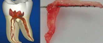
The pulp is an important part of the tooth, thanks to which the dental tissue receives all the necessary substances. When the pulp is removed - complete or partial - the process of mineralization of dental tissue is disrupted, which naturally affects the strength of the tooth. A pulpless tooth becomes more fragile over time and, sooner or later, begins to collapse. Removal of the dental nerve is indicated in the following cases:
- Deep caries;
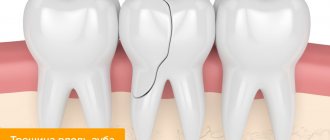
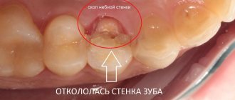
- Destruction of most of the tooth;
- Dental prosthetics (sometimes);
- Periodontitis, chronic pulpitis;
- Pulp necrosis.
As a rule, dentists at the Eurodent clinic decide to remove the dental nerve only if there are irreversible changes in the pulp. In most cases, with timely consultation with a doctor and properly selected treatment, removal of the dental nerve can be avoided.
Removing the nerves of the front teeth: what to pay attention to
A beautiful snow-white smile always attracts attention and encourages communication. That is why during preventive examinations you should pay special attention to the health of the front teeth, which are located on the so-called. "smile lines"
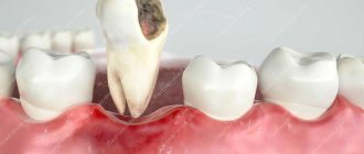
Removing the nerves of the front teeth is not a technically difficult procedure; it is comparable in complexity to similar operations on other teeth. In this case, it should be taken into account that a pulpless tooth becomes a “dead” tooth - without receiving the necessary elements, it darkens and becomes fragile over time. Naturally, in this case it will be necessary to restore the beauty of the dentition over time.
Aesthetic dentistry at the Eurodent clinic offers several options for restoring the appearance of teeth:
- Teeth whitening (including pulpless teeth)
- Installation of veneers (lumineers, ultra-veneers);
- Artistic restoration of teeth;
- Installation of crowns (metal-ceramics, zirconium oxide).
In order to avoid the unpleasant consequences of removing the nerves of the front teeth, you should undergo regular preventive examinations by the dentist. The specialist will be able to notice “problems” before the appearance of alarming symptoms and eliminate them without any problems.
In order to find out how much it costs to remove a nerve in a tooth , you can come for a free consultation at the Eurodent clinic.
With us you can undergo high-quality diagnostics and receive qualified dental care. Based on: 19 votes