The process of baby teeth emerging causes many problems. Baby teeth grow in children in infancy, up to one year. But after five years, the process of their loss and replacement with molars begins to occur. Baby teeth also have roots, which begin to simply dissolve at the age of five or six.
The process of changing baby teeth
Loss of baby teeth in children begins to occur between the ages of five and six years and can continue up to fifteen years. The roots of baby teeth dissolve and they begin to fall out. The molars growing behind them simply push them out of the socket.
Teeth usually change in the same order as they appeared. This process may take a long time, but if a permanent tooth does not appear in place of the lost tooth within a year, then you need to take the child to the dentist.
Primary teeth are necessary for the normal development of facial bones and muscles for the chewing process in a child. In addition, baby teeth help maintain space for permanent teeth, creating spatial balance. Therefore, you need to take care of temporary teeth until they fall out on their own. To do this, you need to follow the rules of hygiene, eat well and take care of your oral cavity. But this is not always possible and baby teeth often have to be removed.
Tooth loss: causes and methods of restoration
The method of restoration will be determined by why the tooth fell out.
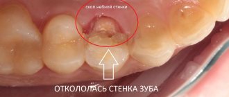
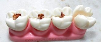
Advanced caries or pulpitis. With insufficient hygiene and lack of treatment for caries, it turns into pulpitis, and hard tissues begin to collapse. The process is very painful, accompanied by inflammation, the crown gradually cracks, and the infection spreads to the pulp and root. When a tooth falls out, it feels like a relief: it will finally stop hurting. In practice this is almost always not the case:
- inflammation can affect periodontal tissue. Without treatment, the infection will continue to spread, affect surrounding tissues, and provoke new complications;
- Teeth destroyed by pulpitis do not always fall out completely. Collapse of the crown walls may occur, leaving the infected root in place.
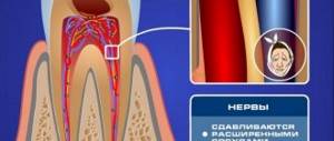
Even if the pain goes away after a loss, this does not mean that the problem is solved. The absence of pain indicates the death of the nerve. It is accompanied by necrosis, which provokes the appearance of new foci of inflammation.
What to do: You need to see a doctor as soon as possible. It is better not to wait until the crown is completely destroyed. If it remains above the gingival margin, there is a chance to preserve the living root and perform restoration or prosthetics. If the crown is completely destroyed, the doctor will remove the remaining root and begin preparation for prosthetics. When the crown is destroyed by caries or pulpitis, diagnostics is needed: the dentist must evaluate the condition of the root, periodontal tissues, and nerves in order to prevent the development of inflammation and complications.
Periodontitis or periodontal disease. This is gum disease. They have different courses, but the result is the same: teeth become loose and fall out. However, they can be quite healthy. The problem is the condition of the gum tissue. Long before the loss, symptoms should appear: loosening, drooping of the gingival margin (tooth enamel is exposed), possible bleeding of the gums, itching, discomfort, and pain. If a tooth has already fallen out due to gum disease, the doctor must evaluate the condition of the gum and bone tissue in order to offer the patient implantation or prosthetics.
What to do: it is better to consult a dentist when the first symptoms appear (the gums begin to bleed, its edges droop, itching appears, a reaction to temperature stimuli). If the patient has not done this, more complex treatment will be needed. The periodontist must restore the normal state of the gums (relieve inflammation, normalize blood circulation, etc.). If such treatment is possible, implantation is performed next. If not, a removable or bridge prosthesis is installed.
Injury. A lost tooth may be healthy and your gums may be in normal condition. The recovery tactics will be determined by how exactly the crown was damaged and whether the root was preserved.
What to do: You need to urgently consult a dentist. If possible, a fallen crown should be preserved. If a patient breaks and swallows a tooth (or loses it or throws it away), prosthetics will be needed to restore the dentition. If a tooth falls out completely and is preserved, there is a chance that it can be returned to its place (it is placed in the hole and secured with a splint).
If a tooth is completely knocked out, you need to:
- stop bleeding in the hole (press with a sterile swab);
- quickly find the tooth, do not touch the root, grab only the crown;
- rinse it carefully in water (do not wipe it, do not wash it under running water);
- put in place (in the hole), fix (bite through gauze or tampon);
- contact your dentist immediately.
Healing is possible if the tooth can be returned to its place (put in the hole) half an hour (before the nerve dies). If you can’t do this yourself, it is placed in whole milk (not water) and delivered to the dentist. It is impossible to leave the tooth dry (not in milk), the nerve will die in a few minutes.
Prosthetics, implantation, restoration.
Leave a request, we will call you back within 5 minutes
+7
Features of milk teeth removal
If you need to remove a baby tooth for a child, then this procedure has several differences from removing a molar tooth. Factors such as the structural features of a growing child's jaw, bite and the possibility of the presence of emerging sprouts of permanent teeth play an important role in carrying out the necessary manipulations. The dentist must approach the removal process with great care, because the walls of the alveoli are very thin and the divergence of the roots is very significant. To avoid damaging molars that are beginning to grow, the dentist must carry out the extraction process with extreme caution.
In order to prevent atrophy of the alveolar margin or the creation of a bone scar, which can result from the early removal of primary teeth, the dentist needs to make a decision about their removal after carefully weighing these negative consequences, which will impair the growth of molars. If the growth zones are injured, the process of formation of the child’s jaws will be disrupted and it will be difficult for him to chew food due to improper growth of the molars, since the load on the teeth is unevenly distributed.
Do I need to restore a broken or lost tooth?
Dentists at the DentoSpas clinic do not recommend leaving gaps in the dentition. It is better to plan and perform restoration, implantation or prosthetics as quickly as possible. Without treatment, bone tissue atrophy will begin, the load on adjacent crowns will increase, and the chewing load will begin to be distributed unevenly. This will lead to displacement of neighboring teeth and may cause their loss. If left untreated, a broken or chipped crown will continue to deteriorate and repair will become impossible. With advanced caries, periodontal disease, and periodontitis after tooth loss, the disease does not go away on its own; it continues to develop until complete edentia.
Main indications for removal
Temporary teeth can be removed long before it falls out in the following cases:
- When its root does not resolve in time, the baby tooth is removed so that it does not interfere with the growth of the molar;
- A temporary tooth can contribute to the process of gum inflammation;
- A loose temporary tooth can prevent a child from chewing food and cause discomfort in the mouth;
- The picture shows that the root of the tooth has already dissolved and the tooth should have already fallen out, having exhausted all the deadlines;
- Due to caries, the tooth was partially destroyed and filling it was not possible;
- A cyst was discovered at the root of the tooth;
- The molar has begun to break through, but the baby tooth does not fall out;
- The gums or jaw become inflamed due to a temporary tooth;
- Crack, fracture or chip of a tooth;
- Fistula;
- Periodontitis, phlegmon and sinusitis
Pulpotomy of primary molars
In 1991, Yacobi et al wrote about the then-evolved techniques and materials for performing pulpotomy of primary molars. A year later, Croll and Killian described a technique for performing pulpotomy of primary molars using a mixture of zinc oxide powder and eugenol as a material to fill the pulp cavity. After performing a pulpotomy, the authors recommended covering the teeth with stainless steel metal crowns. This approach was modified from the original technique of Kaare Langeland, who described the toxicity of the use of formocresol in pulpotomy, and stated that the pulpotomy procedure itself was effective without the use of formocresol, and not because of it. If there is a need to achieve hemostasis, the author suggested using ferrous sulfate. Later, the use of zinc oxide eugenol (ZOE) for pulp capping and filling the pulp chamber space was reported to be successful. MTA is another alternative material that can be used for a similar purpose. MTA is a derivative of Portland cement and is currently most often used during pulpotomy.
A systematic review and meta-analysis by Lin et al found MTA to be the first choice material for pulpotomy of primary molars. In this article we will present three cases of pulpotomy of primary molars with their further covering with metal crowns. In one clinical case we performed the pulp cavity with COE, and in the other two with MTA.
Clinical case 1
A 39-month-old female patient and her mother sought advice regarding an acute carious lesion on the occlusal surface of a right lower second primary molar (Figure 1). The patient had already previously obtained an orthopantomogram, which was performed on the recommendation of the pediatric dentist (photo 2).
Photo 1. Occlusal caries of the lower second primary molar in a patient aged 39 months.
Photo 2. Orthopantomogram demonstrating an extensive cavity.
No other pathologies were found in the oral cavity. Their pediatric dentist recommended contacting a specialist to perform general anesthesia, perform a pulpotomy, and then cover the tooth with a metal crown. But at the appointment, the patient behaved absolutely adequately, was somewhat shy, but cooperated with the doctor, and based on this, it was decided to perform a pulpotomy under sedation with nitrous oxide, which was administered through a nasal mask. After the mother signed the informed consent, the patient was scheduled for the procedure the next day after the initial visit. During sedation, nitrous oxide was administered at a ratio of 20% nitric oxide/80% oxygen. The child also listened to music on headphones in order to avoid irritation from the sound of the drill. The area of the lower second molar was anesthetized by infiltrating a 4% solution of articaine hydrochloride with epinephrine in a ratio of 1:200,000. After the rubber dam was installed, access to the pulp was created using a cylindrical diamond bur (photo 3).
Photo 3. Exposure of the pulp chamber area.
During the formation of the access, it was confirmed that caries had penetrated into the area of the pulp chamber. After removal of carious dentin, the pulp tissue was removed to the orifice area using a sterile, low-speed No. 6 bur. Bleeding from the root canal area indicated that the pulp in these areas remained vital. A cotton ball soaked in water was placed over the orifice area and pressed, which allowed good hemostasis to be achieved without the use of ferrous sulfate solution or any other hemostatic agent (Figure 4). After this, the COE powder was mixed until it reached the consistency of a paste, and packed into the pulp chamber area, carefully removing the excess (photo 4).
Photo 4. COE-pulpotomy.
An orthodontic strip was placed around the tooth to stabilize the position of the rubber dam. The area where COE was applied was covered with modified glass ionomer cement, which was then polymerized and prepared for further fixation of the metal crown (photo 5). After 4 years and 8 months, the patient again sought help for the restoration of a permanent maxillary molar, in the area of which there were signs of carious lesions. During the examination, the successful result of the previously performed pulpotomy and crowning of the lower second primary molar was confirmed. However, given the level of root resorption, the degree of eruption of the second permanent premolar and the presence of minor inflammatory changes in the mucosa, it was decided to remove the primary molar under local anesthesia (photo 8).
Photo 5. View after preparation for a crown.
Photo 6. View 4 years and 8 months after the intervention.
Photo 7. Orthopantomogram 4 years and 8 months after the intervention.
Photo 8. View of the tooth after removal.
Clinical case 2
A 9-year-old boy was referred by his family dentist for treatment of an extensive carious lesion in the area of the right mandibular second molar (Figure 9). The area of the adjacent first primary molar was also noted to have caries, as was the area of the maxillary first primary molar. The radiograph showed signs of penetration of carious pathology into the pulp space (photo 10).
Photo 9. Caries of a primary molar in a 9-year-old patient.
Photo 10. X-ray showing an extensive carious cavity.
After obtaining informed consent from the parents, it was planned to perform a pulpotomy in the area of the lower second primary molar of the mandible on the left and perform a composite restoration in the area of the first primary molar. The patient contributed in every possible way to the intervention, and after anesthesia with a 4% solution of articaine hydrochloride with epinephrine 1:200,0000, a rubber dam was installed (photo 11). All further manipulations were performed similarly to those described in clinical case 1. Using a cylindrical bur, the cavity was cleared of caries (photo 12), the space of the pulp chamber was reached and hemostasis was ensured by means of sterile rollers, which were inserted into the pulp space under compression (photo 13-14). The dense MTA mixture (NeoMTA, NuSmile) was then packed using an amalgam condenser over the root canal orifices (Figure 15). Once the material had cured, the excess was removed with a cylindrical diamond bur, and preparation of the first molar began immediately for further restoration (Figure 16).
Photo 11. Insulation using rubber dam.
Photo 12. Removal of carious tissue.
Photo 13. Achieving hemostasis using cotton rolls.
Photo 14. View after achieving hemostasis.
Photo 15. Packaging MTA in the tooth cavity.
Photo 16. View after pulpotomy.
A bioactive composite (ACTIVA BioACTIVE-RESTORATIVE, Pulpdent) was introduced into the cavities of both teeth, after which it was photopolymerized (photo 17-18). After polymerization, a 5% fluoride varnish was applied to the mesial surface of the permanent first molar. When the contour of the restoration on the first primary molar was completely recreated, the rubber dam clamp was reinstalled on the permanent first molar and the preparation of the second primary molar for a metal crown began (photo 19-20). The latter was contoured, cut and polished according to the usual algorithm, after which it was fixed with modified glass ionomer cement. Excess cement was removed using a Hollenback instrument (Figure 21), and in the area of interproximal contacts - using dental floss. The maxillary first primary molar was restored at the next visit. Photographs and radiographs obtained 22 months after treatment are shown in Figures 22 and 23. After 22 months, the patient no longer returned for monitoring.
Photo 17. Filling the cavity with a bioactive composite.
Photo 18. Polymerization of a bioactive composite.
Photo 19. View after contouring the restoration in the area of the first primary molar.
Photo 20. View after preparing the tooth for a crown.
Photo 21. Removing excess cement.
Photo 22. View 22 months after treatment.
Photo 23. X-ray 22 months after treatment.
Clinical case 3
An 8-year-old boy was referred to the dentist due to the need for extraction of the second primary molar of the lower jaw on the left and restoration in the area of the first primary molar on the same side of the jaw (photo 24-25). Taking into account the initial parameters of the clinical situation, the child’s parents were offered a treatment option with a pulpotomy procedure and further covering with a metal crown. Using the same approach as Case 2, the pulp chamber space was completed with MTA in the orifice area, the tooth was then capped with a metal crown, and the first primary molar area was restored with a restoration (Figure 26). Hemostasis was achieved using sterile moistened cotton swabs, and the dental cavity was filled with a modified glass ionomer material over the MTA. 33 months after treatment, clinical examination and radiological monitoring data indicated a successful outcome of the intervention (photos 27-28). 33 months after treatment, both primary teeth of the lower jaw on the left side were also removed according to orthodontic indicators (photo 29).
Figure 24: An 8-year-old patient was referred for extraction of the mandibular left mandibular second primary molar.
Photo 25. X-ray of the patient before treatment.
Photo 26. View after pulpotomy.
Photo 27. View after 33 months.
Photo 28. X-ray after 33 months.
Photo 29. View of the tooth after extraction.
Discussion
The use of a rubber dam when performing pulpotomy of primary molars is one of the key factors to ensure the success of the manipulation. The rubber dam ensures both safety of the intervention and comfort during the procedure. In cases where the pulp is completely damaged, the pulp tissue shows almost no signs of bleeding, therefore, in such cases, it is necessary to perform a pulpectomy or consider the need for tooth extraction. In cases of extraction, it is necessary to remember the need to manufacture a retainer to replace the space, so as not to compromise the process of eruption of permanent teeth.
In the three clinical cases described above, we were able to achieve hemostasis using conventional cotton swabs without the use of any other additional means. In cases of heavy bleeding, sterile low-speed burs can be used to remove pulp from the orifice. You can also use cotton swabs soaked in a solution of ferrous sulfate, which should be placed into the cavity of the pulp chamber for 30-60 seconds. This procedure is often referred to as a “ferum sulfate pulpotomy,” although in fact this agent is only used to stop bleeding. It is more correct to call the manipulations as COE-pulpotomy or MTA-pulpotomy, depending on the material used to close the orifice area.
Authors: Theodore P. Croll, DDS Constance M. Killian, DMD Rachel L. Bresler, DMD
Contraindications
- Acute inflammation in the mouth, such as candidiasis, gingivitis or stomatitis;
- Pneumonia, tonsillitis, whooping cough;
- Various tumors near the temporary tooth, when it needs to be removed along with it in the hospital.
It is recommended to remove temporary teeth only when the acute stage of dental disease has passed. During removal, it is necessary to take into account possible pathologies of the patient, such as cardiac pathologies, problems with blood vessels and the nervous system, kidney diseases and various blood diseases.
Anesthesia
In cases where the root of a temporary tooth has practically disappeared, it is enough to apply an anesthetic gel to the gums; this method is also called application anesthesia. Anesthesia can be performed using the infiltration method - this is when an anesthetic is injected into the gums from both sides.
In most cases, the child tolerates pain relief well, but if there is a predisposition, a mild allergy may occur. To avoid this, you need to warn your doctor:
- Do any medications cause allergies in him, and what kind?
- When and what painkillers were used and what was the reaction of your child’s body to them;
- Provide information about chronic diseases, if your child has any.
In emergency cases, temporary teeth can be removed under general anesthesia: in case of complex mental illness, purulent inflammation, intolerance to local painkillers.
Process of removing temporary teeth
- The forceps are used to grab the area of the tooth root;
- The dentist moves along the equator of the baby tooth;
- The doctor lightly squeezes the forceps on the tooth;
- And it easily dislocates a tooth. This process is called luxury;
- The further process of removing the tooth from the socket is called traction;
- The doctor must make sure that the roots are removed;
- The dentist uses a tampon to press down on the area where the baby tooth was.
Removal procedure
- Forceps are applied to the crown so that the tooth is fixed without excessive pressure.
- The dentist performs luxation (the so-called luxation of the tooth).
- The tooth is removed from the socket (this manipulation is called traction).
- The dentist makes sure that all roots are removed.
- The hole is closed with a gauze swab.
Read also: Shark teeth in children
Complications
If a baby tooth is removed prematurely, a year before the primary tooth erupts, complications can occur. Those teeth that the child has left may move apart and begin to take up empty spaces, which will lead to problems with the growth of molars, since they will not be able to appear in their proper place.
When removing multiple baby teeth at the same time, be careful to ensure that the child does not develop an overbite. To do this, you can use dentures made of removable plates with artificial teeth inserted into them. This will help prevent the process of teeth moving apart and the molars will be able to erupt in their designated place.
List of possible complications when removing temporary teeth:
- Tooth fracture;
- During removal, soft tissue may be affected;
- Damage to the mucous membrane or gums;
- Dislocation of the jaw;
- Fracture of the alveolar process;
- Nerve damage;
- Aspiration of a root or blood clot;
- Injuries to nearby teeth.
Treatment methods
All therapeutic manipulations are aimed at stopping the pathological process in the dental system and preventing it from spreading to the permanent tooth or disrupting its growth. For this purpose, depending on the causes and characteristics of the identified complication, conservative and surgical methods can be used.
In the first case, treatment is prescribed using local drugs (antiseptics, corticosteroid hormonal ointments, non-steroidal anti-inflammatory gels, etc.), which suppress the growth and activity of pathogenic microbes and stop the inflammatory process. In case of severe bacterial infection, systemic antibacterial drugs may be prescribed.
Sanitation of the oral cavity is mandatory, including the removal of mineralized dental plaque, treatment of caries, gingivitis and other underlying diseases (causing irregular teeth or infection) or complications. When granulation tissue grows, surgical treatment is recommended - removal of excess tissue and gum plastic.
Preparing the child
During removal, it is advisable for one of the parents to be near the baby; he should feel the presence of a loved one nearby.
There is no need to scare your child at the dentist.
It is advisable to take him to the dentist at least once every six months for a preventive examination so that he stops being afraid of the doctor.
To prevent your excitement from being passed on to your child, try not to be nervous yourself. If you go to the dentist, you can take your child with you if you are sure that you will behave calmly. When he sees that the procedures do not cause you pain, he himself will no longer be afraid of going to the doctor.