Author: Brodsky Sergey Evgenievich Deputy Chief Physician, Candidate of Medical Sciences, specialties: dentistry and medical microbiology A hole in a tooth is the result of long-term carious processes. With timely detection and treatment, it is possible to prevent inflammatory processes and save the tooth. Therefore, if you experience pain, discomfort while chewing or brushing your teeth, or darkening or perforation of the enamel, we recommend that you contact the Parter-Med clinic, where experienced dentists will correct the defect and heal the tooth.
Deputy chief physician
Brodsky Sergey Evgenievich
Sign up for a free consultation
+7
Causes of a hole in a tooth
The formation of a hole in the teeth is a complication of untreated caries. Putrefactive processes develop and affect the deep layers of hard tissue, which leads to their perforation. The following factors can trigger the pathological process:
- neglect of oral hygiene;
- the predominance of sweet and starchy foods in the diet;
- low quality of drinking water;
- hereditary predisposition to dental pathologies;
- dental hypoplasia - insufficient development of enamel as a result of rickets suffered in childhood or disruptions during intrauterine development;
- mechanical damage to the enamel after dental procedures, the use of pins to clean the interdental space, jaw injuries;
- decreased protective properties of the body;
- hormonal imbalances;
- smoking;
- diseases of the digestive system with changes in the acidity of saliva.
How does a hole form in a tooth?
Let us make a reservation that a hole in a tooth is not a diagnosis, but a consequence of a dental disease. The phenomenon visually indicates that there is a problem.
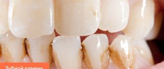
Ideal teeth are straight and smooth. Uncleaned food debris leads to the formation of a special film on the teeth. Dentists call this film plaque. It contains a large number of bacteria. Over time, plaque hardens and becomes strongly attached to the teeth. Pathogenic organisms that are inside produce a specific acid that quickly destroys tooth enamel. It turns out that bacteria and microorganisms gradually begin to destroy teeth. As a result, a person gets a disease such as caries.
Stages of tooth destruction
Initially, small holes are obtained. Later they expand, turning into a hole. The described process can be divided into the following stages:
- First stage: initial. First, a whitish spot appears on the surface, which is the start of the destructive process. Over time, this spot turns from white to brown. At this stage, there are no other (let alone unpleasant) symptoms. Therefore, we people sometimes do not even know about the onset of the disease. At this time, only a dentist can detect it.
- Second stage: development of superficial caries. If the stain is not removed in time, painful microorganisms will affect the very top layer of tooth enamel. A more serious inflammatory process will begin, which is characterized by destruction. Here patients can talk about their complaints. Usually at this time the teeth are very sensitive. They react to temperature changes and acidic foods.
- Third stage: middle. Not only enamel, but also dentin suffers from destruction. Visually you can see a small hole. This is where food regularly falls and is left to rot. In addition to the sensations of the second stage, a reaction to chemical components is added. In addition, a person usually feels pain from inhaling air with an open mouth. If the tooth is not treated, the painful sensations will begin to torment constantly. Even painkillers cannot relieve the symptoms.
- Stage four: deep caries. The hole turns into a cavity and becomes quite large in size. The enamel and dentin itself are made very soft. If you touch the open bottom, you may feel pain. Inflammation of the nerve and vascular tissue begins. As a result, caries turns into pulpitis.
What does white plaque mean after tooth extraction?
Some patients notice that their gums turn white after tooth extraction. In the normal course of events, a whitish coating is nothing more than an “effusion” of fibrin from the blood, and indicates the beginning of epithelization of the wound. White plaque after tooth extraction usually appears on the surface of a blood clot (Fig. 8), as well as on the surface of a severely injured mucous membrane.
White plaque on the gum after tooth extraction -
In this case, the gum surrounding the socket has a pale pink color, when pressing on the gum there should be no purulent discharge (as in the video below), there should be no unpleasant odor from the socket, constant aching pain or pain when responding to cold and hot water (24stoma.ru) .
Reasons for the appearance of holes
A hole in a tooth is a consequence of the carious process. It is formed under the influence of several factors:
Features of people's lifestyle
- improper diets, as a result of which the body weakens and loses essential vitamins.
- poor hygiene or its complete absence.
- insufficient content of such an element as fluorine in the enamel.
External factors
- Having an incorrect bite.
- Bad heredity.
- Floor. It is women who suffer more from caries than men. The above is related to hormonal changes in the female body. And also, it is the weaker sex who eat the most sweets and starchy foods.
- Poor quality of drinking water.
- Particular susceptibility to diseases.
Physiological factor
- Distance between teeth.
- Features of the structure of the jaw.
- Poor quality enamel.
Geographical factor
Surprisingly, the occurrence of caries is influenced by the geographic location in which a person lives. It has been proven that climate, minerals, soil, and topography have a significant impact on human health in general, including teeth. Thus, almost all US residents (with the exception of only 1%) have serious dental problems. In Russia, only 40% of the population has healthy teeth, the remaining 60% suffer from dental problems. And in Norway, only 2% of the population complain of bad teeth.
Nutrition feature
Eating preservatives, carbohydrates, unnatural additives, and large amounts of sweets increases the likelihood of cavity formation many times over.
What symptoms indicate caries in the spot stage?
It is very difficult to suspect caries at an early stage. The patient has no complaints of pain, discomfort while eating, and in fact, white spots are the only thing that indicates the beginning of the carious process. However, white spots are also characteristic of a number of other dental diseases, so the dentist’s goal is to correctly determine the cause.
The following signs help distinguish caries from other dental diseases:
- a white carious spot is usually a single one, whereas in other diseases the lightened areas are located on many teeth;
- A carious spot is white or cream-colored; it does not have clearly defined boundaries and has a smooth matte structure. In other pathologies, the spots most often have a clear border and yellowish pigmentation;
- caries in the spot stage most often forms on healthy enamel, while in other pathologies the tooth may already erupt with white spots.
It is almost impossible to distinguish between caries and other dental pathologies by symptoms without the help of a doctor. Moreover, not everyone is able to independently notice the change in enamel color. Therefore, it is recommended to undergo routine examinations at the dental clinic twice a year.
Hole in a tooth in children
It cannot be assumed that baby teeth are not susceptible to caries. This is wrong. Pathogenic and dangerous microorganisms very often affect baby teeth. During the period of replacement of temporary teeth with permanent ones, microbes that have settled on milk teeth calmly transfer to new ones that have just emerged. As you know, the entire process of changing teeth is quite long. Therefore, already in childhood, caries can be detected in a child’s mouth.
It is necessary to explain to the baby that he must take care of his oral cavity in the same way as his parents. Leftover food gets stuck between his teeth and causes the appearance of microorganisms. This should be avoided. Therefore, it is imperative to teach your child not only to use a brush, but also dental floss.
Installing a seal
The task of the doctor who has discovered a tooth damaged by caries is:
- drilling out dead tooth tissue and darkened parts of enamel;
- expansion of the carious cavity;
- checking the condition of the pulp;
- preparing the tooth for filling.
The filling process consists of:
- in disinfecting the drilled cavity and drying it;
- installing a medicinal inlay into the tooth canals;
- installation of a temporary filling;
- A permanent filling is installed when a tooth is cured and the lost parts need to be restored.
Treatment of holes
Depending on the stage of the disease and the reason for the formation of the hole, a person may simply not be bothered by this cavity or may make itself felt by severe pain with fever. The hole must be healed with the help of a dentist. But if you still can’t get to the dentist’s office, you can relieve the pain at home.
Emergency help at home
If pain occurs, you should immediately brush your teeth. This will remove food debris from the mouth. Next, you should take any analgesic drug. The medicine will also help in case of fever.
If swelling is suddenly observed, it can be reduced by rinsing with soda. Or apply a piece of ice to the sore spot for 15-20 minutes.
Very often people resort to our grandmothers' recipes. The older generation claims that the following will help with initial pain in the tooth:
- A piece of lard, garlic, beets and bee propolis attached to the tooth.
- Normal tears, so you need to cry. Tears will relieve pressure in the jaw and thereby reduce the pain slightly.
- Massage the ear from the side of the diseased tooth.
- Gargling with herbal decoctions.
- Rubbing the sore spot with plantain juice.
Independent measures will only alleviate the condition, but will not cure the tooth. If a tooth stops seriously bothering you, this does not mean that the disease has gone away. The disease simply began to occur hidden. It went to the very depths and became chronic. In any case, you will have to go to the dentist.
Doctor's appointments
A hole in the teeth is treated according to the same scheme:
- First stage: thorough treatment of the oral cavity. Here the doctor removes plaque and tartar from the diseased tooth or from the entire oral cavity.
- Second stage: prick or injection of an analgesic. This kind of pain relief is necessary. Under its influence, the patient’s panic fear of treatment goes away, he does not feel pain and thereby allows the doctor to calmly do his job.
- Stage three: removal of infected tissue. Removal is carried out throughout the entire tooth cavity using special dental devices. Next, the doctor coats the walls of the exposed tooth with a bur or antiseptic. As a result of this action, the growth of bacteria and further relapse of the disease stops.
- Stage four: drilling out the cavity to install the filling. This is necessary so that the new installed material fits tightly into the tooth.
- Fifth stage: application of filling material or protective seal. They try to match the material as closely as possible to the color of the patient’s natural teeth. If suddenly the patient’s tooth has been affected by deep caries, then a therapeutic pad is placed at the bottom of the cavity. It will relieve inflammation from the nerve. Read about how to kill a nerve in a tooth here.
- Stage six: adjusting the filling to the shape of the tooth. This action helps the patient not to experience discomfort when chewing in the future.
Remineralizing therapy
White spots during caries are areas of enamel demineralization. It is possible to remove them if you restore the balance of minerals in tooth enamel. This can be done with the help of remineralizing therapy. It can be applied or carried out using a special mouth guard. In the first case, the tooth surface is covered with a concentrate for remineralization, in the second, a sealed mouth guard is used, individually made from a jaw cast. As prescribed by the doctor, the patient himself will fill the mouthguard with a remineralizing composition and put it on his teeth. Most often, solutions of calcium gluconate or glycerophosphate, as well as gels with calcium phosphate, are used for remotherapy.
Remotherapy helps remove caries in the form of stains, helps strengthen tooth enamel, replenishes the lack of minerals (mainly phosphates and calcium), reduces tooth sensitivity, prevents further development of the carious process, and preserves the natural healthy appearance of teeth.
A filling is the main protection for a hole in a tooth.
Filling material may vary. Let's look at the main ones.
Metal fillings
- From amalgam. It is a special metal combined with zinc, silver, tin and mercury. Currently, copper and silver amalgams are used. Such fillings are quite strong, plastic, they are notable for their low price and good fit to the edges of the cavity. The service life of amalgam fillings is 15 years. The material is able to accurately replicate the shade of the tooth. But a tooth with such a filling stands out from the entire dentition. Such fillings take too long to harden and are difficult to fit into the cavity. In addition, mercury vapor released during work is harmful to the health of the dentist.
- Made of gold alloy. They have enviable strength and a very long service life. But they are practically not used because of their visual visibility. In addition, a gold filling is an expensive pleasure.
Plastic fillings
They are used to temporarily protect the hole. They have good resistance to chemical components, harden quickly and do not irritate the patient’s oral cavity. However, such fillings quickly shrink and change their original shade over time. Plastic fillings can be made:
- Made from acrylic oxide. The material is resistant to any external influence, retains its original shade for a long time, fits tightly to the tooth and gives only slight shrinkage. The disadvantages of such fillings include their toxicity and the ability to start an inflammatory process under the installed filling.
- Made from carbondent. Compared to the filling described above, it has high density and low toxicity. However, such material quickly darkens and breaks.
Ceramic fillings
They are used quite often. They have the necessary advantages: the ability to maintain their size, do not shrink, and have increased hardness. At the same time, the shade of the filling does not change over time. The load under such a filling is distributed evenly over the entire tooth. In addition, ceramic fillings have excellent aesthetics, but high cost. This type of filling can be made of pressed ceramics or metal ceramics.
Composite
These fillings instantly harden under the influence of a photopolymerization lamp, or rather its light. They are distinguished by good strength and the required aesthetics. They are quite easy to polish. Such light fillings are practically indistinguishable from natural teeth. Therefore, reflective materials are often placed on the front teeth. As for shrinkage, they are not strong.
Cement
- Zinc phosphate. Used only to secure other fillings or as a spacer before a regular filling is installed.
- Glass ionomer. The material is quite durable and can last a long time. Such fillings are non-toxic and have excellent adhesive properties. During wear, the filling releases the necessary fluoride, which protects teeth from caries. However, the filling made from the cement in question is fragile and wears out very quickly. Therefore, it is usually used as gaskets and securing inlays in the hollow of a tooth.
- Silicate-phosphate. This is a very cheap filling material. It is often used in public dentistry. Such fillings do not stick to the surface of the tooth cavity. They do not conduct heat at all. However, these fillings are quite hard and transparent in appearance. Their disadvantage: they are toxic to the pulp.
- Silicate. This is long outdated material. It used to be placed after placing a zinc phosphate cement liner on the pulp tissue. Time has shown that this material has a negative effect on the pulp. Silicate cements are toxic and dissolve over time when exposed to saliva.
Toothache during pregnancy: what to do
So, what can you do to relieve pain if your tooth hurts during pregnancy... Remember that even approved medications should be taken by pregnant women only after consulting a doctor.
- Nurofen 200 mg or 400 mg (Fig. 7-8) - it can only be used in the 1-2 trimesters, but not in the 3rd trimester. Nurofen is available in dosages of 200 and 400 mg. Pregnant women can use both dosages. But we recommend that if the pain is not very severe, use a dosage of 200 mg (no more than 4 tablets per day). If the pain is very severe, then the dosage is 400 mg, but not more than 3 tablets per day.
- Paracetamol 500 mg - can be used throughout pregnancy, preferably only after consulting a doctor. No more than 2 tablets per day.
Important: you must understand that analgesics are only a temporary solution to the problem. If you have a toothache, you will still need to see a doctor. There are periods of dental treatment for pregnant women, during which dental treatment is quite safe for both the mother and the unborn child.
Preventive measures
To prevent a hole from appearing in a tooth, you must:
- Adjust your diet. For these purposes, you need to exclude or greatly limit sweet foods, and include foods with fluoride, phosphorus and calcium in your daily consumption. Keep your consumption of carbonated drinks to a minimum.
- Visit the dentist. It is necessary to seek the help of a specialist even for minor problems in the oral cavity. Or go for preventive examinations twice a year.
- Maintain proper and regular oral hygiene. It is best to use fluoride toothpastes. Always rinse your mouth after each meal if you are not brushing your teeth.
- Clean your mouth at least 2 times a day.
- Choose the right brushes. Use medium to soft bristles. The nose of the tool should be rounded.
- Change toothbrushes every 4 months.
- If possible, buy an electric toothbrush and use it to clean. Studies have shown that it removes plaque very well.
- Clean properly. To do this, you need to hold the brush at an angle of 45° and make rotational movements with it in a circle, as well as back and forth. When brushing, do not put too much pressure on your teeth and gums. You should also try to clean the oral cavity from all sides.
- Be sure to clean your tongue. This is what often causes bad breath.
Prevention
To maintain healthy teeth and prevent the formation of carious holes, Partner-Med dentists recommend:
- Brush your teeth twice a day with toothpaste and a brush;
- after eating, rinse your mouth with clean water;
- floss is used to clean the interdental space;
- undergo annual preventive examinations at the dentist.
When you detect the first signs of carious processes, you should contact your dentist. Our clinic specialists will examine the oral cavity and, if there are problems, prescribe the necessary treatment. If a carious hole is detected, they will treat and install a high-quality filling in the tooth hole with high wear resistance and a guarantee.
Request a call back or dial our number!
+7
This phone call does not obligate you to anything. Just give us a chance and we will help you!
Just pick up the phone and call us!
+7
We will definitely make you an offer that you cannot refuse!
Complications from the hole
It has been said that caries is the starting point for the formation of a hole. If it is not treated in time, pulpitis will begin, then the disease will develop into periodontitis. Pulpitis and periodontitis are often treated using surgical methods.
A feature of pulpitis is that the pulp chamber is opened. The person begins to suffer from severe pain. In this case, the occurrence of pain does not depend on irritating factors. The pain wears him down the most at night. Pulpitis can exist in two forms: acute and chronic. If this disease is not treated, it will develop into periodontitis.
With this disease, an abscess forms. If suppuration affects the underlying tissue, osteomyelitis will occur. If a lesion containing pus breaks through, blood poisoning may occur.
How long does it take for gums to heal after tooth extraction - timing
How long it takes for the gums to heal after tooth extraction depends on many factors: the degree of trauma of the removal, whether sutures were applied, the possible addition of infectious inflammation of the socket, and the age of the patient. Healing of the hole after tooth extraction can be divided into partial and complete.
Partial epithelization of the wound occurs on average in 12 days (Fig. 5), but complete epithelization of the surface of the clot is observed in 20 to 25 days (Fig. 6). However, if inflammation of the socket occurs or after a complex tooth extraction, which is usually accompanied by major bone trauma, the healing time may increase by several days.
Reasons for slow healing –
- significant trauma to the bone and gums during removal (both due to the doctor’s indifference and as a result of sawing out the bone around the tooth with a drill during difficult removal),
- when a clot falls out of the socket (empty socket),
- development of alveolitis of the socket,
- the doctor left fragments or inactive fragments of bone tissue in the socket,
- if sharp bone fragments protrude through the mucous membrane,
- if the gum mucosa around the hole is very mobile, and the doctor did not apply stitches,
- antibiotics were not prescribed after a complex removal,
- patient's age.
Do they give anesthesia to children?
Many parents are concerned about how to painlessly treat teeth in children under 5 years old.
Non-invasive methods are absolutely painless. If a cavity needs to be prepared, anesthesia may be required. Children most often undergo two-stage anesthesia. First, the gum area is treated with an anesthetic gel, which numbs the injection site, and then an anesthetic injection is given.
In some cases, it is advisable to use sedation - when the child is introduced into a relaxed state of half-asleep using a special gas. Choosing the optimal method of pain relief and treatment is the responsibility of the dentist.
Prevention of childhood caries of primary teeth
Caries of primary teeth can be prevented if you listen to the recommendations of dentists and follow preventive measures for this dental disease.
- Take care of your oral cavity from the moment the first tooth appears.
Previously, it was believed that young children needed to brush their teeth from about one year old, when they grew eight front incisors. Modern dentistry has revised this approach, and today it is recommended to brush children’s teeth as soon as the first baby tooth has erupted. This usually occurs between 6-9 months of age. It is clear that a baby cannot take care of the oral cavity on his own, so this task falls on the shoulders of the parents. The first teeth are cleaned with special fingertips or a baby brush without toothpaste. The brush should have a small cleaning head and ultra-soft bristles. As they approach the age of one year, they begin to brush their teeth with fluoride-free toothpaste. As the child gets older, the size of the head and the stiffness of the bristles may change. For example, for a child aged 5 years, you can purchase a toothbrush with medium-hard bristles.
- Follow the rules for brushing your teeth.
A child may develop caries even with regular brushing if this process is left to chance. Children under about 7 years old cannot brush their teeth properly, so parents should help them and monitor the process. Proper oral care involves brushing your teeth twice - in the morning after breakfast and in the evening before bed. The hygiene procedure should not take less than 2-3 minutes. It is important to ensure that the child thoroughly cleans each tooth on both sides and cleans the chewing surfaces. A large number of bacteria accumulate on the tongue and the inside of the cheeks, so at the end of cleaning you need to teach your child to remove plaque from these surfaces.
- Routine visit to the dentist.
Few children like to treat tooth decay. Modern methods make it possible to do this quickly, painlessly and without drilling, but only if the disease is detected at an early stage. This is why it is so important to take your child to the dentist twice a year, even if the child has no complaints. The doctor will check the condition of your teeth and give recommendations for care. Regular visits accustom your child to visiting the dentist and help get rid of fear. In the future, such children sit in the dental chair without any problems and have their teeth treated.
Follow these preventive measures and you may not have to treat your child’s caries.
Ozone therapy in the complex treatment of caries
This method is rarely used on its own; more often it is used in the complex treatment of caries at the white spot stage. The dentist treats tooth enamel with ozone, which has an antimicrobial effect. The disinfecting effect of ozone helps stop the spread of bacteria, after which remineralization or deep fluoridation is carried out to restore the mineral composition of the enamel.
The dentist determines how to treat carious white spots on the enamel based on the patient’s age, medical history and other factors.
Good immunity reduces the risk of developing caries
The bacteria that cause tooth decay are part of the normal microflora of the oral cavity. But with weakened immunity and other accompanying factors, they can cause caries. At home, everyone can prevent this dental disease by strengthening the immune system. This is facilitated by hardening, reducing stress levels, quality sleep at night, proper nutrition, giving up bad habits, and maintaining a normal weight.
The fewer factors that weaken the immune system, the lower the risk of dental caries.
How does fluoridation treat caries in the spot stage?
The method helps provide the enamel with a sufficient amount of fluoride, a substance that maintains its strength and durability. The simple fluoridation method will require from 4 to 15 procedures. During each procedure, tooth enamel is coated with fluoride gel or varnish and left for 20 minutes. You must not eat or drink for 60 minutes after each procedure.
Deep fluoridation is considered more effective. To carry it out, you will need two drugs: a composition with fluorine, magnesium, copper and calcium hydroxide. When both compounds react, crystals are formed that seal the enamel structure. Thanks to the procedure, the concentration of fluoride ions in teeth increases approximately 5 times. Deep fluoridation is performed in one, less often - in 2-3 procedures.
With the help of fluoridation, enamel becomes more resistant to the destructive effects of organic acids, the activity of plaque bacteria and, accordingly, the rate of caries development is reduced, the healthy mineral composition of the enamel and the normal structure of hard tooth tissues are restored.
Often, when treating dental caries in the staining stage, methods of remotherapy and deep fluoridation are combined to achieve the best result.


