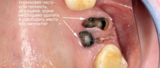
Possible reasons
| The exact cause of this disease has not been identified. Experts believe that the cause is a violation of the formation of tooth buds in the embryonic period. For example, this can happen if a woman took certain medications during pregnancy, had bad habits, or suffered from certain infectious diseases. |
There is also a version that the pleasant appearance of abnormally located teeth is atavism. The essence of this theory is that in past centuries people had a larger number of teeth than today, so some patients show signs of returning to the original structure of the dentition. According to statistics, this anomaly occurs in 7% of cases.
Symptoms
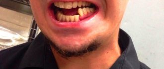
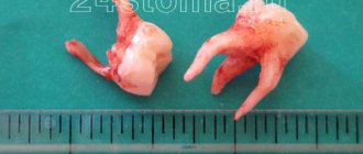
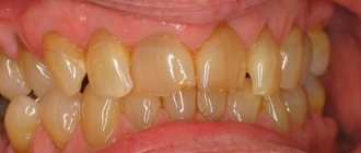
Photo: false hyperdontia
The main symptom of this pathology is the presence of supernumerary teeth, which cause a lot of inconvenience and interfere with chewing and speech function.
But in children and adults, the clinical picture of the anomaly manifests itself slightly differently.
Read the following review to see how edentia manifests itself in children.
In a separate article we will look at how shortening of the upper dentition is corrected.
Here https://orto-info.ru/ortodonticheskoe-lechenie/podgotovitelnyiy-period/metodyi-diagnostiki.html we will talk about methods for diagnosing anomalies of the dentofacial system in orthodontics.
In children
Extra teeth may appear immediately after birth or in the first few months of a child's life.
In this case, the normal feeding process is disrupted, since :
- pain during breastfeeding;
- the appearance of wounds and cracks on the chest;
- improper breast grip;
- sucking function is difficult.
If extra teeth appear at normal times for eruption, then the child experiences the same symptoms as when regular teeth appear, but in a more intense and pronounced form.
Such manifestations may be:
- swelling and soreness of the gum tissue;
- excessive salivation;
- slight increase in temperature, up to subfebrile levels;
- difficulty in nasal breathing due to swelling of the nasal mucosa.
If a child has an abnormal number of teeth, the following symptoms are observed :
- regular formation of wounds as a result of injury to the mucous membrane;
- violation of diction;
- formation of malocclusion;
- incomplete closure of the main row of teeth.
In adults
In adults, the anomaly has the same symptoms as in children, but it is also characterized by additional symptoms :
- presence of dystopic teeth . The growth of an extra tooth with an already formed bite can be stopped at the eruption stage, due to insufficient free space for it;
- predominantly palatal or lingual eruption ;
- incorrect position of the teeth: their rotation, change in the angle of inclination, curvature;
- loosening of neighboring ones;
- protrusion of the jaw bone in the area of the abnormal tooth;
- frequent inflammation of the gums;
- pathologies and deformation of the jaw bone.
In what cases is it necessary to remove supernumerary teeth?
Removal of supernumerary teeth is recommended in 90% of cases. You can refuse surgical intervention in the following cases:
Simply put, if a supernumerary tooth causes inconvenience, pain or discomfort, it is removed. |
What it is?
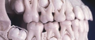
Normally, a child with a primary malocclusion erupts 20 teeth. After changing milk teeth to permanent teeth, there are already 32 of them. Hyperdontia represents an excess of the total number of erupted teeth, which are otherwise called supernumerary teeth.
Most often, hyperdontia is observed in the late period of mixed dentition, when the bulk of the teeth have already erupted.
In isolated cases, extra specimens may appear during the formation of the primary occlusion. This anomaly occurs predominantly in the male half of humanity; in women it is diagnosed only in 35% of cases of the total number.
In 85% of recorded cases, the pathology appears with the eruption of one or two supernumerary teeth, the remaining patients had 3 or more extra teeth, which were located both along the gum line and along the hard palate, filling its entire area.
Most often, with limited hyperdontia, extra teeth are localized in the area of the lateral and central incisors of the upper jaw, and less often - in the lower jaw. They may have a different shape, structure and shade, which indicates their pathological formation.
Basically, they are characterized by a cone-shaped shape and irregular development of enamel . In size, such teeth are usually slightly smaller than others.
How is removal carried out?
If the abnormal tooth is single and it is not difficult to get to it using surgical instruments, then there will be no problems, and the tooth will be removed using a standard method. If there are several teeth, they are located in hard-to-reach places, have several roots, or have not fully erupted - in this case, removal is carried out as follows:
- The doctor administers an anesthetic so that the patient does not experience pain during the procedure.
- A small incision is made on the mucous membrane.
- If the roots of the tooth are deep, the doctor may make a small hole using a special bur to provide access to the root system of the tooth.
- The tooth and root are removed using forceps.
- Next, the area is disinfected using antiseptic agents.
- Stitches are applied.
- To avoid complications, antibiotics, anti-inflammatory drugs, etc. may be prescribed.
Please note that before surgery, an X-ray diagnosis is required. In this way, the doctor assesses the condition of the root system and other features. Only after the diagnosis is carried out is the issue of tooth extraction decided.
Causes for concern
However, in some cases, irregular teething requires medical intervention. Firstly, children have so-called natal and neonatal teeth: if your child was born with teeth or they erupt in the first month of life, you should immediately consult a dentist. As experts explain, these teeth can be additional, or supernumerary, or complete, but erupted very early. They are distinguished by great mobility. If the dentist determines that the teeth are supernumerary, he or she may recommend removing them.
Also, a visit to the dentist may be necessary in case of congenital absence of teeth, or hypodontia. According to researchers from the Kazan State Medical Academy, the absence of incisors or first molars of the upper jaw is extremely rare; Heredity plays a significant role in the development of this deviation.
As soon as your baby's first teeth emerge, start using a special baby toothbrush with a small head and very soft bristles designed to gently but effectively clean baby teeth. Teach your child good oral care from an early age, and be sure to schedule a visit to the dentist between the eruption of the first tooth and your baby's first birthday.
Other treatments
| Another way to solve the problem is orthodontic treatment. This is true if the abnormal unit is located close to the dentition. Braces are usually used to straighten it, since this method is the most effective. The duration of treatment is determined individually, depending on individual characteristics. Orthodontic treatment may also be prescribed after the removal of a supernumerary tooth in order to normalize the bite. |
Anomalies in the size and shape of teeth (macrodentia and microdentia)
Vestibular position of teeth
The lateral incisor can be removed if it is inferior (hypoplasia, caries, periodontitis, abnormal position) and in a situation where the canine is located directly above it. The canine itself is removed in exceptional cases when it is abnormally developed or occupies a particularly abnormal position.
The main methods for correcting the vestibular position of the canines can be: distal movement of the lateral teeth, mesial movement of the incisors, expansion of the dentition, removal of individual teeth (usually the first premolars) and displacement of the canines in the distal direction to the vacant space. The movement of the canine into the dentition can occur through self-regulation, the use of massage, or with the help of orthodontic equipment (braces, plates with arm-shaped springs, etc.).
An important point is preventive tooth extraction. Even before the eruption of the canine, if there is apparently no room for it, but after the eruption of the first premolar, it is advisable to remove the last one and the canine will erupt on its own. In a situation where there is partially not enough space (grade 12) for the canine, but in the presence of the first premolar, it is advisable to remove the second primary molar, which will create a certain reserve of space, since the primary molar is on average 2.3 mm wider than the first premolar. After removing the second primary molar, it is necessary to move the first premolar distally and the canine will be installed in place.
Palatoglossus or oral position of teeth
Depending on the patient’s age and the form of the anomaly, a careful analysis of clinical symptoms is necessary, which play an important role in choosing a treatment method:
- availability of space (there is, not enough, completely absent);
- the degree of overlap of the lower teeth with the upper ones (deep, medium, minimal or completely absent, that is, direct closure);
- position of the lower teeth of the antagonists (dense or with gaps, with a vestibular inclination).
In the primary and first period of mixed dentition, orthodontic intervention is reduced to removing the blockade from the lower teeth and creating conditions for the unhindered development of the upper jaw. This can be achieved by breaking the bite with crowns, aligners or removable appliances. Depending on the clinical characteristics, a bracket system, a pückl apparatus, and A. Katz guide crowns can be used to correct the palatal position.
If there is sufficient reverse incisal overlap and there is space in the dentition, a crown with an extended incisal edge can be used. A crown is made as follows: on a palatally located tooth, the vestibular side is modeled in such a way as to create an inclined plane at an angle of 45-50°. This inclined plane separates the bite by 23 mm. Further production of the crown is usual.
With minimal overlap, it is more advisable to use removable plates with protraction springs. When manufacturing such springs, one should keep in mind the correct choice of wire diameter. The often used diameter of orthodontic wire of 0.6 mm does not have the required rigidity, so it is better to use a diameter of 0.8 mm. In this case, elasticity can be compensated by increasing the length of the spring. The latter can be given a variety of configurations as necessary (spiral, single, double, with an additional loop), but care should be taken that the spring acts in the desired direction and does not slide towards the occlusal surface. It is necessary to attach a ring with a stopper to the tooth or glue a bracket with a button. If there is a slight movement of one central incisor and, especially if it is necessary to tilt it, a plate with a Topel lever can be applied to the upper jaw.
Mesial, distal teething
To correct mesially displaced teeth, if there is space and a free path, you can use springs and rubber traction, in particular the A.I. Pozdnyakova. And for distally displaced teeth, the same principle is used, but the device must act in the opposite direction. In this case, the group (chain) principle should be observed. Currently, various bracket systems are successfully used for these purposes.
Transposition of teeth
If transposition of teeth does not cause functional and aesthetic disturbances, then orthodontic treatment is not indicated. In other cases, therapy is carried out on the principle of mesial or distal tooth movement. With early loss of a primary canine, closure of the gap can occur due to mesial shift of the lateral teeth, which causes crowding of the anterior teeth and incorrect position of the premolars. However, W.R. Proffitt believes that this occurs primarily due to distal displacement of the incisors due to active contraction of the interdental ligaments and pressure from the cheeks and lips.
Much earlier, approximately the same explanation was given by D.A. Kalvelis, speaking about chain mesiodistal movement of teeth. In orthodontics, when individual teeth are removed to create space, for example due to crowding, the remaining teeth are moved as a group. The teeth located in the dentition are connected by interdental ligaments and form a single system. In addition, there are so-called supra-alveolar ligaments, which are attached to the tooth on one side and grow into the gum on the other. And thus, when a single tooth is loaded, the entire group of teeth moves in the mesiodistal direction. Thanks to this chain movement, the interdental septa thicken and the teeth are spaced more sparingly, which is important, for example, in case of crowding. The design of the orthodontic apparatus must be such that the fulcrum point is significantly more powerful than the point of application of force, that is, the tooth being moved.
Supraocclusion and infraocclusion (high and low position of teeth)
Most designs of orthodontic appliances for the vertical movement of individual teeth (infraocclusion of the lower and supraocclusion of the upper) are designed to “stretch” semi-impacted or impacted teeth, most often canines and incisors. Correction of infraocclusion of the upper teeth and supraocclusion of the lower teeth is carried out using medicinal bite plates and mouthguards. Under the influence of increased functional load, the bone tissue of the alveolar process in the area of these teeth is rebuilt and they are placed in the correct position.
Tooth anomaly
For treatment, a removable plate with a retraction arc and protracting springs can be used, but with a slight rotation (10-20°). Tooth anomaly of more than 35-40° is difficult to treat with hardware. To rotate a tooth, it is necessary to apply a couple of forces, that is, two parallel, equal in magnitude, but opposite in direction, forces. The points of application of forces should be as far as possible from the axis of rotation. Most often they are located on contact surfaces. Sometimes even the rotation arm is extended in the form of a lever. Springs and rubber rods are used as acting forces.
You can use a removable plate with a vestibular arch made of wire with a cross-section of 0.6-0.8 mm, adjacent to the vestibular edge of the tooth being moved and shaped loops in the canine area. A pushing spring is bent from the same wire, which rests against the oral edge of the same tooth. The base of the plate is brought to the distal surface of the canines or to the necks of the anterior teeth, but in the latter case you can do without a spring. Arc devices can be used.
When a tooth rotates along its axis, tension occurs in the periodontal fibers and interdental ligaments, which tend to contract. Therefore, a long retention period is required. Some clinicians suggest cutting the supraalveolar ligaments to prevent recurrence of “rotated teeth.” The most optimal period for correcting a tortoanomaly is a mixed bite. If orthodontic treatment is impossible, the rotated tooth is prepared and its shape is changed through prosthetics.
We should not forget about the surgical method of rotation. This is the so-called method of forced redressing (Redressstrong force), that is, rotation using forceps. It was used in the area of the front teeth, mainly in the upper jaw. Such accelerated rotation of the tooth can be carried out only in cases where the mesiodistal diameter (width of the tooth) does not exceed the size of the gap available for it, and its root is completely straight (radiography). The slightest curvature of the root is a contraindication. The cheeks of the forceps that turn the tooth are covered with rubber tubes to protect the enamel from injury. The tooth should be rotated extremely slowly, for example, a 90° rotation should last 10-15 minutes. This method, the main disadvantage of which is the possible rupture of the neurovascular bundle and pulp necrosis, is used only in extreme cases.
A fixing splint should be prepared in advance to secure the new position of the tooth. From a historical perspective, the spring proposed at the beginning of the 20th century for fixing a tooth after surgical rotation is of interest. A ring with finger-like processes on adjacent teeth is used as a retention device.
Postoperative complications for wisdom teeth
Dry sockets are one of the most common complications encountered during wisdom teeth removal. After a tooth is removed, a blood clot forms in the empty socket. Formation of a blood clot is an important part of the socket healing process. If for some reason a blood clot does not form, it results in a dry socket.
Dry sockets usually form after difficult extractions associated with significant trauma. Dry sockets appear as dry or sharp pain after wisdom tooth removal, 3-4 days later. There is a feeling that the pain on the wisdom tooth spreads from the extraction site towards the ear. Dry socket often causes an unpleasant taste or smell in the mouth.
To treat a dry socket, the dentist places a cotton swab with a medicine on the extraction site that reduces and soothes the pain. You should change your tampon every 24 hours until all symptoms disappear. This procedure may take several days.
Paresthesia is the most rare complication during the wisdom tooth extraction procedure. Paresthesia is a long-lasting numbness of the lip, tongue and chin. This happens because wisdom teeth may be located very close to the jaw nerves. And during the removal process, sometimes these nerves are touched or damaged. Usually the paresthesia lasts for several days, weeks or months: however, unfortunately, if the nerve is severely damaged, the paresthesia can become permanent.
Diagnostics
To diagnose hyperdontia, a visual examination of the patient and instrumental examination are sufficient. To clarify the diagnosis and identify unerupted rudiments, they resort to x-ray examination of the jaw.
Panoramic diagnostics are most often used. If the picture is not completely clear, then a computed tomography scan is prescribed.
Complications with polyodontia
Polyodontia can provoke a number of complications that significantly worsen a person’s quality of life.
Such problems could be:
- violation of the physiology of occlusion;
- the formation of speech defects that cannot be corrected by a speech therapist;
- damage to the mucous membrane;
- deterioration in the quality of chewing food and, as a result, disruption of the gastrointestinal tract;
- curvature and displacement of the roots and natural crowns of the main teeth.
Age indications for wisdom tooth removal
There are no rules determining the optimal age for wisdom tooth removal. However, practice shows that the sooner you get rid of these teeth, the fewer complications you can expect from them. If there are very serious reasons for removing a wisdom tooth, it needs to be removed quickly. It is preferable to remove wisdom teeth when the roots of these teeth have not yet formed, and the bone surrounding the wisdom tooth is not yet very dense - this is late adolescence or early adolescence.