Caries: what is it?
The crown of the tooth, i.e. that part of it that protrudes above the gum is covered with enamel. Tooth enamel is almost as hard as diamond. It performs a protective function, covering the tooth tissue from the effects of the active environment existing in our oral cavity. Violation of the integrity of the enamel can lead to the destruction of the entire tooth.
Caries
is a disease that leads to the destruction of the hard tissues of the tooth - first the enamel, and then the dentin underneath it.
Causes of caries
There are more than 400 theories explaining the causes and mechanism of caries. Currently, dentists mainly adhere to the following concept.
Plaque
, which inevitably occurs in the oral cavity, accumulates in those places where it is not naturally removed by chewing. If oral hygiene is not carried out properly, plaque accumulates and, thanks to the activity of its constituent bacteria, hardens over time. A so-called dental plaque is formed. Bacteria concentrated in such a plaque release lactic acid, which begins to destroy tooth enamel.
In the structure of dental plaque, a significant proportion is formed by dextran, produced by bacteria from sucrose. This is why sugary foods increase the likelihood of tooth decay.
However, susceptibility to tooth decay varies from person to person. This is largely due to the properties of enamel - with a decrease in the amount of calcium in the surface layer of enamel (or rather, a decrease in the calcium/phosphorus ratio), the vulnerability of teeth increases. How well the immune system works is also important. Our saliva contains immunoglobulin A. Normally, its amount is sufficient to prevent microbes from attaching to tooth enamel, preventing the formation of plaque. If the immune system is not healthy or the composition of tooth enamel is compromised, tooth decay can affect the teeth at a young age or even in childhood. Over the years, the activity of the immune system decreases, metabolic processes change, which means the likelihood of developing caries increases.
Factors contributing to the development of caries
Factors contributing to the development of caries are:
- weak immunity;
- poor nutrition (in addition to excess sweets, a lack of vitamins is also important, as is the lack of fruits and vegetables in food);
- drinking water with insufficient calcium, fluorine and phosphorus;
- diseases suffered in childhood that can affect the formation of dental tissue (rickets, tuberculosis, etc.);
- hereditary predisposition.
Symptoms of caries
Symptoms of caries depend on the stage of development of the disease
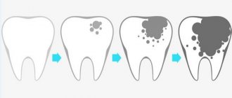
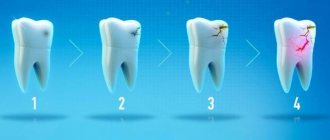
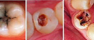
There are four stages of caries development: carious spot, superficial, medium and deep caries.
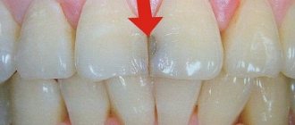
Carious spot
Carious spot
looks like a patch of enamel that has been stripped of its natural shine. Carious spots can be either pigmented or matte (white). The appearance of such a spot means that the process of destruction of the enamel has begun in this place. At this stage, the tooth does not yet respond to stimuli.
Superficial caries
In the stage of superficial caries
the enamel softens. The tooth begins to sense chemical irritants (sweet, sour and salty foods). Sometimes the tooth also reacts to heat or cold. The pain disappears as soon as the irritating factor stops.
Average caries
For average caries
destruction reaches dentin (dentin is the hard tissue that forms the bulk of the tooth; it is located under the enamel). A carious cavity appears in the tooth. However, the layer of softened dentin does not yet come into contact with the pulp (soft tissues inside the tooth), so the pain caused by physical, chemical and temperature effects on the affected area is not as acute as with deep caries.
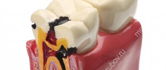
Deep caries
Deep caries
characterized by significant destruction of tooth tissue. A deep carious cavity is formed. A layer of softened dentin reaches the pulp. As a result, any food entering the carious cavity causes acute pain.
If the process of tooth decay continues, the exposed pulp may become inflamed and pulpitis will begin. Pulpitis is characterized by attacks of painful toothache, often at night. In advanced cases, tooth loss is likely.
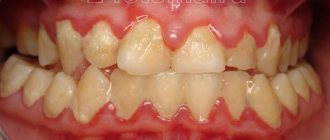
Caries
Classification of caries Dental caries is the most common human disease. Anatomical and physiological characteristics, reactive properties and general resistance of the body in childhood leave their mark on the course of caries. Caries of primary teeth under the age of 2 years is localized mainly on those tooth surfaces that formed in the antenatal period (smooth surfaces of the incisors of the upper and lower jaw), especially if it was unfavorable for the development of the fetus (hypoxia of various etiologies, malnutrition, chronic extragenital diseases of the mother, anemia, toxicosis of pregnancy, etc.). After 3 years, caries affects the chewing surfaces of molars, and after 4 years, the contact surfaces of temporary molars. It should be noted the high incidence of caries on the chewing surface (80.8%) of the first permanent molars. A feature of the carious process is its occurrence during the period of teething (6-7 years - first permanent molars, 11-13 - second permanent molars) and rapid progression due to incomplete mineralization. The largest percentage of the occurrence of initial forms of caries occurs precisely during the period of tooth eruption. An increase in intensity is observed at a later age and is due to the progression of existing foci of initial caries. In accordance with the International Classification of Diseases (ICD-10), the following are distinguished: - By 02.0. Enamel caries. — By 02.1. Dentin caries. — By 02.2. Cement caries. — By 02.3. Suspended dental caries. — By 02.4. Odontoclasia. — By 02.8. Other dental caries. — By 02.9. Dental caries, unspecified. Stain stage (macula cariosa) Focal demineralization of enamel, depending on the nature of the course, is divided into slow and fast-flowing. A differential diagnosis between these forms can be made on the basis of anamnesis, clinical picture (color, size, shape of the lesion), and data from staining of teeth with a methylene blue solution. The clinical picture indicates that demineralization of tooth enamel goes through at least three stages. The early stage is a white spot measuring 1-3 mm. In the 2nd, developed stage, distinctive signs of slow and rapid demineralization of the enamel appear. Slow-flowing demineralization is characterized by uniform changes in the enamel surface: on several teeth one of the stages of development of focal demineralization of the enamel predominates, which suggests the possibility of the simultaneous occurrence of foci of demineralization. The rapid demineralization of enamel in the 2nd stage is characterized by the activity of the process. Foci of demineralization lose clear boundaries, their edges become blurred. The enamel surface is rough, matte. The probe easily gets stuck in the demineralization area. The enamel loses its density and is easily scraped off with an excavator. The intensity of staining is on average 60 points. Increased staining is associated with an increase in enamel porosity. Rapid demineralization moves into the 3rd stage - the defect stage. At this stage, characteristic signs for both forms of damage are also noted. Summarizing the above, G. N. Pakhomov et al. propose the following classification of dental lesions with focal demineralization. Focal demineralization of tooth enamel: 1. Slow: - initial stage; - developed stage; — stage of the defect. 2. Fast-flowing: - initial stage; - developed stage; — stage of the defect. In children who frequently consumed sweets, the slow-moving form of enamel demineralization was 1.7 times more common and the fast-moving form was 3.5 times more common than in children who consumed sweets in moderation. After removing plaque from the entire surface of the tooth, an area of dull white or pigmented (from gray to black) enamel is discovered; the surface is smooth, sometimes rough, but painless and dense. Lesion in the stain stage on the vestibular and cervical surfaces of the tooth most often appears in children with the third degree of caries activity on a large group of teeth, up to the defeat of all teeth. It can occur in children of any age. The slow-onset form of enamel demineralization most often affected the incisors of the upper jaw (54.9%). In 2nd place in terms of the frequency of detection of a cervical slow process was the group of lower incisors (17.9%), followed by the group of small molars of the lower (8.7%) and upper (6.7%) jaws. Foci of rapid demineralization of enamel were more common on the upper incisors (45.8%) than on the lower jaw (21.5%). The canines of the upper (7.2%) and lower (7.4%) jaws, as well as the small molars of the upper (9.1%) and lower (9%) jaws were affected equally. Superficial caries (caries superficialis) . It is characterized by softening of the affected enamel, which is removed with a little effort by an excavator. Most children at this stage do not complain. Some of them indicate pain from sweets and sours, and children 1-3 years old refuse sour fruits. Upon examination, an enamel defect is detected, usually round in shape. When the process is chronic, its edges are flat, and when it is acute, they are steep. Exposure to cold and chemical irritants is often painful. Average caries (caries media) . Depending on the activity of the process and age, average caries has some clinical differences. In children 1-3 years old: in most cases it is very active, but they still cannot localize their sensations and express them, so average caries is detected during preventive examinations of children by a dentist, less often by parents. A feature of the clinical course of caries in children is the so-called planar form of caries, when the process of tissue demineralization spreads faster over the surface of the tooth than in depth. Occasionally, planar, slow-moving caries does not progress without treatment, but “stabilizes.” The affected enamel is erased when chewing, the exposed dentin has a color from light yellow to dark brown, dense, shiny, painless when probing. This form is called arrested caries. It is most often found on the chewing surfaces of the first temporary molars in children 4-7 years old. The slow progression of caries in children is observed relatively rarely: carious dentin is brown, dry, and difficult to remove with an excavator in the form of scales; at the bottom of the cavity the dentin is dense and often pigmented. In preschoolers and schoolchildren, there is an intermediate course, when both decalcification and pigmentation of carious tissues are moderately expressed. Deep caries (caries profunda) In temporary and permanent teeth with incomplete root formation, this form of pathology practically does not occur. Due to the morphofunctional characteristics of dentin and pulp, deep destruction of dentin is always accompanied by pronounced reactive and dystrophic changes in the pulp. These changes, under the influence of irritations caused by the treatment of the carious cavity with a drill and medications, easily turn into inflammation and necrosis. Each case of deep destruction of tooth dentin should be thoroughly examined using clinical (response to temperature stimuli, probing, percussion, etc.), electrodiagnostic and radiological research methods. During the active course of caries in children aged 1.5-3 years, replacement dentin is practically not formed, the dentin of the bottom of the carious cavity is deeply infected, there are changes in the pulp characteristic of developing forms of chronic pulpitis or pulp necrosis, even in the absence of complaints from the child on the day of the visit doctor In the differential diagnosis of deep caries and complicated caries, it is necessary to take into account the anatomical features of the teeth. The process is most active in children aged 1-3 years, when the activity of the pulp is reduced, there is little functional ability to produce replacement dentin, and the protective properties of the pulp are minimal. The process progresses rapidly, causing in most cases complicated forms of caries. In relation to permanent teeth, the diagnosis of deep caries is justified for any activity of the process. With regard to primary teeth, this diagnosis is made with great caution, mainly in older preschool children and in cases of compensated caries. Temporary teeth are smaller than permanent teeth and their enamel layer is thinner. When determining a carious cavity, i.e. proximity of the pulp, it is necessary to take into account the group affiliation of the teeth, their size, the age of the child, and the location of the cavity. For example, on the contact surface of the lower incisors in children 2-3 years old, a cavity with a depth of 1 mm is considered deep, and in schoolchildren 12-15 years old on the chewing surface of the molars, a cavity with a depth of 3-3.5 mm can be considered medium. With active caries, not only is there no or almost no replacement dentin, but also the protective sclerosis in the dentin of the cavity bottom is weakly expressed. Dentinal tubules remain wide, the cytoplasmic processes of odontoblasts are destroyed, the tubules are filled with mixed bacterial flora, therefore, irreversible changes in the pulp of temporary teeth often occur in shallow carious cavities. Despite the fact that caries in temporary teeth develops in accordance with the same patterns as in permanent teeth, the clinic identifies a number of features in the manifestation of the main symptoms of the pathology. These features, in turn, are determined by the degree of maturity of the tooth in which caries develops, as well as risk factors predisposing to a certain localization of the carious cavity, the intensity of destruction of tooth tissue, pulp reaction, etc. As already noted, the main feature of the carious process in children is the rapid course of the pathological process. Children more often than adults experience acute, or “blooming” caries (CC), which in a short time (from several weeks to 3 months) can completely destroy the tooth crown. “Blooming” caries is an acute carious process that affects many or all erupted teeth, quickly destroys coronal tissues, often localized on surfaces that are usually not susceptible to caries, with early involvement of the dental pulp in the process. In one recent study, individuals with active CD were defined as having 5 or more new carious lesions per year. "Blooming" caries usually affects primary teeth in the order of their eruption, with the exception of temporary incisors on the lower jaw. These teeth are likely to be resistant to caries because they are in close proximity to the submandibular salivary glands, which secrete secretions, and because of the cleansing action of the tongue during pacifier sucking. The first carious lesions are usually found on the labial surface of the maxillary incisors near the gingival margin as an area of whitish decalcification or softening on the enamel surface shortly after their eruption. These lesions quickly become pigmented and acquire a light yellow color; within a short time they reach the proximal surfaces and the cutting edge of the incisors. Less commonly, areas of decalcification may initially be localized on the palatal surfaces, and in some extreme cases even near the incisal edges of the incisors. As the process progresses, it often spreads around the circumference of the tooth, leading to pathological fracture of the crown with minimal trauma. Gradually, other teeth are involved, namely the first and second primary molars and, ultimately, the canines. Blooming caries can also affect permanent teeth due to children's tendency to frequently consume cariogenic breakfasts and sugary drinks between meals. Typical “blooming” caries in adolescents is characterized by carious lesions of the buccal and lingual surfaces of molars (buccal and lingual caries), the proximal and labial surfaces of the mandibular incisors (proximal and labial caries). A specific form of “blooming” caries can develop in children and adolescents whose saliva production has significantly decreased under the influence of radiotherapy undertaken to treat cancer in the head and neck area, or due to surgical excision of tumors in the oral cavity. In such patients, fillings fall out soon after treatment, teeth are often covered with plaque, saliva is viscous and scant. Children suffering from acute dental caries have a history of acute infectious diseases, chronic concomitant diseases - rheumatism, chronic tonsillitis, etc., and a tendency to colds. A dental examination reveals caries damage to a large number of teeth, often exceeding 12-14. All groups of teeth are affected, including the canines and lower incisors, which are usually resistant to caries. As with acute caries, symmetrical teeth are often affected. Acute (“blooming”) caries is characterized by a multiplicity of carious defects (up to 3-4 defects on the crown of each affected tooth). So-called “bottle” caries develops quickly in a group of children when they are bottle-fed at night. Usually occurs in 2.5-15% of children. It is characterized by rapid damage to the anterior teeth of the upper jaw, which later spreads to the chewing teeth of both jaws. Due to late eruption, canines are less affected than first molars. “Bottle” dental caries is typical for all socio-economic groups of the population and is often an indicator of the social level of the family. The same system for the occurrence of caries with indiscriminate and prolonged breastfeeding. The cause of this pathology is prolonged exposure to a cariogenic substrate, which comes into contact with the vestibular surface of the upper anterior teeth for 8 hours. In addition, at night there is a low level of salivation and a reduced buffer capacity of saliva. The peculiarity of the course of acute caries manifests itself at different depths of the defect. Thus, initial caries in its most acute form is characterized by the formation of a dirty gray spot (spots) or area(s) of enamel clouding with unclear contours. Such lesions are usually detected by a painful reaction when exposed to mechanical, temperature or chemical stimuli. In case of superficial caries, when the defect is localized in the enamel or reaches the enamel-dentin junction, the enamel appears heterogeneous, fragile, brittle; such defects are usually extensive, with uneven edges, as the process quickly spreads in breadth, along the plane. This picture is especially often observed in temporary teeth, with a characteristic cervical localization, encircling the neck of the tooth. In such cases they talk about “circular” caries . In the most acute cases of superficial caries, there may be complaints of pain associated with eating sweet, sour, and salty foods. When such a defect is localized on the approximal surface, as with other forms of caries, complaints about food getting stuck come to the fore. In the most acute course of moderate caries, a cavity (cavities) with uneven contours, undermined edges, formed by brittle whitish enamel is discovered. Usually there are complaints of pain from chemical, temperature irritants, and when localized on the proximal surface - of food getting stuck. In the most acute cases of moderate caries, there may be complaints not only about the effects of cold. Sometimes pain occurs from hot foods, which may be due to the involvement of the pulp in the chronic inflammatory process. Deep damage to primary teeth in the most acute course of caries has to be diagnosed extremely rarely, since the progression of the process is complicated relatively early by inflammation of the pulp. A detailed diagnosis, reflecting both the depth of the lesion and the nature of the course of caries, is the basis for complex treatment and is of great importance for the practical use of preventive means. Complications of caries Without timely and proper treatment, caries can develop into more severe forms of tooth disease (pulpitis, periodontitis) and lead to tooth loss.
Treatment methods for caries
In order for the disease to be treated in a timely manner, it is necessary to undergo preventive examinations, visiting a dentist at least once every six months. This makes it possible to detect existing caries at an early stage.
The earlier treatment of caries is started, the less medical intervention is required. You spend a minimum of your time. Treatment turns out to be more economical. Timely treatment of caries allows you to save the tooth.
The medical offices of the Family Doctor dental network are equipped with modern equipment. The experience and qualifications of dental therapists allow caries treatment to be carried out effectively and painlessly. The treatment uses light-curing composite materials with a chameleon effect, ensuring that the color of the filling matches the natural color of the tooth enamel. As a result, the visual integrity of the tooth is preserved.
Treatment of carious spots
Treatment of caries at the carious stain stage consists of careful oral hygiene using medicated toothpaste. It is also recommended to treat the affected area with special preparations based on fluoride and calcium. Procedures such as fluoridation and remineralization of teeth are carried out. Children can have their baby teeth silvered.
Treatment of caries
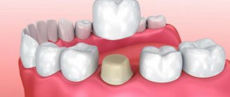
Treatment for carious destruction of enamel and dentin includes removal of the affected tissue, after which a filling is placed that protects the tooth tissue from environmental influences.
Prevention of caries
Treatment of caries necessarily includes measures to prevent the disease. By taking care of the health of natural teeth, we maintain the comfort of our daily lives for as long as possible. By following these simple rules you can significantly reduce the likelihood of developing caries:
- do not abuse flour and sweet foods. The basis of the menu should be fresh vegetables and fruits;
- be sure to brush your teeth at least twice a day (morning and evening) with a paste and toothbrush selected in accordance with the recommendations of your dentist;
- undergo preventive examinations and remove tartar (advanced technologies such as AirFlow and ultrasonic teeth cleaning can be used).
Make an appointment Do not self-medicate. Contact our specialists who will correctly diagnose and prescribe treatment.
Rate how useful the material was
thank you for rating
Treatment of multiple caries
In both children and adults, treatment of multiple caries is carried out comprehensively. Local therapy in Doka-Dent dentistry includes preparation and filling of teeth:
- under local anesthesia, the affected tissues are drilled using a drill;
- the cavities are washed with an antiseptic solution;
- the anatomical shape of the teeth is restored: cements containing fluorides are used for baby teeth, and light-curing composites are used for permanent teeth.
With multiple caries, it is important to eliminate the very cause of the disease. Therefore, in addition to the dental clinic, the patient is recommended to visit an immunologist, endocrinologist, or gastroenterologist. Treatment also includes taking vitamins and immunomodulators.