In medical terminology, mucositis (this article discusses one of its subtypes - oral) - means an abnormal condition of the external soft tissue, accompanied by external manifestations in the form of foci of inflammation, ulcerative wounds, and in severe cases, painful bleeding.
As a disease, it can be diagnosed not only in the oral cavity, but also in the digestive system and intestines. At the same time, it is not so much the disease itself that is scary, but its relapse - acute sepsis and the spontaneous spread of infection in the case of treatment started too late.
Description of the pathology
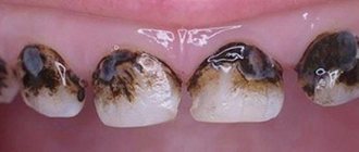
Oral mucositis is an inflammatory process in soft tissues, in which painful bleeding ulcers appear on the mucous membrane. Erosions form on the gums, inner surface of the cheeks and tongue.
This disease causes severe discomfort. It becomes painful for the patient to drink and eat. The person begins to avoid food and liquid intake, which leads to severe weight loss, exhaustion and dehydration.
Mucositis of the oral mucosa is a dangerous disease. If left untreated, ulcerative inflammation can spread to the throat and gastrointestinal tract. In addition, bacteria and fungi can get into bleeding wounds. This leads to worsening of the inflammatory process, suppuration, and sometimes to sepsis.
conclusions
Immediate implantation has the same capabilities as delayed implantation, and, subject to the conditions of adequate stabilization of the implant, is completely compatible with operations such as sinus lifting or osteoplasty.
At the same time, by preserving the soft and hard tissues surrounding the tooth, immediate implantation can easily achieve a satisfactory clinical result. Also, this technique greatly facilitates the positioning of implants, with the exception of cases of removal and subsequent implantation of abnormally located teeth.
The timing of osseointegration of implants is quite comparable to the timing of tooth socket regeneration, therefore these processes occur simultaneously and in parallel. The period at which we begin prosthetics after tooth extraction and implant installation is, on average, 6-10 weeks, which makes immediate implantation very desirable for prosthetics of dentition defects in an aesthetically significant area.
The incidence of complications with immediate implantation does not exceed that with a delayed technique. This is due to the fact that the bulk of complications of implantation in general are associated with a deficiency of bone tissue and mucous membrane, and with the immediate implantation method this problem is absent in principle.
However, there are disadvantages:
- Not every brand of implant is suitable for installation in the socket of a newly extracted tooth. Preference should be given to screw implants with small, non-aggressive threads.
- The immediate implantation technique is very demanding on the technique and quality of tooth extraction. From this point of view, low-traumatic tooth extraction with preservation of all alveolar walls represents the most difficult task for a dental surgeon.
- Despite the fact that in my practice there were two successful cases of tooth extraction and immediate implantation during exacerbation of chronic periodontitis, I would not recommend using this technique in the area of acute inflammatory process.
- The roots of an extracted tooth are not always an accurate guide for selecting an implant of the required length and optimal diameter. Therefore, the immediate implantation technique does not replace the need for computed tomography (and, in some cases, the production of a surgical template) for diagnosis and planning of surgical intervention.
- the decision to install an implant is made only after the tooth is removed, after a visual and instrumental examination of the alveoli. Therefore, before the operation begins, it is impossible to guarantee the patient that immediate implantation will be carried out in his case - and this must be warned about.
- work in an initially infected tooth socket requires increased attention to the postoperative regimen and prescriptions. Vitamin therapy is also recommended.
Etiology
Mucositis most often develops after chemotherapy. This is the body’s reaction to the action of drugs used in oncological practice. High doses of anticancer drugs negatively affect not only cancer cells, but also healthy tissue. Mucositis may also appear after radiation therapy for cancer.
In more rare cases, the following factors can trigger the development of mucositis of the oral mucosa:
- decreased immunity after infections;
- chronic stomatitis;
- oral thrush;
- caries;
- metabolic disorders;
- pathologies of the gastrointestinal tract;
- complications after bone marrow transplantation;
- frequent irritation of the mucous membrane with hot food and liquid;
- smoking.
Mucositis can also develop after taking certain medications. We have already mentioned that taking antitumor drugs can lead to inflammation and ulceration of tissues. However, any cytostatics and immunosuppressants can provoke the occurrence of mucositis. Such drugs are prescribed, for example, for autoimmune diseases. These drugs weaken the body's defenses and reduce the number of beneficial bacteria in saliva.
Mucositis usually occurs within 2-3 weeks after starting medication. If the patient does not have any symptoms in the mouth within a month, then most likely the pathology will not develop.
Risks of implant rejection in the postoperative period
After successful dental implantation and after the implants have fully fused with the bone (that is, after six months to a year), you still shouldn’t relax. It is important to maintain good oral hygiene and maintain your health. It is clear that no one is immune from developing any disease in the future, but if you have chronic problems, you need to ensure that they do not develop into an acute stage. For example, if you have diabetes, you need to follow a diet and take all medications prescribed by your doctor, and monitor your blood sugar levels.
Immediately after installation of the implants, it is necessary to strictly follow the doctor’s recommendations in terms of therapeutic treatment. If antibiotics have been prescribed, then you must take them for a strictly specified amount of time or at least ask your doctor if you can stop taking them.
If the prosthesis was installed immediately, then you can also eat right away. But the load should be moderate - you can’t go to a restaurant on the first day and eat a hard steak, snacking on nuts and drinking a glass of whiskey.
Read our useful article about how to behave after dental implantation >>>
After 1.5 years, Alexander went to the clinic to replace the adaptive prosthesis with a permanent one made of ceramic composite. During this time, he regularly underwent preventive examinations and paid attention to oral hygiene, so there were no complications after implantation.
View review
“It’s been a year since I lost my teeth. I am happy with everything! My situation was very difficult and serious; I had to remove eight teeth at once. Thanks to the doctors for returning me to normal life. I eat, chew normally, and smile!” – our patient shared his impressions.
View review
ICD code
According to the International Classification of Diseases, oral mucositis refers to pathologies of the oral cavity, jaw and salivary glands (divisions K00-K14). This disease is considered as a lesion related to stomatitis (code K12). The full ICD-10 code for mucositis is K12.3.
This only applies to oropharyngeal mucositis. If the ulcerative process has already spread to the gastrointestinal tract, then a different code is used. The code for mucositis of the gastrointestinal tract is K92.8. This disease is considered as one of the pathologies of the digestive organs.
Positioning and selection of implants by size
One of the advantages of immediate implantation is the almost complete absence of atrophic changes in the area of operation, as well as the presence of a socket of the extracted tooth, which greatly facilitates the positioning and selection of implants by size.
The length and diameter of the implant should correspond as closely as possible to the length and size of the tooth being removed. In my own practice, I am guided by the following criteria (Table 2):
Table 3. Example of positioning implants in the sockets of extracted teeth
Maxillary central incisors:
The implant with a diameter of 4.5 mm is adjacent to the palatal wall of the alveolus, its axis extends to the intended cutting edge of the future crown.
Maxillary lateral incisors:
An implant with a diameter of 3.5 mm is adjacent to the palatal wall of the alveolus
Canines of the upper and lower jaws:
The implant with a diameter of 4.0 mm is adjacent to the palatal wall of the alveolus, its axis extends to the intended cutting edge of the future crown.
Premolars of the upper and lower jaws:
An implant with a diameter of 4.5 mm is positioned along the interradicular septum (in the case of a double-rooted premolar) or the palatal wall of the alveolus for a single-rooted tooth. The axis of the implant extends onto the expected palatal tubercle of the future crown.
Molars of the upper and lower jaws:
An implant with a diameter of 5.5 mm is installed in the center of the interradicular septum.
Mandibular incisors:
An implant with a diameter of 3.0 mm is adjacent to the palatal wall of the alveolus, the axis of the implant extends onto the lingual surface of the future crown.
Below, using examples from my patients, I will try to show the features of immediate implantation surgery in different clinical cases.
Symptoms
The first sign of the disease is redness of the mucous membrane. Then rashes appear on the affected areas, resembling herpes blisters. Over time, they become ulcerated. The pathology is accompanied by other symptoms:
- redness of the mucous membrane;
- severe itching;
- dry mouth;
- pain during talking and eating;
- the appearance of white spots on the affected areas;
- a slight but constant increase in temperature.
If a patient experiences dyspeptic disorders, this may be the first sign of gastrointestinal mucositis. What it is? As already mentioned, the pathological process can spread to the digestive organs; this is considered a gastrointestinal form of pathology. The following symptoms indicate this complication:
- diarrhea;
- the appearance of bloody and mucous impurities in feces;
- frequent abdominal pain.
With such a clinical picture, the patient needs emergency help. In advanced cases, diarrhea gives way to intestinal obstruction, and the patient may die from peritonitis.
Gastrointestinal mucositis during chemotherapy can develop as an independent disease. Pathology does not always begin in the oral cavity. In some cases, inflammation and ulcers immediately form in the gastrointestinal tract, and no damage to the oral cavity is noted.
Clinical case #3
The above clinical situations are resolved relatively simply. We worked with single-rooted teeth under conditions of relative preservation of the volume of bone tissue and mucous membrane. However, this is not always the case.
Clinical situation: the upper sixth tooth was previously treated with the resorcinol-formalin method. An attempt to perform endodontic retreatment ended unsuccessfully:
The tooth must be removed. The situation is complicated by the low location of the bottom of the maxillary sinus.
Together with the patient, a decision is made to combine tooth extraction, immediate implantation and sinus lift into one procedure.
The 16th tooth is removed. In parallel, the gum former is fixed onto the 15th tooth implant installed two months earlier:
The reference point for positioning the implant is the interradicular septum. Despite the fact that installing an implant in one of the alveoli would avoid sinus lifting, it is highly undesirable to do this, since such positioning will create a number of serious problems during prosthetics and further operation of the prosthetic structure
An additional lateral approach is used for sinus lift surgery. The subantral space is filled with Bioss xenograft mixed with autologous bone shavings in a 2:1 ratio. Also, this material is used to fill the sockets of the roots of the teeth, although there is no great need for this. A Friadent Xive implant with a diameter of 5.5 and a length of 11 mm is installed.
The implant holder is removed:
In this case, the primary stability of the implant is extremely low. Therefore, the decision was made to abandon the installation of a gum former.
The tooth socket is closed with a BioGide collagen membrane, which is fixed to the implant with a plug through a pre-made hole.
Unlike the previous case, the surgical wound is completely sutured. This is associated with certain technical difficulties, but is necessary because, as mentioned earlier, the stability of the implant is relatively low, and, moreover, the use of a collagen membrane and a xenograft, in order to avoid infection, requires hermetically suturing the wound.
The postoperative regimen and prescriptions do not differ from those for delayed implantation and sinus lifting.
After 10 weeks, you can begin to open the implant and install the gum former.
After resection of the mucous membrane covering the former, we refer the patient to an orthopedic dentist (Arthur Makarov) for the manufacture of temporary prosthetic structures.
This clinical case clearly shows that the low primary stability of the implant and the low location of the bottom of the maxillary sinus are not an obstacle to immediate implantation. However, it is necessary to carry out a number of additional procedures to avoid infection of the surgical area, namely: hermetically suturing the wound, refusing to immediately install a gum former, etc. In the postoperative period, special attention is paid to antibacterial therapy, it is advisable to prescribe vitamins C and D3 in therapeutic dosages.
Stages of oral lesions
There are several stages of mucositis. What it is? The pathological process in the oral cavity develops in stages. The stage of the disease depends on the degree of tissue damage and the severity of pain. Each stage of pathology development is characterized by its own symptoms:
- Zero stage. This stage of the disease is considered the incubation period. The pathology is already developing, but the patient does not yet feel any symptoms.
- First stage. There is redness in some areas of the oral cavity. Single ulcers may be observed. There is no pain while eating yet.
- Second stage. Multiple erosions appear in the mouth. While eating, a person feels discomfort, but this does not prevent the patient from eating.
- Third stage. The ulcers increase in size and become deeper. Sometimes minor bleeding is observed. Large pockets of inflammation form in the mouth. At first, a person experiences discomfort when speaking. Then there is severe pain while eating. Because of this, the patient can only take liquid and pureed food. Eating solid food becomes impossible.
- Fourth stage. Most of the mucous membrane is covered with ulcers and inflamed. The patient experiences pain even when drinking liquid. There is severe weight loss and dehydration. Ulcers often bleed.
Advanced stages of mucositis of the oral mucosa pose a danger to human life and health. The patient may die from lack of food and dehydration. In such cases, the patient is transferred to feeding through a tube.
Preoperative preparation and planning
In general, preoperative preparation is no different from that for delayed implantation.
Despite the fact that the presence of a socket and the removed tooth root makes it easier for us to select the size of the implant and position it, in some cases it is necessary to make surgical templates. This is especially true for situations where, for some reason, the teeth being removed have moved (for example, due to the absence of adjacent teeth or antagonist teeth).
is mandatory for immediate surgery
, since it allows you to assess the condition of the bone tissue around the tooth socket, accurately determine the localization of the anatomical structures surrounding the surgical area, measure the parameters of the tooth socket and make a preliminary decision on the possibility of immediate implantation.
At the same time, it is important to know
that the decision to install an implant largely depends on the quality and low-traumatic nature of tooth extraction, and therefore is made by the doctor only after the tooth is completely removed from the socket. The patient is warned about this without fail.
In 2012, in two cases out of 183, we were unable to complete the immediate implantation operation due to bone loss during extraction, so we limited ourselves to tooth extraction only. Subsequently, patients underwent delayed implantation.
In some cases, it is advisable to prescribe antibacterial prophylaxis. This is to reduce the number of antibiotics taken and reduce the risk of infectious and inflammatory complications in the postoperative period.
Laboratory tests are prescribed only according to indications and, as a rule, involve consultation with doctors of the relevant specialty (cardiologist, endocrinologist, allergist, etc.). There is no point in prescribing laboratory tests for everyone, since the result of implantological treatment is noticeably affected only by severe general somatic pathology, which, in any case, requires the attention of both doctors and the patient himself.
Degrees of damage to the gastrointestinal tract and toxicity criteria
Mucositis of the mucous membrane of the gastrointestinal tract also develops in stages. The following stages of development of the pathological process can be distinguished:
- Stage 1. There are no pronounced symptoms of gastrointestinal tract damage. Mild diarrhea may occur occasionally.
- Stage 2. Abdominal pain occurs. Stools become frequent (4-6 times a day) and liquid, blood and mucus appear in the feces.
- Stage 3. The frequency of stool reaches 8 or more times a day. The patient is constantly experiencing severe abdominal pain. The temperature rises sharply. In some cases, initial signs of intestinal obstruction are observed.
- Stage 4. Dead areas appear on the gastrointestinal mucosa. Frequent bleeding is observed; in severe cases, perforation of the intestinal wall and the development of peritonitis are possible. Without emergency medical care, the patient dies.
Doctors also consider the degree of toxicity associated with mucositis. What it is? If an ulcerative process has developed after taking cytostatics, then it is necessary to assess the level of negative effects of the drugs on the organs. In this case, the main criterion is the severity of diarrhea:
- 1st degree. The volume of feces ranges from 500 ml to 2 liters per day.
- 2nd degree. The volume of feces is more than 2 liters per day. Blood and mucus appear in the stool.
- 3rd degree. Intestinal obstruction develops, which requires surgical intervention. There are signs of severe colitis with frequent bleeding.
Mucositis after chemotherapy is especially dangerous. After all, the patient’s body is greatly weakened by cancer and the use of cytostatics. Without treatment, advanced forms of pathology often lead to death.
Clinical case #2
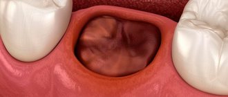
Central incisor, recurrence of the root cyst after cystectomy. Unsatisfactory aesthetics. The tooth must be removed for orthopedic and therapeutic reasons.
The immediate implantation technique is of particular importance when making dental prosthetics in an aesthetically significant area. The natural appearance of the prosthetic structure largely depends on preserving the volume of soft tissue and bone surrounding the implant. The timing of treatment is also important - few patients will agree to remain without central teeth for a long time.
As in clinical case #1, an incision is made (namely an incision, not peeling) along the periodontal sulcus, plus additional vertical loosening incisions, after which the periosteum is peeled off to a normally visible surgical field:
And one of the problems is immediately revealed - a significant loss of height of the vestibular wall of the socket, associated with long-term chronic inflammation.
The tooth is removed using forceps, after which the root cyst and granulations are removed through the hole.
The tooth socket is prepared for the implant using a standard surgical protocol, similar to clinical case #1.
The quality of socket preparation and positioning is checked using an implant analogue. In this case, the implant is positioned palatally, tightly adjacent to the inner wall of the alveoli.
A Friadent Xive implant with a diameter of 4.5 and a length of 13 mm is installed:
In this situation, it is important to avoid excessive immersion of the implant - ideally, its neck should be located at the level of the edge of the palatal wall of the socket, or 1-1.5 mm lower. In case of excessively strong immersion, we risk not only worsening the aesthetic result, but also getting complications in the form of gum recession in the area of neighboring teeth.
The implant holder is removed:
We are left with only the problem that was already mentioned earlier - loss of height of the vestibular wall of the socket. It should be taken into account that, despite implantation, slight atrophy of bone tissue occurs in the area of the extracted tooth socket, which can lead to a deterioration in the aesthetic result. Therefore, in this case, a decision is made about osteoplasty using a collagen membrane and a xenograft mixed with autogenous bone chips.
BioGide collagen membrane 16x22 mm is fixed palatally. The space between the implant and the vestibular wall of the socket is filled with Bioss xenograft mixed with autogenous bone shavings in a 2:1 ratio.
To avoid material getting into the implant shaft, it is closed with a plug during filling.
The main task of this stage is to prevent “subsidence” of the bone tissue in the implant area and maintain its volume.
The membrane is fixed with a gum former through a pre-made hole.
To avoid losing the appearance of the gums, when preparing for suturing, you should avoid releasing incisions under the periosteum.
Stitches are applied. Non-resorbable suture material is used.
Recommendations and prescriptions in the postoperative period are practically no different from those for delayed implantation.
If the primary stability of the implant is high, after 1-2 days a temporary crown can be made and fixed to it. But, in this case, implantation is complicated by the presence of a root cyst and partial loss of the vestibular wall of the socket. That is why a gum former is installed on the implant, and the dentition defect is replaced with an adhesive crown made by the direct method.
Clinical situation after 3 days:
As in the first clinical case, we pay attention to the condition of the mucous membrane in the area of the postoperative wound, the stability of the implant and the sensations when pressing on it.
After 6 weeks, the temporary adhesive crown, masking the dentition defect, is removed, and the orthopedic dentist (David Akhinyan) begins making a temporary composite crown for the implant.
A temporary composite crown is made for the implant. In parallel, the 12th tooth is restored with a temporary crown.
Well, or in the general picture like this:
One of the features of the Friadent Xive implant system is that there are only two diameters of healing abutments, which does not always allow achieving the desired “pink” aesthetics. Therefore, to individualize the gums in an aesthetically significant area, it is better to use temporary crowns or, in extreme cases, composite Estetic Base abutments, which are easily selected and adjusted to the size of a specific tooth.
Diagnostics
The dentist treats this disease. If the pathology arose as a result of chemotherapy, then it is necessary to report the occurrence of undesirable symptoms to the treating oncologist. If the gastrointestinal tract is affected, consultation with a gastroenterologist will be required.
Mucositis in its manifestations resembles many other dental pathologies, for example, aphthous stomatitis, which is also accompanied by the appearance of ulcers. For the purpose of differential diagnosis, the patient may be prescribed the following examinations:
- examination of the oral cavity;
- blood test for leukocytes and ESR;
- swab from the mouth for bacterial culture;
- MRI or CT scan of affected tissues.
Prescribed drugs
In domestic medical practice, the following drugs are used to treat mucositis:
- iodine-containing components included in liquids for irrigation of the mucous membrane (chlorhexidine, benzydamine) - reduce the pain threshold at the local level, reduce the possible risk of secondary infectious lesions;
- biological group of agents that stimulate tissue restoration at the cellular level - filgastrim, palifermin. The drugs perfectly neutralize foci of inflammation, but require hospital treatment and intramuscular administration of drugs, the cost of which is quite high;
- glutamine is an amino acid capable of carrying out the synthesis of proteins, pushing them towards self-renewal;
- zinc softens the course of the disease , constricts blood vessels, improves blood flow, increases coagulation, which has a positive effect on the external manifestations of inflammation;
- aloe extract is used both externally (ointments, creams, suspensions) and internally. It has a pronounced antiseptic effect and is rich in vitamins and minerals. Effective and absolutely safe.
Therapy
Treatment of mucositis must be comprehensive. It's not just taking medications that is necessary. It is very important to carefully care for the oral mucosa, otherwise the ulcers may become infected. The patient experiences great difficulty while eating, so it is necessary to organize gentle nutrition for him.
When the mouth is affected, the following groups of medications are prescribed:
- Antiseptics: Chlorhexidine solution, Oralsept and Tantum Verde sprays. These medications are used for rinsing and irrigation. They reduce pain and also prevent bacteria from entering wounds.
- Ointments with aloe extract. They are used for compresses and applications. Aloe has wound healing and anti-inflammatory properties. This is an effective and safe remedy.
- Amino acids. Glutamine-based medications are prescribed. This substance stimulates the formation of protein and promotes the healing of ulcers. These medications are available in tablet form.
- Protein preparations: “Filgrastim”, “Kepivance”. These medications are given intravenously or subcutaneously. They can only be used in a hospital setting. They promote tissue healing and restoration.
- Corticosteroid ointments and solutions. The most commonly prescribed drugs are dexamethasone. Locally acting steroid hormones can quickly relieve inflammation. However, such drugs should be used with caution, as they reduce immunity.
- Local anesthetics: Lidocaine, Trimecaine. Prescribed for severe pain.
If the patient has signs of damage to the gastrointestinal tract (diarrhea, abdominal pain), then the following medications are indicated:
- Antibiotics and antiprotozoal drugs: Vancomycin, Ciprofloxacin, Trichopolum. These remedies help prevent the development of infection in the intestines.
- The drug "Sulfasalazine". This medicine has a strong anti-inflammatory effect.
- Antacid "Omez". The drug relieves pain.
- Adsorbents. The most effective drug is Neointestopan. It quickly reduces diarrhea and removes harmful substances from the body.
Conservative methods help only in the early stages of gastrointestinal mucositis. If the patient experiences severe bleeding and intestinal obstruction, surgical intervention is indicated.
Progress of surgery
The clear sequence of actions in the process of bone grafting during dental implantation completely depends on the type of surgical procedure. The average plan of action for a dental surgeon is as follows:
- Providing access to the operated area of the gums of the upper or lower jaw (alveolar process);
- Preparation of the bone bed for graft installation;
- Fixing the bone block, immersing it in tissue or placing special shavings (depending on the type of bone grafting);
- Installation of barrier membranes (not required everywhere);
- Suturing soft tissues.
This is followed by the process of graft engraftment and the formation of one’s own bone tissue, which lasts about six months. Healing times depend on the grafts used, the amount of bone grafting and the type of surgery. Transplants are of the following types:
- Own bone tissue taken from certain areas of the body (autograft);
- Human bone tissue taken from cadavers (allograft);
- Bone tissue of animal origin (xenograft);
- Artificial bone tissue (alloplant).
Each of the listed options has its own advantages and disadvantages, so they are all widely used in modern dentistry. An autograft implanted into spongy gum tissue full of blood vessels and biologically active substances takes root best.
Diet
During treatment for oral mucositis, it is very important to follow a gentle diet. The patient experiences severe pain while chewing food. Therefore, all irritating foods should be excluded from the diet:
- solid food;
- alcohol;
- hot and sour dishes;
- too hot and too cold food;
- high-calorie foods.
You should give preference to light foods that are quickly digested. It is recommended to include steamed vegetables and meat in the menu. They should be eaten pureed. Food should be at room temperature.
If it is difficult for the patient to eat even gentle food, then feeding through a tube or parenteral administration of solutions is indicated. Such procedures are carried out in a hospital setting.
Features of choosing implants for immediate implantation
Currently, some manufacturers (Nobel Biocare, AlphaBio, Biohorizont, etc.) produce separate lines of implants designed for installation in the socket of a newly extracted tooth. As a rule, they are distinguished by large aggressive threads, the purpose of which is to ensure high-quality stabilization of the implant in the conditions of the socket of a newly extracted tooth.
However, as my own practice has shown, implants with aggressive threads behave less predictably (Table 1). In my opinion, this is due to two nuances: firstly, large pronounced coils exert strong pressure on the bone tissue surrounding the implant, disrupting the microcirculation so necessary for normal healing; and secondly, loose contact of the implant surface to the wall of the socket complicates osseointegration and contributes to its infection.
In other words, the most optimal from the point of view of prognosis are screw implants with small, non-aggressive threads: on the one hand, due to the contact area, high-quality stabilization of the implant is ensured, and on the other, due to the tight fit, conditions for infection of the implant surface disappear.
Oral care
Mucositis is often complicated by bacterial inflammation. Mouth ulcers are entry points for pathogenic microorganisms. Therefore, it is very important for the patient to observe the following hygiene rules:
- Use a brush with very soft bristles to clean your teeth.
- Temporarily stop wearing dentures, as they further injure the oral cavity.
- Rinse your mouth with antiseptics several times a day.
- Teeth must be cleaned of plaque and food debris after each meal.
- Treat the skin of your lips with special balms to moisturize. This will help prevent cracks from appearing.
In addition, during treatment you should try to drink more clean water (at least 2 liters per day). This will help remove toxins from the body. If the patient has difficulty swallowing, then the liquid should be consumed in small portions, but as often as possible.
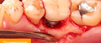
Why do complications occur after implantation?
Implant rejection occurs, as a rule, for two reasons: violations are made by the doctor or the patient. Failure to comply with oral hygiene rules is the easiest way to develop peri-implantitis or mucositis. It is important not only to properly care for your teeth, but also to visit the dentist at least once every six months. Immediately after implantation, you must strictly follow the doctor’s recommendations and do not overload the implant.
The success of implantation depends 90% on the actions of the doctor. An implantologist is like a sapper: make one mistake and everything is lost, the implant will not take root. Contraindications to implantation are not taken into account, the location for the implant is chosen incorrectly, the load on the jaw is incorrectly calculated - mucositis or peri-implantitis develops.
Prevention
How to prevent the occurrence of unpleasant manifestations of mucositis? If the patient is to undergo chemotherapy, it is necessary to treat caries in advance. It is also very important to carefully monitor the cleanliness of the oral cavity while taking cytostatics. Any neglect of personal hygiene while taking anticancer drugs can cause an inflammatory and ulcerative process.
During chemotherapy, trauma to the oral cavity should be avoided. You should not chew hard pieces of food, consume hot food and drinks, or use sharp toothpicks. Damage to the mucosa during the course of treatment can lead to the appearance of ulcers and the development of mucositis. During treatment with cytostatics, you must inform your doctor about any inflammation or discomfort in the oral cavity.
Risk factors for developing mucositis
Weakened immunity may be a risk factor for the development of mucositis
- Failure of metabolism, disturbances in the gastrointestinal tract;
- weakened immunity due to past illnesses;
- stomatitis in the chronic stage;
- untimely treatment of dental diseases;
- allergies to medications: antitumor antibiotics (Mitomycin, Vinblastine, Dactinomycin);
- antimetabolic drugs (Thioguanine, Fluuracil, Cytarabine);
- alkylating drugs (Cyclophosphamide, Cisplatin, Temodal).