Central lower incisor
Average age of eruption: 6-7 years
Average age of root formation: 9 years
Average length: 20.7 mm
Narrow and flat in the labial-lingual direction, the lower central incisor is the smallest adult tooth. Radiologically it is visible in only one projection and thus appears more accessible than it actually is. The crown, narrow in lingual projection, has a limited area for access. For gentle access formation, a fissure bur and a spherical bur No. 2 are used. The access cavity should be oval and performed on the lingual side.
The lower central incisor often has two canals. One study reported that 41.4% of mandibular central incisors studied had two separate canals, of which only 1.3% had two separate apical foramina [1]. After completing the access formation, the doctor must examine the tooth cavity to identify an additional canal. Endodontic treatment failures in the lower incisor area are most often associated with an unidentified canal, usually in the lingual location. To create conditions for the straight entry of endodontic instruments into the additional canal, access can be expanded in the incisal direction.
There is a great danger of perforation of the vestibular wall, but it can be avoided if the doctor remembers that it is almost impossible to perforate in the lingual direction, since the bur axis is in contact with the incisal edge. The slit-like lumen of the canal is so common that it can be considered a normal variant, and this requires special attention when cleaning and shaping.
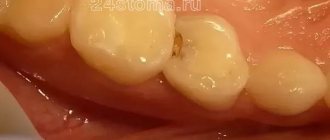
Lateral perforations and pulp anatomy will be discussed in an illustration of the lower second molar.
Tooth numbering systems: ISO (FDI), universal, Zsigmondy-Palmer (+ visual illustrations)
There is probably no student of the Faculty of Dentistry who at one time would not have been confused by various tooth numbering systems. Where did they come from? For what? And how to convert tooth numbers from one system to another? We will answer the first two questions in text, and the illustrations given in this material will help you with the third.
Lateral lower incisor
Average age of teething: 7-8 years
Average age of root formation: 10 years
Average length: 21.1 mm
Very similar to the lower central incisor, so access cavity preparation follows the same principles.
Their similarity can cause rare but serious errors. Hasty installation of a rubber dam, identical fillings and carelessness can lead to preparation of the access cavity on the wrong tooth. This error can be prevented by marking the vestibular surface of the tooth with a felt-tip pen before applying the rubber dam.
Trauma, periodontal disease, carious lesions and malocclusion can lead to obliteration of the canal. When moving apically to identify the orifice, great care must be taken to prevent unnecessary destruction of the crown and root. Labial perforations were discussed in Illustration IX. If the bur is not oriented along the long axis of the tooth, there is a risk of lateral perforation. The situation becomes more complicated with the traumatic loss of an anatomical crown. Without anatomical landmarks, a lateral perforation can be easily made when moving in a coronal direction. To prevent this, access is carried out without a rubber dam so that the root can be palpated.
Lateral perforation with endodontic files and Gates-Glidden burs is facilitated by the presence of slit-shaped canals with a narrow, hourglass-shaped cross-section. To avoid vertical fracture along the approximal root wall, minimal expansion and preparation of the space for the abutment pin is indicated.
Apical curves and accessory canals are common in the lower incisors.
Mandibular canine
Average age of teething: 9-10 years
Average age of root formation: 13 years
Average length: 25.6 mm
The canine of the lower jaw is more powerful and significantly wider than the incisors in the mesial-distal direction. It rarely causes treatment problems. The atypical form with two roots can be problematic, but is rare.
The access cavity is oval and can be expanded in the incisal direction to facilitate vestibular-lingual access. In the cervical region the canal is oval, in the middle third it is rounded. To completely clean its walls, directed instrumental action is necessary.
If there are two roots, one of them will always be easier to instrument. The other canal must also be opened and funnel-shaped in accordance with the first to prevent dentine filings from entering it and impairing access. Pre-bending the instruments during the initial approach will allow the clinician to walk along the walls of the buccal or lingual root until the tip of the instrument enters the orifice. Once a difficult-to-reach canal has been identified, every effort must be made to shape and create a funnel-shaped orifice to keep access open.
What does the dental formula look like in a medical record?
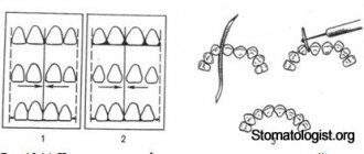
The dental formula of an adult, as well as a child, in the medical record of a dental patient looks in the form of a schematic table (Fig. 5), which will reflect only the serial numbers of permanent or baby teeth. Directly in this formula, the doctor will mark the missing teeth (in this case, the number is crossed out with a cross), which teeth are affected by caries, pulpitis or periodontitis, as well as which of them have crowns or bridges.
Dental formula in the form of a table in the medical record –
First lower premolar
Average age of teething: 10-12 years
Average age of root formation: 12-13 years
Average length: 21.6 mm
Often considered a mystery to endodontists, the mandibular first premolar, with its two canals separating at different levels of the root, can be very difficult to machine.
The crown consists of a well-developed buccal cusp and a small or almost non-existent lingual enamel protrusion. The approach is performed buccally from the central sulcus and directed along the long axis of the root to the central cervical region. The oval-shaped pulp chamber is opened using fissure burs with a cutting apex and elongated spherical burs No. 4 or 6. In teeth with one canal, the pulp cavity in the neck area has an almost circular cross-section, and in teeth with two canals it is oval.
One study reported that “at least 23% of first mandibular premolars have a second or third canal”[17]. The canals can split almost anywhere in the root. Due to the lack of direct access, cleaning, shaping and filling these teeth can be extremely difficult.
In a recent study, Vertucci [13] showed that the first lower premolar has one canal at the apex in 74.0% of cases, two canals in 25.5% and three canals in the remaining 0.5% of cases.
Classification and frequency (%) of canal types in first and second lower premolars
By Vertucci, F. J. Am. Dent. Assoc. 97:47, 1978.
Reasons for creating a dental formula
Human teeth, despite their visual similarity, have certain differences. Each tooth is individual and designed to perform its function. Thanks to numbering in the dental field, examination of the patient's oral cavity is optimized to the maximum.
Clear and concise markings help to accurately record information in the patient's chart. In this case, the use of the same numbering system is observed. This is necessary so that the new doctor can decipher the medical history and past treatments that were performed by another dentist. Different dentists cannot use an individual calculation system, as this will change the patient's hospital history.
International two-digit Viola system
In practical dentistry, such designations are rarely used, and the teeth of a person’s jaws are simply numbered from incisors to molars (from 1 to 8).
International two-digit Viola circuit
The scheme was adopted by the World Health Organization in 1971 and is used throughout Europe [3]
All teeth are divided into 4 sectors (counterclockwise when viewed from the inside):
- The teeth of the upper jaw are on the right (respectively, the central incisor is 11, the second incisor is 12, the canine is 13, the first premolar is 14, the second premolar is 15, the first molar is 16, the second molar is 17, the third molar or wisdom tooth is 18).
- The teeth of the upper jaw are on the left (21, 22, 23, 24, 25, 26, 27, 28, similar to the right side) [4].
- Lower jaw teeth on the left (31, 32, 33, 34, 35, 36, 37, 38).
- Lower jaw teeth on the right (41, 42, 43, 44, 45, 46, 47, 48).
For children's teeth, a similar numbering is used from 51 to 85, or they are indicated in Latin numerals.
Haderup scheme
This scheme also uses the division of the jaws into segments, and individual numbers are assigned to the teeth. These numbers are calculated as in the international system - from the center of the jaw to its edge and have digital values from “1” to “8”. However, in this case, instead of the digital designation of segments, the signs “+” and “-” will be used. Here, a positive value indicates the upper jaw, and a negative value indicates the lower jaw. Showing the left and right sides of the jaws is very simple. You just need to place the desired sign to the left (for the left side) or to the right (for the right side) of the number that indicates the tooth. [3]
Haderup numbering scheme
| permanent teeth | |||||||||||||||
| top right | top left | ||||||||||||||
| 8+ | 7+ | 6+ | 5+ | 4+ | 3+ | 2+ | 1+ | +1 | +2 | +3 | +4 | +5 | +6 | +7 | +8 |
| 8− | 7− | 6− | 5− | 4− | 3− | 2− | 1− | −1 | −2 | −3 | −4 | −5 | −6 | −7 | −8 |
| bottom right | bottom left | ||||||||||||||
| baby teeth | |||||||||||||||
| top right | top left | ||||||||||||||
| 05+ | 04+ | 03+ | 02+ | 01+ | +01 | +02 | +03 | +04 | +05 | ||||||
| 05− | 04− | 03− | 02− | 01− | −01 | −02 | −03 | −04 | −05 | ||||||
| bottom right | bottom left | ||||||||||||||
| baby teeth (alternative system) | |||||||||||||||
| top right | top left | ||||||||||||||
| V+ | IV+ | III+ | II+ | I+ | +I | +II | +III | +IV | +V | ||||||
| V− | IV− | III− | II− | I− | −I | −II | −III | −IV | −V | ||||||
| bottom right | bottom left | ||||||||||||||
Zsigmondy-Palmer scheme
The system was first used in 1976. It is also called square-digital.
To indicate that a tooth belongs to any segment, a special icon is placed next to the number (in one of the four corners) - “corner”. It seems to indicate a piece of the crown, showing how it grows - down or up. [3]
Zahnschema nach Zsigmondy
| permanent teeth | |||||||||||||||
| top right | top left | ||||||||||||||
| 8 | 7 | 6 | 5 | 4 | 3 | 2 | 1 | 1 | 2 | 3 | 4 | 5 | 6 | 7 | 8 |
| 8 | 7 | 6 | 5 | 4 | 3 | 2 | 1 | 1 | 2 | 3 | 4 | 5 | 6 | 7 | 8 |
| bottom right | bottom left | ||||||||||||||
| baby teeth | |||||||||||||||
| top right | top left | ||||||||||||||
| V | IV | III | II | I | I | II | III | IV | V | ||||||
| V | IV | III | II | I | I | II | III | IV | V | ||||||
| bottom right | bottom left | ||||||||||||||
Second lower premolar
Average age of teething: 11-12 years
Average age of root formation: 13-14 years
Average length: 22.3 mm
The second lower premolar, which is very similar in crown shape to the first premolar, has a less complex root.
Its crown has a well-developed buccal cusp and a much better formed lingual cusp than on the first premolar. The access is made slightly oval, wider in the mesial-distal direction. They begin to form access in the central sulcus with a fissure bur with a cutting apex, and then expand and form the contour of the burr hole with spherical burs No. 4 and 6.
Researchers reported that only 12% of mandibular second premolars studied had a second or third canal [17]. Vertucci [13] also showed that second premolars had one apical foramen in 97.5%, while only 2.5% of the teeth examined had two foramina.
An important circumstance that should not be forgotten is the anatomical location of the mental foramen and the vessels and nerves passing through it. Due to the proximity of these structures, an acute inflammatory process in the area of the lower premolars can cause temporary paresthesia. The exacerbation of the pathological process in this area is more severe and resistant to conservative treatment than in other areas.
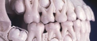
Structure of primary and molar teeth - differences
The posterior and anterior temporary and permanent teeth of a person undergo changes in structure throughout their lives. But, despite the difference in quantity, they have similar functions and external shape. The main difference between baby teeth and molars is size—baby teeth are smaller than adult teeth.
Other differences include the following:
- In childhood there are no premolars.
- The roots and crowns of molars are large.
- Milk teeth have a white-blue tint, molars are yellow.
- Baby teeth are more susceptible to caries because their protective layers - enamel and dentin - contain small amounts of minerals.
- Most of a child's teeth is occupied by pulp, which increases the risk of pain.
- In children, tooth roots have a more round shape.
- The first roots are destroyed and dissolved during the change of the milk dentition to the permanent one.
- Children have fewer teeth, which is due to the small size of the jaw arch.
The first teeth are characterized by a not very long and not very well developed root system. This structure facilitates the process of replacing baby teeth with molars.
It is necessary to take care of your oral cavity throughout your life. The negative condition of baby teeth can affect the health of primary teeth. Following simple hygiene rules will ensure that you maintain a beautiful smile for many years.
First lower molar
Average age of teething: 6 years
Average age of root formation: 9-10 years
Average length: 21.0mm
The mandibular first molar erupts earlier than other permanent teeth and most often requires endodontic treatment. It usually has two roots, but sometimes three roots are found, with two canals in the mesial root and one or two canals in the distal root.
The distal root is easily accessible for endodontic cavity preparation and mechanical treatment.
The doctor can directly see the opening(s) of the canal. The distal root canals are wider than those of the mesial root. Sometimes the mouth is wider in the buccolingual direction. This indicates the presence of two channels or a slit-like channel with a complex network-like configuration that can complicate cleaning and shaping.
The mesial roots are usually curved, with the greatest curve in the mesiobuccal canal. The orifices of the canals at the bottom of the pulp chamber are usually clearly separated from each other and located buccally and lingually relative to the upper tubercles.
The tooth often undergoes extensive filling. It almost always experiences a strong chewing load, so the coronal pulp cavity can be obliterated. It is easiest to identify the mouths of the distal canals.
Then the mouths of the mesial canals are found, which will be located in the above locations in the same horizontal plane.
Since the mouths of the mesial canals lie under the mesial tubercles, they may not be detected during normal cavity preparation. In this case, to determine their location, it is necessary to remove the hard tissue of the tubercle or filling. During access preparation, overhanging molar cusps need to be ground down [15]. Remember that this tooth, like all other lateral teeth, after endodontic treatment requires complete restoration of the entire occlusal surface area. Therefore, to identify anatomical landmarks and orifices, it is better to make a wider access cavity than to skip one or more channels for the sake of “sparing” preparation, which may cause failure.
Skidmore and Bjoradal [11] found that approximately one third of the mandibular first molars examined had four root canals. If there are two canals, “they either remain separate with separate apical foramina, or unite and form a common apical foramen, or communicate with one another by transverse anastomoses partially or completely... If the tooth, instead of the usual triangular shape, had a more rectangular shape, this would allow better vision and explore a possible fourth canal in the distal root.”
In the area of bifurcation of the roots of the lower molars there are several orifices of additional canals [9]. They are usually impossible to clean and shape, and are rarely visible except incidentally on radiographs when they are filled with root cement or heated gutta-percha during treatment. It would be correct to assume that if irrigation solutions tend to clean the canal from protein decomposition products, then the area of root bifurcation in the pulp chamber must be thoroughly cleaned (remove denticles, etc.) so that the solutions can reach the small mouths of the canals.
All infected dentin, leaking fillings, and pulp denticles must be removed before endodontic treatment begins. It is recommended to completely cover the cusps with a restorative structure after endodontic treatment.
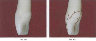
Anatomical structure of teeth with pictures
External anatomical structure of a human tooth:
- The crown is the visible area covered with enamel, the hardest part of the tooth. The surface layer performs a protective function and is designed to preserve dentin.
- The root part is a complex of nerve fibers, arteries and veins that provide nutrition and innervation to the tooth. The second function of the root is retaining.
- The neck is the area between the root and the crown, which performs a connecting function. Differs in a narrowed shape.
Diagram: external structure of a human tooth
How does a human tooth work at the tissue level?
All human teeth have a similar internal structure. The diagram below shows all the dental layers - enamel, dentin, cement, pulp.
The figure shows the histological structure of the tooth
Enamel covers the teeth like a cover and makes up 25% of the total mass of all dental tissues. The hardness and strength of this tooth layer is ensured by a high mineral content of 397 kg/mm.
The composition of the enamel layer includes:
- minerals – 96%;
- organic substances – 1.2%;
- liquid, water – 2.3%.
The enamel is covered by an outer shell, the cuticle, which covers the chewing surface of the tooth. Most of the tooth is occupied by dentin, located immediately under the enamel. It has less strength, its hardness is 58.9 kg/mm.
Cement – covers the root part and in most respects is similar to bone tissue. Depending on the type of structure, there are two types of cement:
- acellular or primary - consists of collagen fibers;
- cellular or secondary - the composition includes cemenoblasts.
The internal dental cavity is occupied by the pulp - soft tissue, including:
- numerous nerves;
- blood vessels;
- connective tissue.
Sectional structure of a tooth
Despite the differences in the purpose and structure of the root system, all teeth have the same internal structure:
- Enamel. A very hard surface layer that protects teeth from environmental influences. It is designed as a structure of glued prisms. Thanks to the large amount of mineral salts in its composition, enamel is the strongest tissue in the human body. Its main task is to preserve the dentin of the crown. The presence of a pellicle ensured the tissue's resistance to the effects of acids.
- Dentine. It consists of coarse fibrous tissue, the basis of which is collagen fiber. The structure is similar to porous bone. There are two types of dentin: superficial and internal. The first has a higher density and prevents infection from entering the dental cavity.
- Cement. Consists of collagen fibers saturated with limestone salts. Layer thickness - from 50 to 150 microns, depending on location. There are two types of cement: cellular and acellular.
- Cavity. It has a shape identical to the crown and is located under the dentin. The entire area of the cavity is filled with pulp, a type of tissue responsible for nourishing the tooth and performing a connecting function. The presence of blood and lymphatic vessels ensures high sensitivity of the pulp.
Photo: this is what a tooth looks like in cross-section
Second lower molar
Average age of teething: 11-13 years
Average age of root formation: 14-15 years
Average length: 19.8 mm
The crown is somewhat smaller in size than the first molar, and the more symmetrical second lower molar is characterized by a close arrangement of roots. The roots gradually converge distally and have closely spaced apices.
Exposure is made at the mesial portion of the crown with access extending only slightly distal to the central sulcus. After trephination of the cavity with a fissure bur with a cutting apex, an elongated ball-shaped bur is used to expand it until free access is achieved. The distal root angles often allow for a smaller opening than the lower first molar.
Particular attention should be paid to the shape of the mouth of the distal canal. A narrow oval orifice indicates the presence of a slit-like lumen in the distal canal, which requires more careful treatment.
All carious tissue, leaking fillings and denticles should be removed and replaced with a suitable temporary filling prior to endodontic treatment.
The lower second molar is most susceptible to vertical fractures. After preparing the access, but before starting endodontic treatment, the clinician should examine the floor of the pulp cavity using fiber optics. For mesial-distal crown or root fractures, the prognosis is extremely unfavorable.
To avoid vertical fractures after endodontic treatment, it is necessary to completely cover the cusps with a restorative structure.