Age groups for monitoring the causes of caries
From the point of view of the most complete information content during the examination, age groups 1 2, 15, 35-44 years .
In the age group of 12 years, the incidence of dental caries is assessed, and at the age of 15 years, the condition of the periodontium is assessed, and this makes it possible to assess the effectiveness of preventive measures.
The age group of 35-44 years is studied to identify the intensity index, which allows us to judge the quality of dental treatment. Research shows that the following tendency towards an increase in caries is expressed: 22% in 6-year-olds, 99% in 65-year-olds.
Classification of caries
In dentistry, dental caries is classified according to several criteria: the depth of the lesion, the type of tissue involved in the pathological process, the localization and severity of the process.
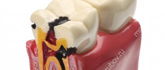
Thus, depending on the depth of damage, uncomplicated dental caries can be distinguished in the form of spots, superficial, medium and deep caries. In the case of complications, we will talk about pulpitis and periodontitis.
Classification of caries according to the type of tissue affected allows us to distinguish caries of enamel, dentin and cementum of the tooth. By localization - fissure, interdental, cervical and basal caries. Well, according to the severity of the process, compensated, subcompensated and decompensated forms of caries are distinguished.
At the first appointment, the doctor at the Apex dental clinic will make a diagnosis and prescribe the required amount of treatment, agreeing on its timing and cost.
What is Case's Shamrock? List of factors for the occurrence of caries
To visually represent the essence of the occurrence of caries, the “Case Trefoil” is used, which is depicted by three circles overlapping in the center.
This peculiar symbol shows that only when 3 conditions coincide, caries occurs, namely:
- in the presence of cariogenic flora
- when eating a diet with a large amount of easily digestible carbohydrates
- in the presence of low enamel resistance.
To date, it has been clearly established that the process of dental caries begins with demineralization, which is caused by organic acids , mainly lactic acid. There is no such thing as sterile caries. The appearance of a carious cavity is the result of an imbalance in the confrontation between the acid-forming microflora of the mouth and the tooth enamel on which this flora lives.
Thus, several factors are present in the development of dental caries, and it is their interaction that leads to the emergence of a focus of demineralization.
Factors contributing to the development of caries include:
- oral microorganisms;
- nature of nutrition (presence of carbohydrates);
- diet;
- quantity and quality of salivation;
- changes in the functional state of organs and systems of the body;
- the amount of fluoride entering the human body;
- extreme effects on the body.
Deep caries. Clinic, differential diagnosis and treatment.
Clinical picture of deep caries. Patients complain of short-term pain from mechanical, chemical and temperature stimuli, which quickly passes after the stimulus is eliminated. When examining the tooth, a deep carious cavity is discovered with overhanging edges of the enamel, filled with softened and pigmented dentin. Probing the bottom of the cavity is painful. The tooth pulp responds to a normal current of 2-6 µA. but there may be a decrease in excitability to 10-12 μA. Pain occurs from chemical and temperature irritants, but, as a rule, quickly calms down after the cessation of the irritants. If the carious cavity is located in such a way that it is difficult to remove and wash out food debris, the tooth may hurt for a longer time until these irritants are removed. Percussion of the tooth is painless.
Differential diagnosis of deep caries is carried out with those dental diseases that have a clinical picture similar to deep caries, namely: with medium caries, which is characterized by a less deep carious cavity located within its own dentin. The bottom and walls of the cavity are dense, probing is painful at the enamel-dentin junction, while with deep caries the cavity is within the peripulpal dentin, probing the bottom is painless, temperature stimuli cause pain that quickly passes.
Deep caries must be differentiated from focal pulpitis, which is characterized by acute spontaneous paroxysmal pain, intensifying in the evening and at night. Probing the bottom of a carious cavity is painful at one point, often in the area of projection of the source of pulp inflammation. In case of deep caries, probing the bottom of the carious cavity is painful evenly over the entire surface of the peripulpar dentin; there are no spontaneous or paroxysmal pains.
A differential diagnosis should also be made with chronic fibrous pulpitis; it is characterized by the presence of a deep carious cavity filled with softened dentin. When probing the bottom of the carious cavity, a connection with the pulp chamber can be detected; probing this area is sharply painful, the pulp bleeds, and there is a decrease in pulp excitability to μA current. With deep caries, probing is painful along the entire bottom; the tooth pulp reacts to a current of 2-12 μA.
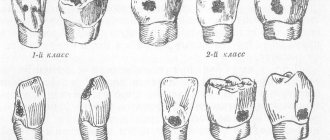
Treatment of deep caries. When treating deep caries, preparation of the carious cavity is a prerequisite. The dentist is required to strictly adhere to all principles and stages of preparation: anesthesia, opening, expansion, necrectomy, formation of a carious cavity - only in this case can a positive result be expected. Any formed cavity must have an optimal number of retention points that would provide the best fixation of the filling, and also serve as a counterforce to the antagonist tooth. Preparation of the bottom and walls of the carious cavity is carried out before cripitation. If you leave softened dentin at the bottom of the carious cavity, the process of demineralization under the filling will continue. You can leave pigmented, creeping dentin in cavities of the 1st and 2nd Black classes according to the principle of biological expediency, which does not work in cavities of the 3rd, 4th and 5th Black classes, since pigmented dentin, visible through the enamel, will not allow achieving an ideal cosmetic effect with filling a tooth with composite filling materials. Next, antiseptic treatment of the existing cavity is carried out. Warm physiological antiseptic solutions of 0.02% furatsilin are used; 0.06% chlorhexidine; as well as 0.02% solution of ethacridine lactate; 5% dimexide solution; 1% etonium solution; enzymes with 1% novocaine solution. Physiological warm antiseptic solutions do not irritate the dental pulp and do not overcool the hard tissues of the tooth (cold solutions can lead to overexcitation of axons). Then it is necessary to dry and degrease the prepared carious cavity. Sterile cotton swabs are used. The use of alcohol and ether for drying and degreasing the cavity is unacceptable, as they are highly irritating substances. It is advisable to use EDTA-based preparations. Next, a medical pad is applied, always warm and only on the bottom of the prepared carious cavity, no more than 0.5 mm thick. The treatment pad should:
- stimulate the reparative function of the dental pulp;
- have a bactericidal and anti-inflammatory effect on the dental pulp;
- act as an analgesic;
- do not irritate the dental pulp and oral mucosa;
- have good adhesion;
- be plastic;
- withstand pressure after hardening.
Domestic and foreign preparations containing calcium hydroxide have all of the above properties:
- calmecin,
- dental lining material,
- Dycal from Dentsply,
- Calcipulpe from Septodont,
- Life by Kerr,
- Calcimol from Voco,
- Reocap from Vivadent. Plastic pastes containing eugenol are successfully used to treat deep caries: • biodent,
- zinc eugenol cement,
- Cavitec from Kegg,
- Eugespad from SPAD.
A thin layer of a therapeutic pad is applied to the bottom of the prepared and medically treated carious cavity, then a thin layer and only to the bottom of the carious cavity is applied to an insulating pad made of glass ionomer cement, covering the therapeutic material. This sequence is only possible if the therapeutic lining material is adapted to the permanent filling material. After applying therapeutic and insulating pads, we transform the topographically deep carious cavity into a medium-depth carious cavity. Further, all stages of filling a deep carious cavity correspond to the treatment of medium-depth cavities: total etching of enamel and dentin; washing off phosphoric acid; drying the carious cavity; application of dentin adhesive (primer) - 2-3 layers; applying enamel adhesive to the walls, bottom and finished enamel - 2-3 layers successively (each layer polymerizes within 20-30 s); adding light-curing filling material; polymerization of each layer; grinding and polishing the filling.
Did you like the article? Share with friends
0
Similar articles
Next articles
- Examination of a surgical patient
- Anesthesia
- General anesthesia
- Conducting general anesthesia in a dental clinic
- Nitrous oxide anesthesia
Previous articles
- Flowable composites
- Average caries. Clinic, differential diagnosis and treatment.
- Superficial caries. Clinic, differential diagnosis and treatment.
- Initial caries. Clinic, differential diagnosis and treatment.
- "Herculite XRV Ultra", "Premise" and "NanoPaq"
Add a comment
Is it possible to avoid dental caries through diet?
The idea that the development of caries is influenced by a certain diet, namely a lack of protein, vitamins A, C and D, has not been confirmed. Only a gross nutritional deficiency, and over a long period of time, can be the cause of the development of caries disease. Because it is this factor that affects the buffering changes in the capacity of saliva and the concentration of electrolytes in it, and this is what reduces the remineralizing potential.
Local factors for the development of caries
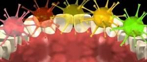
Presence of dental plaque
Poor hygiene, irregular and poor-quality teeth brushing lead to the accumulation of deposits. They contain a large number of cariogenic bacteria.
Over time, soft bacterial plaque turns into hard tartar, which adheres tightly to the enamel. It can no longer be removed with a regular toothbrush, so you have to go to the dentist.
Violation of the composition, properties and pH of saliva
An adult produces approximately 2 liters of saliva per day. This liquid washes away plaque from the enamel surface and neutralizes the effect of acids due to the alkaline environment. It also contains beneficial immunoglobulins (proteins that destroy viruses and bacteria).
Various functional disorders of the salivary glands lead to insufficient production of saliva, changes in its composition or pH level. All this increases the caries susceptibility of the tooth surface.
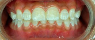
Malocclusion
Crowded teeth make it difficult to maintain proper hygiene and create conditions for food particles to accumulate in the mouth. In addition, improper occlusion (closing of the jaws) leads to increased chewing load on individual teeth, which accelerates the carious process. You can also add the habit of chewing food on one side of the jaw.
Features of the structure of teeth
Teeth with flask-shaped fissures are more susceptible to caries; these are natural grooves on the chewing surface. In such pits, food particles quickly accumulate and microbial plaque forms.
Carbohydrates (carbohydrate leftover food)
Remember the formula: carbohydrates + bacteria = acids. As noted above, cariogenic bacteria (streptococci, lactobacilli, actinomycetes) feed on carbohydrates. As a result, the fermentation process in the mouth begins.
The most intensive fermentation of sucrose occurs; it is a simple carbohydrate found in confectionery products and sweet carbonated drinks. Starch and fructose are less dangerous.
Does the occurrence of caries in a person depend on his genetics and diseases?
The fact of the direct influence of past and concomitant diseases on the occurrence of caries has also not been established. Of course, this influence is not completely excluded, because diseases suffered during the maturation of the body and the formation of teeth make them less resistant to the effects of cariogenic factors.
A focus of demineralization that exists on the enamel for a long time leads to the dissolution of its surface, more stable layer. The process of carious disease can be stopped and last longer. However, this is only possible at the stage of a white spot before the formation of a carious enamel defect: this manifests itself in the form of a pigmented (brown or black) spot.
The intensity of caries disease varies widely in different regions of the world, and even in those population groups with low or high levels of caries intensity, significant deviations from the average are observed.
CHAPTER 19. BIOFILM OF THE DENTAL SURFACE AND PATHOGENESIS OF DENTAL CARIES
19.1. FEATURES OF DENTAL SURFACE BIOFILM
Dental plaque is a variant of a multilayer biofilm covering the supra- and subgingival parts of the tooth (the latter plays a major role in the development of periodontal diseases). Biofilm forms both on the enamel and on the cement of the tooth, penetrating into natural depressions - fissures and pathological formations - carious cavities.
Numerous studies have shown that in dental plaque, biofilms have a highly ordered structure, and microorganisms closely interact, distributing their functions.
Dental plaque begins to form within the first minutes after brushing the teeth, and significant changes in the nature of the microbiocenosis occur in the dynamics of its formation. The general trend is a change in the composition of the microflora from the dominance of aerobic and facultative anaerobic forms, predominantly gram-positive cocci, to obligate anaerobic gram-negative rods and convoluted forms.
• The first phase - the formation of dental plaque - the first 1-4 hours after thorough brushing of the teeth (or ultrasonic treatment on the Piezon-Master apparatus). It mainly consists of cocci (streptococci, neisseria, veillonella) and short rods (diphtheroids). This is the so-called early dental plaque.
• The second phase - dynamic plaque - up to 4-5 days. It is characterized by a decrease in the proportion of gram-positive cocci and an increase in the proportion of gram-variable filamentous forms - leptotrichia, as well as gram-negative veillonella and fusobacteria. In persons with good adaptive abilities, with the so-called high natural sanitation, the microbiocenosis of dental plaque can be maintained in this state for significant periods of life. Such a dental plaque is called equilibrium, since the ratio of microorganisms is maintained even in the absence of systematic tooth brushing.
• The third phase - mature dental plaque - from 6-7 days or more. The dental plaque takes on its final form in terms of the composition of symbionts, although quantitative changes in it occur constantly. This is a mature dental plaque. The number of aerobic species in it sharply decreases: neisseria, rotium, facultative anaerobic streptococci. Filamentous bacteria and rods forming chains predominate. Such a plaque can remain in equilibrium for a long time, and it should be called a mature stable dental plaque.
Stages of carious lesions
To understand how quickly caries develops, you need to know the main stages of the disease.
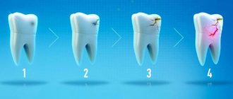
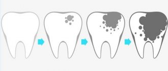
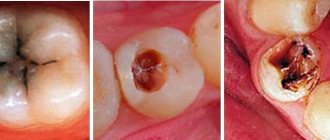
The occurrence of carious lesions is characterized by several stages:
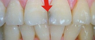
- Formation of a barely noticeable white or milky spot. At this stage, the pathogenic process can still be stopped using conservative methods, thereby prolonging the health of the tooth.
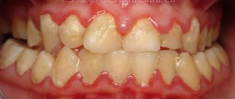
- Darkening of dental tissues, especially in the recesses, interdental spaces, near the gums.
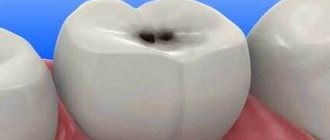
- The appearance of cavities. This stage requires specialist intervention.
- Complete tooth destruction as a complication of the disease. The pathological process spreads to the canals, roots, and gums.
The price of treatment depends on at what stage of caries development the patient came to us. It is important to monitor the health of the oral cavity in order to recognize the pathological process in time.
Not only a doctor, but also an ordinary person can distinguish healthy teeth from diseased ones. The stages of caries development are clearly presented in the photo gallery below.
Caries therapy
Therapeutic measures for caries include:
- Remineralization through fluoridation, as well as taking calcium and fluoride supplements. They provide saliva with a natural balance between demineralization and remineralization.
- Treatment of caries with ultrasound allows you to stop the pathological process at the initial stages.
- The surgical method involves mechanical removal of necrotic tissue and filling of carious cavities.
Instructions for caries treatment are created by the dentist during the visit.
Local factors for the development of caries
Presence of dental plaque
Poor hygiene, irregular and poor-quality teeth brushing lead to the accumulation of deposits. They contain a large number of cariogenic bacteria.
Over time, soft bacterial plaque turns into hard tartar, which adheres tightly to the enamel. It can no longer be removed with a regular toothbrush, so you have to go to the dentist.
Violation of the composition, properties and pH of saliva
An adult produces approximately 2 liters of saliva per day. This liquid washes away plaque from the enamel surface and neutralizes the effect of acids due to the alkaline environment. It also contains beneficial immunoglobulins (proteins that destroy viruses and bacteria).
Various functional disorders of the salivary glands lead to insufficient production of saliva, changes in its composition or pH level. All this increases the caries susceptibility of the tooth surface.
Malocclusion
Crowded teeth make it difficult to maintain proper hygiene and create conditions for food particles to accumulate in the mouth. In addition, improper occlusion (closing of the jaws) leads to increased chewing load on individual teeth, which accelerates the carious process. You can also add the habit of chewing food on one side of the jaw.
Features of the structure of teeth
Teeth with flask-shaped fissures are more susceptible to caries; these are natural grooves on the chewing surface. In such pits, food particles quickly accumulate and microbial plaque forms.
Carbohydrates (carbohydrate leftover food)
Remember the formula: carbohydrates + bacteria = acids. As noted above, cariogenic bacteria (streptococci, lactobacilli, actinomycetes) feed on carbohydrates. As a result, the fermentation process in the mouth begins.
The most intensive fermentation of sucrose occurs; it is a simple carbohydrate found in confectionery products and sweet carbonated drinks. Starch and fructose are less dangerous.