Features of the procedure
The canines on the upper jaw have one root located laterally. In cross section, part of the element resembles the outline of a triangle. In 30% of cases, the apex of the canine root has a curved structure. The outer part of the root is thicker than the inner one, but both sides of the alveoli are thicker in width than the incisors. Due to the listed features of the structure of the fangs, some difficulties arise when removing them from the socket.
During the operation, the doctor should place the fingers of the left hand in the same way as when removing incisors on the upper jaw. When removing an element from the right side, the patient's head should be slightly turned to the left and vice versa. This position of the patient will be more comfortable at the time of surgery.
The canines on the upper jaw are removed from the socket using wide forceps. During the operation, the element swings alternately towards the lips and palate with rotation around the longitudinal plane of the fang. The doctor makes the first movement of the instrument towards the outer wall of the alveoli, since it has a thinner structure.
Preparation and stages of tooth extraction
The next dislocation is performed towards the palatine alveolus, and then the specialist performs rotations. The procedure consists of successive rocking and rotation of the element until the nerve fibers and tissues surrounding it are completely ruptured.
Due to the anatomical features of the canines on the upper jaw, when extracting them, a specialist has to make a lot of effort.
When Removal is Required
Indications for the removal of fangs on the upper jaw are divided into local and general. The last group of prescriptions includes chronic endogenous intoxication of the body due to rheumatism, sepsis, endocarditis and connective tissue pathologies.
Local recommendations for canine removal are divided into relative and absolute. Tooth extraction can be performed urgently if there are special indications for the procedure:
- suppuration of periodontitis;
- acute osteomyelitis;
- periostitis;
- phlegmon;
- other dental pathologies that cannot be treated with medication.
Fangs with a longitudinal fracture, severe damage to the coronal part and exposure of the pulp are urgently removed.
What are the dangers of tooth extraction under anesthesia?
Elements of bone tissue are removed as planned in the following cases:
- if treatment of the inflammatory process in periodontitis and bone structures is unsuccessful
- in case of improper treatment, causing damage to the tooth root or its cavity;
- in case of complete destruction of the crown of the element and the impossibility of using its remaining part for prosthetic purposes;
- with significant mobility of the canine and its protrusion relative to the remaining elements of the row;
- with incorrect anatomical location of the tooth, as a result of which permanent damage to the mucous membranes of the oral cavity occurs;
- in the presence of unerupted or partially manifested elements that cause inflammation of the soft tissues.
In addition to the indications for surgery, the doctor must establish any restrictions on the procedure. Tooth extraction may be delayed due to chronic diseases or poor health of the patient.
Experts' opinion
Question: Is it possible to rinse your mouth after tooth extraction? If yes, then with what?
Answer : During the first 24 hours after a molar tooth has been removed, doctors at the Optimal Choice clinics do not recommend rinsing your mouth at all. Active rinsing of the mouth, even if its goal is to cleanse food debris, can lead to a number of negative consequences. The main danger is the destruction and washing out of the formed blood clot in the socket, which interferes with rapid healing. This can also lead to the development of alveolitis. In the first few days, instead of rinsing, it is better to use baths with a decoction of medicinal plants (sage, chamomile, oak bark), a weak solution of salt or baking soda (or a mixture of them in equal proportions), furatsilin, miramistin, chlorhexidine, if prescribed by a doctor.
Question: After tooth extraction, the gums hurt and swelling is noticeable. What to do?
Answer: Slight tissue swelling and pain are very often consequences of the removal of molars. For relatively tolerable pain, it is recommended to take analgesics prescribed by your doctor. If the pain is severe, then you urgently need to visit the dentist again for a check. A cold compress applied to the cheek on the side of the extracted tooth for 15-20 minutes will help relieve swelling.
Contraindications to the procedure
The list of contraindications for removing a fang on the upper jaw includes:
- Cardiovascular pathologies – myocardial infarction (suffered earlier six months ago), bacterial endocarditis, hypertension.
- Diseases of the urinary system - pyelonephritis in the acute stage, glomerulonephritis, renal failure.
- Diseases of an infectious and viral nature - hepatitis, acute respiratory infections, influenza and other pathologies transmitted by airborne droplets.
- Exacerbation of psychological problems - manic psychosis, epilepsy.
- Pregnancy at 1, 2 and 9 months.
- Poor clotting .
- Cancer – acute leukemia.
- Malignant tumors located in the maxillotemporal zone.
Progress of the operation
Before removing a fang on the upper jaw, local anesthesia of the treated area is mandatory. Dentistry offers a wide selection of anesthetics and methods of their administration, after which the patient will not feel severe pain during surgery.
The most popular drugs used for tooth extraction are Ultracaine, Ubistizen, Articaine. If the patient is contraindicated with anesthetics belonging to the group of vasoconstrictors, then pain relief is performed with Lidocaine or Scandonest.
Elements of bone tissue located on the upper jaw are often removed using bilateral infiltration anesthesia, less often using palatal, tuberal or infraorbital anesthesia.
What are the risks of removing dystopic and impacted teeth?
It should be remembered that the therapeutic effect of the drugs used is reduced if there is inflammation at the location of the canine. Therefore, if the doctor advises urgent removal of an element, you should not refuse the operation and postpone it to a later date.
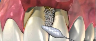
Extraction of the maxillary canine from the socket is performed using special tools - forceps and elevators. The equipment used has various designs.
When removing elements on the upper jaw, bayonet-shaped forceps are used. Elevators are used when extracting the roots of fangs or if forceps cannot be used to grasp the tooth tightly.
The operation can be performed according to two schemes: it all depends on the condition of the root. When saving the tooth crown, the procedure is performed according to the following algorithm:
- Peeling off the canine ligaments for better grip with forceps.
- Perform swinging movements.
- Fixing an element.
- Dislocation.
- Extracting a tooth from its socket.
This type of canine removal is called simple because it is performed using one instrument. The tactics of the operation change if the coronal part is preserved and only the root needs to be extracted from the socket. This procedure is considered complex because the doctor uses several tools - elevators to dislocate the roots and forceps to remove fragments from the hole.
Removing fangs using elevators requires certain skills and abilities from the doctor. The basic rule when working with a tool is to correctly find the fulcrum for its working part.
If the alveolar wall is thick, the doctor tries to place the equipment between the root of the canine and the wall of the socket, while performing rotational movements. Such manipulations can loosen the tooth.
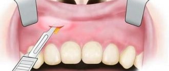
Sometimes during the process of removing a fang, the doctor needs to make an additional soft tissue incision. This type of operation is called atypical. The technique is used to remove unerupted fangs or those that have not completely appeared above the surface of the gum.
Why do fangs erupt incorrectly?
In children, under the milk teeth in the jaw bone in the second row there are the rudiments of permanent teeth, and even deeper, in the third row, permanent fangs. Therefore, during the period of change in occlusion, the “three” canines erupt last, and the first permanent teeth become “sixes”, at the site of the eruption of which there were no milk teeth at all.
- If for any reason a child prematurely loses a milk “four” or “five,” the sixth tooth will move into the vacant space, and there will simply be no room left for the canine to erupt. He will have to crawl out either outside the row or on the palatal surface.
- The cause of pathology can also be crowding of teeth, caused by the small size of the jaw, when there is simply no room in it for the physiological placement of large complete teeth.
Possible complications
After removing the fang from the socket, various complications may develop, which in the vast majority develop due to infection of the wound.
Dry socket syndrome
The problem arises due to the patient’s failure to comply with the doctor’s recommendations or too active treatment of the oral cavity with antiseptic solutions. The actions lead to the fact that a blood clot does not have time to form in the wound, protecting the soft tissues from the penetration of pathogenic microorganisms.
The condition is not life-threatening, but can cause a number of consequences, for example, inflammation of a postoperative wound.
Alveolitis
The disease is an inflammatory process that spreads to the mucous membranes of the socket. Alveolitis causes infection to spread to bone structures. Pathology rarely develops after the removal of fangs on the upper jaw; more often, this condition appears after the extraction of molars located in the lower row.
Nerve injury
If the canine is positioned incorrectly or has a curved root, then during surgery damage to its nerve endings may occur. Characteristic symptoms of this condition are loss of sensitivity in the operated area, numbness of the cheek, and the appearance of “running goosebumps.”
Alveolar exposure
Even with a normal course of the postoperative period, problems with wound regeneration may occur. If pain occurs at the site of the hole when consuming hot or cold food, this indicates that the area of the bone is not covered with soft tissue.
Cyst on a tooth
The development of a pathological formation after canine removal is a rare consequence. A cyst is a growth filled with fluid. In this way, the human body independently separates infected soft tissue from healthy areas.
The tumor, increasing in size, can spread to neighboring areas and therefore requires immediate surgical intervention.
The occurrence of odontogenic phlegmon
The pathology develops against the background of osteomyelitis of the jaw structures. The problem manifests itself as pain and swelling of the cheek in the upper jaw area. It becomes difficult for a person to open his mouth and consume food. The pathology is accompanied by a deterioration in health and a rise in temperature.
Odontogenic periostitis
It develops against the background of alveolitis or osteomyelitis and is an inflammation of the periosteum. The condition is manifested by a number of the following symptoms:
- rise in temperature;
- toothache ;
- swelling on one side of the cheek.
Removal of the maxillary canine is carried out taking into account the structure of this element of bone tissue. The tooth has only one root, which in 30% is located in the wrong position. The stages of surgery for this type of intervention are no different from removing the remaining elements located in the top row. Depending on the location of the canine and the condition of its crown, the doctor performs simple, complex or atypical tooth extraction.
Preventive measures
In order to avoid serious complications after molar tooth extraction, you need to follow a number of recommendations:
- 20 minutes after the end of medical procedures, the gauze swab should be removed from the socket of the extracted tooth;
- In order for healing to occur faster, you should not drink or eat for the first 2-3 hours after surgery;
- for several days you should avoid eating solid foods, as well as spicy and hot foods and drinks;
- hot water procedures and physical activity are contraindicated until the hole is completely healed;
- You cannot wash the hole yourself or try to clean it with foreign objects that have not been disinfected;
- During the first 24 hours after surgery, you should not rinse your mouth;
- A cold compress on the right side of the cheek will help prevent swelling and reduce the likelihood of bleeding.
If the hole does not heal for a long time or it seems that there are remains of a tooth in it, then you should contact your dentist again and under no circumstances try to solve these problems yourself.