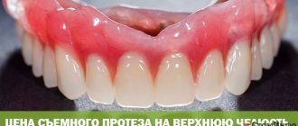
A removable denture is a compensatory orthopedic structure for replacing lost teeth; it has a movable attachment to the jaw and is maintained by the patient independently (using special cleaning agents). A removable denture completely restores chewing function and returns the aesthetics of a healthy smile.
Structure of a removable complete denture for teeth
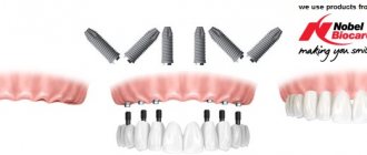
This orthopedic design completely models the dentition of the upper or lower jaw. The base of a complete removable denture imitates part of the soft tissues of the oral cavity and serves as a mount for an artificial dentition. The boundaries of a complete removable denture end at the point where the gum tissue meets the mucous membrane of the lips. Unlike a partial denture, a complete denture does not have clasps (hooks for attaching the denture to natural teeth), so the fixation of the denture is ensured by the base that covers the palate. The entire structure does not have metal components, which is why among specialists such a system is called a “complete removable plate denture”. One should not confuse a removable laminar denture, which is used in cases of complete absence of teeth, with conditionally removable dentures, which are also installed in cases of edentia. These structures are placed on implants, and only a doctor can remove them, while a regular plate prosthesis can be removed at any time independently for cleaning or oral hygiene.
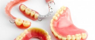
Complete dentures for the lower and upper jaws.
News
Anatomical and physiological features of the boundaries of a complete removable denture of the upper jaw
When prosthetizing patients with complete secondary edentia, they are taken into account, which are of great practical importance.
ANATOMICAL AND PHYSIOLOGICAL PREREQUISITES FOR THE CONSTRUCTION OF THE BOUNDARIES OF COMPLETE REMOVABLE DENTURES The vestibule of the oral cavity. The vestibule has the appearance of a narrow horseshoe-shaped gap; on the outside it is limited by the mucous membrane of the lips and cheeks, and on the inside by the labial and buccal surfaces of the alveolar process of the jaw. On the mucous membrane in the vestibule area there are frenulums of the upper and lower lips and bucco-alveolar cords (folds). On the upper jaw there are four buccal-alveolar cords, which are located in the area of the transitional fold at the level of the canines and premolars. More often they are single, less often - multiple. On the lower jaw, the buccal alveolar cords are located in the canine area. As the alveolar processes of the jaws atrophy, the place of attachment of the frenulum changes. With slight atrophy of the jaw bones, the vestibule of the oral cavity has a high arch, the frenulum of the upper and lower lips are located at the base of the alveolar process. The size of the frenulum of the lower lip is, as a rule, somewhat smaller than the frenulum of the upper lip. The frenulum of the upper lip can be shortened and then a gap appears between the central incisors - a diastema. With significant atrophy, the arch of the vestibule flattens, the frenulum extends onto the alveolar process, and with deep atrophy it can be located at the level of the top of the crest of the alveolar process of the jaw. In cases of its high attachment, during contraction of the facial muscles, the frenulum can be stretched and displaced, which leads to the prosthesis being thrown off. To prevent shedding, a recess should be created in the prosthesis. The size of the recess for the lip frenulum should be minimal, that is, strictly correspond to the height and width of the frenulum, in order to avoid injury to it and maintain the continuity of the circular closing valve in this area.
It follows that the most favorable for prosthetics is a high arch of the vestibule of the oral cavity and low attachment of the frenulum. ANATOMIC AND PHYSIOLOGICAL RATIONALE FOR THE BORDERS OF A COMPLETE REMOVABLE DENTURE FOR THE UPPER JAW The shape of the vestibular slope of the alveolar process of the upper jaw.
Depending on the degree of atrophy, three types of vestibular slope of the alveolar process are distinguished:
1) sloping;
2) sheer;
3) with a “canopy”. The most favorable for creating and maintaining a closing valve on the prosthesis during chewing are alveolar processes with a steep vestibular slope, because with small displacements, the edge of the prosthesis slides along the mucous membrane of the alveolar process and fits tightly to it, forming a closing valve, i.e. contacts of the edges of the prosthesis with the slope of the jaw are not disturbed. Less favorable for prosthetics are sloping slopes, in which the prosthesis is freely applied to the jaw, but with small displacements, contact with the mucous membrane is lost. With this shape of the alveolar process, prostheses with expanded boundaries are indicated to achieve and maintain the valve effect. Unfavorable are processes with a “canopy”, in which the base of the prosthesis can injure the mucous membrane due to the presence of bone protrusions on the alveolar processes that appeared after tooth extraction. In addition, in the presence of such processes, it is difficult to create conditions conducive to good suction of the prostheses. Shape of the vault of the hard palate. Depending on the degree of atrophy of the upper jaw, a high, flattened and flat arch of the hard palate is distinguished. A high vault of the palate is most favorable for prosthetics, because it is a point of anatomical retention and limits the movement of the prosthesis in the transversal direction.
Relief of the palatal suture.
The suture of the hard palate is formed by the connection of two bone plates.
He can be:
1) convex due to the bony prominence - the torus, the size and shape of which vary. This form of the palatal suture complicates prosthetics, because when making a prosthesis, the torus must be isolated;
2) smooth - with atrophy of the maxillary bone of class 3 according to the Kurlyandsky classification;
3) concave - most convenient for prosthetics. Incisive papilla. Along the midline of the upper jaw, slightly distal to the apex of the alveolar process, is the incisive papilla. With atrophy of the alveolar process, it may be located directly at the top of the alveolar ridge. The incisive papilla is a prominence of soft tissue covering the incisive foramen from which blood vessels and nerves emerge. When an impression is taken, it is usually compressed. Subsequently, to prevent irritation of the papilla, which can be caused by the pressure of the prosthesis, it is necessary to ensure unloading of the underlying tissues in this area and avoid their displacement during impression taking. In the absence of teeth, the incisive papilla serves as a guide to determine the midline of the model. Transverse palatal folds. In the anterior third of the hard palate, the incisive papilla is bordered by transverse palatal folds - from 3 to 6 on each side. These anatomical structures must be clearly depicted on the cast. Otherwise, they will be pinched and cause pain when using the prosthesis. The maxillary alveolar cusps are located in the distal parts of the alveolar process. They are points of anatomical retention. In case of terminal defects of the dentition and in the complete absence of teeth, the maxillary cusps are always covered by the base of the prosthesis, but in no case should it go further, because Behind the tubercles there are pterygomaxillary folds, which straighten when the mouth is opened strongly, and therefore can throw off removable dentures.
The blind (palatal) fossae are located on the sides of the posterior nasal spine. They represent the fusion of the excretory ducts of the mucous glands, the openings of which are located in close proximity to line “A”. The blind fossae are a convenient reference point for determining the posterior edge of the prosthesis. Structure of tissues of the mucoglandular zone. E.I. Gavrilov introduced the concept of “buffer zones” back in 1962. He noted that the mucous membrane of the hard palate is pliable not only due to the presence of fibrous structures, but also mucous glands and a dense network of blood vessels. The mucous membrane covering the alveolar process and the palatal suture area has minimal buffering properties. As you approach the base of the alveolar process and the soft palate, the buffering properties increase. The tissues of the posterior third of the hard palate have the greatest buffering properties. Line “A” is the boundary between the hard and soft palate. It can be presented as a zone of varying widths. The vibrating zone “A” is the area of the mucous membrane that is detected when pronouncing the sound “A”. The direction of the vibrating zone usually varies according to the shape of the palate: the higher the palatine vault, the more anterior this line is located and the sharper its bend. With a flat palate, the vibrating zone “A” usually extends posteriorly and is characterized by a smooth bend, thus forming its wide posterior edge. Line “A” (vibrating zone “A”) serves as a guide to determine the boundary of the posterior edge of the removable denture: in the absence of teeth, it should overlap it by 1-2 mm. Forms of the soft palate. The degree of possible lengthening of the distal edge of the prosthesis also depends on the shape and magnitude of the inclination of the soft palate in relation to the pharynx. There are three forms of the slope of the soft palate: steep, gentle and average. With a steep, steep palatine slope, the posterior edge of the hard palate corresponds to the place of transition of the fixed mucous membrane into the moving tissues of the soft palate. In such cases, the palatal valve appears as a narrow strip, and the possibility of lengthening the distal edge of the prosthesis is very limited (the prosthesis is then reset, and the patient has a gag reflex). With a gentle slope of the soft palate, the width of the palatine valve can be maximum, which allows the distal border of the prosthesis to be lengthened to improve its fixation. With an average inclination of the soft palate, the width of the palatine valve is of average size.
Which complete denture is best?
Today there are quite a lot of such designs on the market, so the question of which complete removable dentures are better is quite reasonable. In the case of this type of orthopedic system, the main factor that determines its quality was and remains the material of manufacture. Designs on mini-implants stand apart because they are conceptually different from other designs. We will discuss below what materials are most often used for complete removable dentures.
| Types of complete removable dentures | Description |
| Complete removable denture on implants | These are the best complete removable dentures that are installed after implantation. Fixation is also possible with mini-implants. They are installed not into bone (like classic ones), but into soft tissue. Mini-implantation allows you to achieve optimal load distribution and the most reliable fixation among all types of removable dentures. |
| Complete removable acrylic denture | The most affordable, but at the same time the least high-quality complete dentures, which are made from acrylic plastics. |
| Complete removable nylon denture | A more elastic and comfortable prosthesis that is superior to plastic structures in aesthetics and comfort, but distributes the chewing load worse (does not apply to all models). |
| Complete removable denture Akri Free | The best removable denture for complete absence of teeth in its class. The Akri Free prosthesis is hypoallergenic and comfortable to wear, but has sufficient rigidity for high-quality restoration of chewing function. |
Complete removable denture supported by implants.
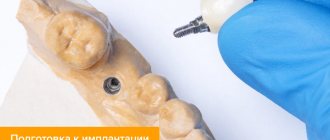
The main stages of conditionally removable prosthetics on implants
Conditionally removable prosthetics on implants is carried out in several main stages, and below we will analyze in detail the features of each stage of treatment.
Diagnosis and treatment
During the patient’s initial visit to the dental clinic, a thorough examination of the oral cavity is required and a number of diagnostic measures are prescribed. Diagnostics is of fundamental importance: it allows us to exclude possible contraindications to conditionally removable prosthetics on implants. If during preparation for the procedure gum inflammation or other dental diseases are detected, therapeutic measures must be taken to eliminate them before prosthetics on implants.
Implantation
The procedure for implantation before installing a conditionally removable prosthesis can be carried out according to the protocol of classical or simultaneous implantation. The second option is more gentle for the patient and also allows you to significantly reduce the overall treatment time. The implants are implanted into the sockets of the extracted teeth, and almost immediately a temporary prosthesis is placed on them, which is replaced with a permanent structure within two to three weeks. However, if the teeth were lost a long time ago, and the bone tissue has lost its height and volume, it is not possible to carry out express implantation. Classical implantation is carried out in two stages, and a permanent prosthesis will be installed only after the artificial roots have completely engrafted; usually the duration of the osseointegration process of implants is several months.
Manufacturing and installation of a permanent prosthesis
After installing the implants and temporary prosthesis, impressions are taken, on the basis of which a conditionally removable prosthesis will be made. The price of a permanent prosthesis will depend on its type, material of manufacture and method of fixation. We will examine in detail the possible options for fixing conditionally removable dentures on implants in the next section of the article.
Complete upper and lower dentures - what are their differences?
As already mentioned, fixation of complete removable dentures has its own characteristics. Since the patient does not have natural teeth, the palate and soft tissue are used to attach the prosthesis.
Complete denture for the upper jaw
A complete removable denture for the upper jaw is placed on the alveolar processes, and special suction cups on the artificial palate make the fixation more durable.
Complete denture for the lower jaw
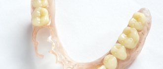
As for the lower jaw, only the alveolar processes are involved in the fastening (the so-called complete removable denture without palate), so the structure on the lower jaw is less stable. That is why mini-implantation is considered the best option (if classical implantation cannot be performed).
If several natural teeth are preserved on one of the jaws, then clasps or attachments provide more reliable fixation, but in this case the denture will be partial and not complete. We should not forget that partial and complete removable dentures have different principles of fastening, so the latter in any case will lose in terms of stability and stability.
Complete denture for the upper jaw.
Complete denture for the upper jaw.
Indications and contraindications for use
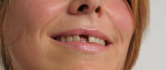
Partial removable dentures are used in cases where the jaw is missing only a few teeth, but there are own healthy teeth that will be used as supporting teeth. Suitable for restoring both chewing and front teeth. The use of removable partial dentures is recommended in the following cases:
- missing several teeth;
- end defects of the jaw;
- atrophied bone tissue;
- the need for temporary prosthetics;
- contraindications to the installation of implants.
There are practically no contraindications to this type of prosthetics, and they are all temporary. These include viral diseases, carious lesions of teeth and periodontal tissue diseases.
Making a complete removable denture
The production of a complete removable laminar denture is quite fast. The design itself does not have complex elements, and the patient does not require a preparatory period (except for the installation of a prosthesis supported by implants). Modern methods for manufacturing complete removable dentures imply receiving a finished structure 10–14 days after the initial visit to the clinic.
Stages of manufacturing a complete removable denture:
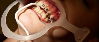
- Initial consultation. Photo of the jaw and choice of prosthesis.
- Taking impressions.
- Casting models.
- Making a wax base.
- Determining the relationship of the patient's jaws, correcting the prosthesis in the articulator.
- Making the final model after successful wax fitting.
- Correction and processing of the finished prosthesis.
- Installation of a complete removable denture.
Making removable dentures in the absence of teeth is usually not accompanied by major mistakes on the part of doctors, but if at the fitting stage the design gives you severe discomfort or pain, report it immediately.
Prosthetics with removable structures
The sequence of actions is determined by the condition of the oral cavity, the number of lost teeth, the type of prosthetic system and the material of manufacture. Basic stages of the process:
- diagnostics of the oral cavity with visual examination, x-rays, selection of materials and type of structure;
- preparing the site for the installation of a prosthesis (periodontal, orthodontic treatment, elimination of caries, depulpation of abutment teeth - if necessary);
- taking impressions to model a plaster jaw and develop an individual dental design;
- fitting of the finished prosthesis with its subsequent fitting (in several visits to the dentist);
- final installation of the prosthesis and detailed consultation on its care.
On a note! To avoid unpleasant consequences of prosthetics (gum recession, irritation of mucous tissue, destruction of supporting crowns), you should visit the dentist every six months. This will help prevent the development of pathology in time and avoid drastic and expensive measures to eliminate it.
Rules for wearing and caring for prostheses
The first time after installation, removable dentures can cause discomfort. As a rule, it goes away by the end of the first week. In some cases, especially when using plate structures, addiction can last up to 3 weeks.
To speed up adaptation, there are general rules and recommendations: rinse your mouth with warm water more often, do not overload the denture with solid food for the first 3-4 days.
Carefully monitor hygiene, rinse your mouth after every meal, and beware of excessive chewing loads. For cleaning, you should not use abrasive cleaners; give preference to special solutions and pastes.
Stages of manufacturing a removable partial denture
Depending on the type of prosthesis, the course of orthopedic treatment may differ slightly, but, in general, prosthetics can be divided into several main stages:
Preparation. After examination and diagnosis, the orthopedic doctor draws up a treatment plan and helps with choosing the optimal design option suitable for your particular case. At this stage, the oral cavity is sanitized. If necessary, teeth that cannot be saved are removed.
Taking impressions. Using impression material and a custom tray, the specialist takes impressions of the teeth on both jaws.
Manufacturing of the structure. In the dental laboratory, wax models are cast from the impressions, from which the technician makes the dentures.
Fitting and fixation. At the last visit, the doctor tries on the structure and fixes it in the oral cavity. If necessary, the prosthesis is adjusted and polished according to the bite.
Lifespan of removable partial dentures
Practice shows that the service life of removable plate structures is on average 3-5 years, after which the structure has to be changed. Bugel products have a longer lifespan - up to 5-7 years.
In general, the duration of wearing depends on several factors:
— the quality of the design and the professionalism of the specialist who carried out the installation;
— characteristics of the body and the condition of the oral cavity, the presence of gum diseases and deformation of the dentition;
— accuracy of compliance with doctor’s recommendations and proper home care.