22.11.2019
After installing implants, many patients forget that they need to pay attention and monitor hygiene no less than healthy teeth. Therefore, they are perplexed why the gums near the crown periodically hurt and bleed. Normally, such bleeding can be observed in the first weeks after prosthetics. But sometimes such a problem is accompanied by mild pain or a burning sensation that radiates to the roots. These are clear signs of inflammation and you should definitely find the cause in order to prevent suppuration.
Saving the tooth of the chief physician of Dial-Dent S.V. Zukora
It so happens that even good dentists themselves need dental treatment. Our chief physician S.V. Zukor had a toothache. Or rather, this tooth had been hurting for a long time. Periodically, once or twice a week, while chewing, a short but very intense painful shooting occurred in the teeth of the upper jaw on the right. The pain was short and very intense. After a couple of seconds everything passed. Nothing was found during the examination. A thorough visual examination of all the teeth in this area, x-rays and computed tomography of the teeth revealed nothing. Our specialists could not find the source of the pain. The cause of pain in the teeth, that is, toothache, can be not only problems with the teeth. Often diseases of the ENT organs or neurological symptoms are disguised as toothache. That is why the Family Dental Clinic employs both a neurologist and an ENT doctor, who are involved in diagnosis in complex cases. Tsukor S.V. was examined by these doctors, who gave an expert opinion that everything was normal with the neurological and ENT systems.
So, after all, teeth! But nothing is visible. The tactics of observation and wait were chosen. Yes, painful impulses were repeated, but nothing could be done until the exact cause of the pain was identified. Monitoring was carried out throughout the year, periodically repeating examinations.
Detecting a crack in a tooth with a microscope
And then at one moment S.V. Zukor felt that the situation had changed a little. The pain became different. From sharp and shooting, it became more constant and pulling and was localized in the area of one tooth, the fourth from the center of the tooth on the upper jaw on the right. After examining the tooth tissue more closely with a microscope, we discovered a crack in the tooth! This is the real cause of pain! They gave me anesthesia. Under anesthesia and with a microscope, they found the beginning and end of the crack.
A cracked tooth is a very common dental problem. The problem is complicated! A crack in a tooth is not always visible to the eye! It’s very good that Dial-Dent has a dental microscope! With repeated magnification with a microscope, many subtle cracks become visible. A dental microscope not only magnifies the image, but also illuminates the working area, which improves visibility.
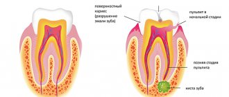
Stages of crown caries
Caries on the crown develops according to the same scenario as other types of this pathology, that is, in four stages. First, a white spot forms on the tooth surface - an area of enamel demineralization, which is destroyed by bacteria. It does not have time to recover naturally and cannot fully protect the tooth from external factors.
Gradually, the carious process penetrates into the internal tissues of the enamel and its superficial stage begins, during which the white spot becomes cloudy. The pathology is still within the boundaries of the enamel surface and does not affect the dentin, but the patient is already experiencing the first subjective symptoms: he develops increased sensitivity of the tooth to sweet foods. After eating foods high in fast carbohydrates, a person feels discomfort or pain in the area of the affected tooth. These signs disappear after eliminating the irritating factor.
The third (middle) stage of caries is accompanied by damage to dentin. A brown carious cavity forms in the tooth, in which cariogenic microbes continue to actively multiply. The patient develops bad breath. Caries can be noticed with the naked eye, especially if it develops on the crown of the tooth. The patient also experiences pain while eating and during temperature changes. The pain is acute, sharp, but quickly passes at rest.
The fourth stage of caries is called deep and is considered an advanced form. A large, almost black hole forms in the tooth; it constantly hurts, especially when in contact with external factors. The soft tissues located inside the diseased tooth are protected from carious cavity only by a thin partition. There is a risk of developing pulpitis - inflammation of the nerve (pulp).
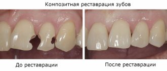
Deep caries of the crown can lead to its splitting into several parts. More often this happens with incisors and canines, which have a finer structure. It is more difficult to treat a front tooth than a chewing tooth, especially at the last stage of the disease.
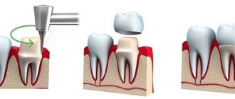
If the pulp had to be removed, it darkens and looks aesthetically unattractive. In cases where the tooth could not be saved or its aesthetic functions need to be restored, the doctor installs a microprosthesis, also called a crown, on its coronal part.
Treatment tactics for a tooth with a crack
Cracks vary in depth and length. A crack in a tooth is like lightning, its path is unpredictable. When it comes to saving a tooth with a crack, you need to determine exactly how the crack runs. It is important to see both the beginning and the end of the crack. If the crack is shallow and short, then you can remove the area broken by the crack and save the tooth. If the crack is deep, through, and passes through the entire tooth, then the tooth is removed and immediately replaced with an Astra Tech dental implant. These implants are specifically designed to replace teeth removed due to cracks.
The problem was that the crack in the tooth ran very deep, going far under the gum and getting close to the bone. The situation was on the brink: remove and replace with an implant or fight for the tooth.
Of course, the fight for a tooth in such cases occurs only with the consent of the patient, if he is ready to carry out treatment without a guarantee and with an uncertain time prognosis for success. If the patient wants reliable treatment and a guaranteed successful result, the tooth is removed and an implant is placed in its place.
Our chief doctor nevertheless chose to fight for the tooth without guarantees! And the fight began!
Solutions to the problem
If the prosthesis is installed according to all the rules, and the patient complies with all medical recommendations, after 10-14 days he usually forgets about the existence of the structure in his mouth and perceives it as his own teeth.
If after some time blood begins to appear from under the crown, you should not self-medicate. To eliminate the pathological phenomenon, you must consult a doctor.
To exclude an allergic reaction or rejection of the prosthetic material by the body, the patient is asked to perform a series of special tests. If the cause is confirmed, the doctor prescribes a course of antihistamines and decides on replacing the prosthesis.
When the cause of bleeding is a worn-out prosthesis or improper manufacturing, experts usually advise taking a course of anti-inflammatory therapy and replacing the structure with a new one.
If bleeding occurs due to the development of an inflammatory process (which happens in most all cases), a comprehensive course of treatment is prescribed, which includes:
- Professional cleaning of all elements of the jaw row.
- Restoring the balance of microflora. The patient is recommended to take probiotics, which will additionally restore local immunity. This could be Acylact, Bifidumbacterin, Lactobacterin.
- Taking antibacterial drugs (antibiotics in the form of a mixture or powder). Biseptol, Tsiprolet, Lincomycin, Gramicidin (for children) are usually prescribed.
- Rinse. Antiseptic solutions Miramistin, Chlorhexidine, Hydrogen Peroxide, Furacilin are recommended.
- The use of folk remedies. Solutions of sea salt or soda, decoctions of sage, oak bark, chamomile and calendula, propolis tincture, freshly squeezed juice of Kalanchoe, lemon, lingonberry and aloe also have good antiseptic effects.
Important! The duration of treatment is determined by the doctor and depends on the cause of the phenomenon, the degree of its severity and the patient’s well-being.
What does the smell from under the crown indicate, and how to get rid of it.
In this publication we will talk about the composition of dental ceramics.
Here https://zubovv.ru/protezirovanie/nesemnyie-p/koronki-np/vse-tonkosti-polu.html read about the technology for making half-crowns.
Indications for removal
In most cases, to eliminate the cause of bleeding, the doctor decides to remove the crown.
These are the following states:
- Pain in a tooth covered with a crown or adjacent elements.
- There are problems with the gum tissue due to an incorrectly placed prosthesis.
- The cement that fixes the crown is destroyed, which is why small particles of food penetrate inside, causing the active proliferation of bacteria.
- Allergy to the material from which the structure is made.
- The appearance of a cystic formation both on the dental element under the denture and on soft tissues.
- Destruction of the unit adjacent to the crown.
- Putrid smell.
In the first case (for toothache), the removal of the prosthesis is not always performed. The dentist may try to treat the problem unit through a hole made in the structure, which will be sealed with a filling after the end of the therapeutic course.
Important! Although this approach to treatment violates the integrity of the crown and reduces its service life, it delays the time for its replacement.
But there is no need to try to avoid repeated prosthetics. As practice shows, it is better to make a new denture and save the tooth underneath than to lose it.
The video provides additional information on the topic of the article.
Removing part of a tooth, treating gums with a laser, treating tooth canals with a microscope
First, the chipped wall of the tooth was removed.
After this, the gum was trimmed with a laser to free the edge of the chipped tooth from under the gum.
As a result of the split, the nerve in the tooth was damaged. The damaged nerve had to be removed.
Removing nerves from a tooth is a complex procedure that requires the highly qualified doctor who performs the procedure and the availability of the necessary equipment, including an operating microscope. At Dial-Dent, such procedures, called tooth depulpation, are always carried out using microscopes by doctors who have at least 10 years of experience. Yu.A. Borisova, a dentist-endodontist at Dial-Dent, a specialist in the treatment of dental canals with extensive experience, knows about all the advantages of dental treatment with a microscope.
Dial-Dent expert in dental canal treatment with a microscope Yu.A. Borisova removed the nerve from the tooth and filled the canals.
The photo shows tooth canals treated with a microscope:
The interval between visits is 2 weeks, since on the first visit an antimicrobial medicine was placed in the tooth and the tooth was closed with a temporary filling. Under no circumstances should you fill the canals right away or rush. The infection could enter the bone through a crack, and if the canals are filled in one visit, there is a high probability of complications, the tooth will continue to hurt, or a cyst will appear near the root of the tooth over time.
During the second visit to the endodontist, the tooth canals were thoroughly washed and hermetically sealed with hot gutta-percha.
Factors provocateurs
A person can complain about the appearance of blood from under the crown at any stage - immediately after its fixation, or after several years.
Doctors note several reasons for this phenomenon:
- The material from which the structure is made is not suitable . This is a common allergy that has resulted in inflammation of the soft tissue around the covered unit. Enlarged gums are compressed by the crown, which is why bleeding appears.
- The height of the edge of the product is incorrectly calculated. An edge that is too high for soft tissues creates increased stress, as a result of which they constantly rub and become deformed. Over time, non-healing bleeding erosions appear on the tissues.
- Poor blood flow after installation . During the procedure, it is very easy to touch blood vessels or deeply injure the mucous membrane.
- Breakage or abrasion of part of the crown. Distortion of the shape of the prosthetic structure also leads to bleeding due to damage to soft tissues.
- Development of the pathological process in the depths of periodontal tissues . Blood from under the crown can also be one of the clearest symptoms of periodontitis, gingivitis or periodontal disease.
- Poorly processed dental canals. Installation of a metal-ceramic crown involves preliminary tooth depulpation.
If the treatment is carried out incorrectly, the seal of the filling is broken, which is why pathogenic bacteria actively multiply in the cavity. An increase in the number of microorganisms provokes the development of an inflammatory process, the symptom of which is bleeding gums.
Important! The appearance of blood may be associated with a serious illness! Only a specialist can determine the exact cause of bleeding.
Strengthening a tooth with a pin insert and temporary prosthetics
After root canal treatment by endodontist Yu.A. Borisova, the patient switched to another doctor - orthopedic dentist A.S. Ivankov for further prosthetics. An orthopedist is a specialist in restoring damaged teeth and prosthetics.
This tooth, significantly damaged by a crack, had to be restored with a pin (pin stump insert) and a ceramic crown. Our chief physician did not want any metal ceramics. He insisted on using the best, most innovative and modern materials and technologies. The restoration of the tooth by the orthopedist also took place in several stages. A pin insert (pin) is installed in the tooth.
A tooth with a pin inlay is prepared for a crown:
Orthopedic dentist A.S. Ivankov put a temporary crown on the tooth, which he was supposed to wear for six weeks.
Why was six weeks needed? It's all in the gum! The gum, damaged as a result of the chip, was bleeding. It was necessary, by recreating the shape of the tooth on a temporary crown, to “deceive the gum”, make it think that there are no problems, and the tooth is in place. The gums around the temporary tooth will heal and take on a normal appearance. After 6 weeks, the final ceramic restoration, a ceramic crown, will be made and will look like a natural tooth, tightly enclosed by the gum.
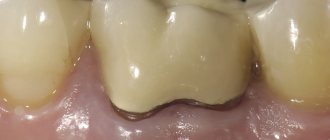
The photo shows the healed gum around the temporary crown after 2 months:
Diagnostic measures
To accurately determine the cause of this phenomenon, you need to consult a doctor. Along with an external examination of the oral cavity, the patient may be prescribed radiography.
Visual inspection
Often, the dentist receives most of the information about the condition of the gums from an external examination.
If damage to the pulp, dentin and enamel coating does not change the condition of the gum tissue, then pathologies of the root membrane will immediately change its appearance:
- The reddened, thickened edge of the gum, the presence of hardened dental plaque indicate gingivitis. Swelling can be easily identified if you press the edge of the instrument onto the gum tissue. After pressing on them, an imprint is formed, which will remain for some time.
- An abscess under the gum is an elongated bulge at the junction of the gum and cheek. Often, severe tissue swelling prevents detection of this formation.
- A fistula is indicated by the presence of a depression with a hole on the gum.
- The discharge of a purulent mass from under the gum margin indicates the presence of chronic gum inflammation.
External examination also reflects the condition of the prosthetic structure. An experienced dentist can determine the degree of wear of the crown, the tightness of its fit to the gum, and the correspondence of the height of the edges in relation to the soft tissues.
Radiography
Allows you to determine the condition of the tooth inside the crown, as well as detect neoplasms and pathological processes in the root.
Making a ceramic crown
The permanent dental crown was made in the dental laboratory of the Dial-Dent clinic by dental technician D.V. Ceramic wolf E.max. Photographs of teeth in different modes were used to give the technician maximum information to recreate the color.
The dental technician will match the color of the crown to make it indistinguishable from natural teeth in your mouth.
Having our own laboratory in our center simplifies many tasks related to dental prosthetics and makes all stages better and more predictable. All necessary corrections and fittings are made in the presence of a technician!
Permanent ceramic crown, material – E.max ceramics:
When to see a dentist
When installing a crown, it is recommended not to overload the jaw with chewing hard food during the first weeks. It is better to temporarily switch to broths, soups and purees, and give up apples and seeds. For a speedy recovery, it is necessary to thoroughly rinse your mouth with special antiseptics and treat with ointment with an antimicrobial effect. If you follow all the rules, after 7–14 days a person completely forgets about the bridges that were installed.
Some patients experience complications and problems. This is mainly due to improper installation and assembly of the bridge. An error can occur at any stage. Sometimes a deviation of 1 mm from the size is enough for the product to begin to rub the mucous membrane. A characteristic sign is that the gums around the crown turn red and bleed, and the patient feels full. When biting, the tooth seems to interfere and prevents the jaw from closing completely.
Another common problem is inaccurate fitting of the bridge over ground molars. Sometimes there is a gap that cannot be seen with the naked eye. Microparticles of food get into it and plaque accumulates. This is an ideal place for bacteria to grow, so you should immediately contact your dentist if:
- a person notices an unpleasant odor or taste in the mouth even after brushing;
- when pressing on the crown, ichor or cloudy exudate appears;
- the mucous membrane aches, reacts to changes in temperature and bothers you when chewing;
- The gums under the crown hurt and bleed.
If the doctor was able to preserve the roots and pulp of the tooth, it can easily become infected and acute suppuration develops. The pain becomes very strong and shooting, increasing in the evening or after eating hot food. Purulent fluid quickly accumulates and an inflamed focus forms in the periodontal tissues. It needs a way out, so a lump appears on the gum filled with whitish contents - a fistula. In addition to the symptoms listed above, a person has:
- high fever, weakness and slight chills;
- enlarged lymph nodes under the lower jaw or behind the ears;
- the cheek may become swollen.
In each case, the help of a specialist is necessary. If you self-medicate, you can seriously delay it until it becomes an acute abscess. Pus easily penetrates inside and spreads throughout the patient’s body, leading to life-threatening sepsis, kidney or liver damage.
Dental prosthetics with a ceramic crown - the result
We tried on the crown and it fit perfectly!
After fitting, the crown is fixed on the tooth.
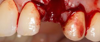
During fixation, due to polishing the seam between the crown and the tooth, the gums were irritated and the photograph shows that the gums are bleeding a little. It will go away on its own in a few days.
Comment by the chief physician of the Dial-Dent clinic S.V. Tsukora: “ I experienced the work of the Dial-Dent team myself! Understanding the problems doctors face in cases like mine, I could only trust our specialists! The treatment was comfortable and went exactly according to schedule! I've never gone without a tooth. There was always a temporary crown that allowed me to chew and smile through all stages of treatment! I am grateful to all participants - doctors and dental technicians of Dial-Dent! And also to our assistants who monitored my comfort during the treatment process, and to the administrators who always reminded me about the visit! If we talk about the cause of the crack, it could have been boxing training, for which I forgot to wear a mouth guard. It always seems that training is not a fight, that there is no danger of injury, but this is not so. Take care of your teeth - always wear a mouth guard!
»
Monitoring of the treated tooth continues. Dial-Dent specialists, like the patient himself, are ready, if necessary, to remove this tooth and install a dental implant. Currently, the tooth has ceased to cause concern and is functioning perfectly.
Galvanic syndrome
You cannot install crowns and dentures made of different metals. This can lead to the development of galvanic syndrome.
It is known that each metal and its alloy has its own potential when immersed in an electrolyte. Saliva acts as such an electrolyte in the mouth. Dentures and crowns made of different metals can create ideal conditions for the occurrence of galvanic current. The use of different metals is only possible if the metal is covered on top with porcelain or plastic. Galvanic syndrome causes a general deterioration in the patient's condition.
Dial-Dent specialists who took part in the treatment:
- Orthopedic dentist A.S. Ivankov – installation of a pin tab, dental prosthetics with a ceramic crown.
- Implant surgeon V.P. Alaverdov – removal of the broken part of the tooth, laser treatment of the gums.
- Dentist-endodontist Yu.A. Borisova – inspection of a crack with a microscope, treatment of tooth canals with a microscope.
- Dental technician D.V. Wolf – making a ceramic dental crown.
- Neurologist E.V. Saxonova – diagnosis of neurological problems.
- ENT doctor A.V. Arkhandeev – diagnostics of ENT problems.
- Dental assistants – A. Antoshkina, M. Erkimbekova.
- Center administrators – coordinating appointments with specialists, reminders about the visit.
See other examples of the work of Family Dental specialists here.
Make an appointment for a consultation by phone +7-499-110-18-04 or through the form on the website. You can ask questions about dental prosthetics to the chief doctor of the clinic, Sergei Vladimirovich Tsukor, at
The treatment regimen includes:
- systematic professional teeth cleaning – removal of plaque and tartar;
- a course of physiotherapeutic procedures to relieve inflammation;
- sanitation of the oral cavity.
The next stage of treatment is home therapy: rinsing with disinfecting solutions (salt and soda, chlorhexidine).
For a purulent infection, the patient is prescribed antibiotics.
At home, you can independently use traditional medicine methods, including decoctions and infusions of medicinal plants:
- oak bark;
- chamomile flowers;
- sage herbs.
You can use these decoctions and calendula tincture as lotions on problem areas of the mouth.
Use of pharmaceutical products:
- fluoridated toothpastes with the addition of medicinal herbal extracts;
- dental anti-inflammatory gels (Cholisal);
- vitamin complexes.