Treatment method
The main stages of treatment for concremental pulpitis are the same as for other types of inflammatory nerve diseases. Thanks to modern techniques and materials, all stages of therapy in most cases take one visit. The general treatment plan for pulpitis is as follows:
- anesthesia;
- preparation of carious cavity and canals;
- instrumental and medicinal treatment;
- obturation (filling with filling material);
- filling a carious cavity.
The differences lie in the complexity of preparing and filling obliterated (deposit-filled) canals with filling material. To pass them through, a special ultrasound device is usually used, but for large stones this does not always lead to a positive result.
The prognosis is favorable; in the absence of a tendency to excessive growth, denticles do not cause problems for the doctor and the patient. Treatment is most often carried out in full, and inflammatory symptoms completely disappear and do not recur.
To avoid the development of complications during treatment, it is necessary to promptly contact the dentist and conduct an examination of the dental system. There is no way to prevent the formation of denticles and petrification, but you can reduce the risk of their occurrence. To do this, it is necessary to treat inflammatory dental diseases in a timely manner, preventing the formation of foci of pulp necrosis, which subsequently mineralize.
The severity of inflammation symptoms depends on the form (acute or chronic) and the severity of pulpitis. Pain due to inflammation of the pulp occurs spontaneously, often at night. In addition, with pulpitis there is an acute reaction to temperature stimuli. The pain syndrome is not always concentrated in the area of the diseased tooth; it often spreads along the trigeminal nerve.
Methods of getting rid of the disease
Therapy for concremental pulpitis cannot be carried out at home without the help of a doctor.
Self-medication is excluded.
Treatment takes place in a hospital under the supervision of a doctor.
The dental method is the only effective way to eliminate pathology. Its essence is to remove the affected parts of the pulp and denticles.
The inner surface of the tooth is treated with special preparations that eliminate bacteria and prevent the recurrence of the disease .
The doctor installs insulating material into the tooth canals and places a filling. If the patient had a crown, a new, undamaged one is put on top.
Treatment takes place in a hospital under the supervision of a doctor.
What are petrificates and why are they formed?
Petrification is the focal or diffuse deposition of calcium salts in the soft tissues of teeth.
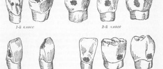
The composition of these formations includes collagen fibers with hydroxyapatite crystals included in them. Calcifications can vary in size from a microscopic inclusion to a formation that occupies the entire tooth cavity. Formations come in the following forms:
- round or oval with a smooth surface:
- indeterminate with outgrowths in different directions.
Mechanism and reasons for development
Salt degeneration occurs along the blood vessels or directly in the inflammatory focus. In this case, calcium salts are not excreted by the body, but are deposited in soft tissues. Examination of the pulp revealed that petrification proceeds quite quickly. As a result of a number of biochemical and physiological processes, a protein matrix is formed, on which calcium salts are deposited.
There are two main theories of the development of petrificates, according to which the main reasons for their formation are:
- Congenital anomalies in the formation of hard dental tissues are an inherited trait that manifests itself in some genetically determined diseases, for example, Alberts-Schoenberg syndrome (marble disease).
- According to experimental studies, petrificates are of bacterial origin - nanobacteria may participate in their formation.
What are denticles, the reasons for their formation
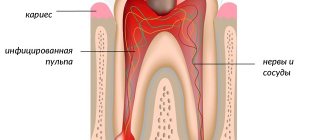
Dentin-like areas formed from root or coronal pulp tissue as a result of the development of replacement dentin are called denticles. The formations are round or irregular in shape and tend to merge into conglomerates, causing obliteration (blockage) of the entire lumen of the canal.
Structurally, the inclusion consists of a core, which is a fine petrificate, surrounded by tissue with dentinal tubules, and its chemical structure resembles natural tooth tissue. The development mechanism is as follows - pulp tissue cells are transformed into odontoblast-like and form dentin-like tissue.
Causes and mechanism of development
The true reasons for the development of denticles are not fully understood, but scientists believe that the factors provoking the development of these formations may be:
- injuries – dislocations and fractures of the roots of both permanent and baby teeth;
- dental manipulations - treatment for crowns, the presence of metal fillings, orthodontic structures;
- various health problems - metabolic disorders, development of thyrotoxicosis, excess vitamin D and lack of vitamins A and C in the body, long-term therapy with hormonal drugs;
- inflammatory and degenerative diseases of the periodontium (tissues surrounding the tooth);
- age-related changes in the dental system – increased wear of teeth, involution (reverse development) of soft tissues;
- hereditary imperfect formation of dentin is the most pronounced example of dysplasia that develops with Stanton-Capdepont syndrome.
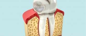
Denticle occupying the entire pulp cavity
Age-related changes in the pulp –
Even after the complete formation of a tooth, with age there is a gradual reduction in the size of the pulp chamber. This occurs as a result of continuous deposition of secondary dentin as well as intermittent deposition of tertiary dentin. Consequently, at a more mature age, the pulp will occupy a significantly smaller volume than at a young age. In addition, the shape of the pulp chamber itself changes (as a result, the pulp horns are smoothed out).
All this also has clinical significance, for example, deep preparation of dentin in the area of the pulp horns in old age will be less dangerous. In addition, with age there is a decrease in the number of cellular elements in all layers of the pulp (up to 50% of the initial level), but at the same time the content of collagen fibers increases 3 times. The blood supply to the pulp deteriorates due to the reduction of a large number of capillaries (especially those located in the capillary plexus in the Weil layer). Loss of nerve fibers also occurs, and in particular, this is associated with an age-related decrease in the sensitivity of the pulp to irritants.
With age, the frequency of formation of calcifications in the pulp also increases. Although, if calcification occurs not locally, but diffusely, then this process is called petrification. Petrifications most often form in the root part of the pulp (along the periphery of vessels, nerves, and also inside the vascular wall), sometimes causing disturbances in the blood supply to the pulp and pain. We hope that our article was useful to you!
Sources:
1. Higher professional education of the author in dentistry, 2. The European Academy of Paediatric Dentistry (EU), 3. “Anatomy of human teeth” (Gayvoronsky, Petrova). 4. “Periodontology” (Danilevsky N.F.), 5. “Pulpitis of temporary and permanent immature teeth” (Mamedova).
Types of denticles
The table shows the differences between true and false denticles.
| № | Characteristic | True denticles | False denticles |
| 1. | Location | Upper part of the root canal | Along the entire length of the canal and tooth cavity |
| 2. | Structure | Similar to natural dentin, have tubules and are surrounded by odontoblasts | They are mineralized areas that do not have a dentin-like structure |
| 3. | How are they synthesized? | Directly by the pulp | On one's own |
| 4. | Diagnosis frequency | Very rare | In most cases |
Depending on the location, the following types of formations can be distinguished:
- parietal ones are fixed near the walls of the canal or tooth cavity;
- free-lying are located in the thickness of the pulp;
- interstitial are surrounded by dentin.
Features of the course, diagnosis and treatment
The disease is characterized by long-term, sluggish development and the absence of clinical manifestations in the anamnesis. During a dental examination, a change in the coloration of the crown of the tooth may be observed. As the inflammatory process develops, paroxysmal pain appears, more pronounced at night. Mostly, the patient can accurately identify the causative tooth.
Clinical manifestations
In most cases, the formation of denticles is not accompanied by clinical manifestations; they are discovered accidentally during an X-ray examination of the teeth, which is carried out in connection with the treatment of other dental diseases. Painful sensations occur if the nerve bundle is compressed by denticles or petrification, when formations are displaced, as well as during the development of the inflammatory process.
Radiologically, the formations look like multiple or single dense shadows, having a round or irregular shape, located inside the cavity of the tooth and its canals.
Diagnostic features
The clinical manifestations of concremental pulpitis are quite similar to the symptoms of trigeminal neuralgia, so the disease is differentiated from it. The table shows the main differences:
| № | Characteristic | Concrete pulpitis | Neuralgia |
| 1. | Pain | Increasing attack of dull pain | Sudden onset of acute pain |
| 2. | Time of attack | Mostly nocturnal | Any time of day |
| 3. | Novik's test | Pain occurs with sudden changes in position | Negative |
| 4. | Percussion | Sharp pain when tapping on the crown of a tooth | Absent |
| 5. | Causal tooth | Determined | Not determined |
| 6. | X-ray result | Dense formations in the tooth cavity are clearly visible | No tissue changes |
| 7. | Development | The disease develops slowly, in which the intensity and number of attacks constantly increases | Painful attacks are constant and do not change intensity. |
| 8. | Manifestations from the autonomic nervous system | No | Eat |
Treatment method
The main stages of treatment for concremental pulpitis are the same as for other types of inflammatory nerve diseases. Thanks to modern techniques and materials, all stages of therapy in most cases take one visit. The general treatment plan for pulpitis is as follows:
- anesthesia;
- preparation of carious cavity and canals;
- instrumental and medicinal treatment;
- obturation (filling with filling material);
- filling a carious cavity.
The differences lie in the complexity of preparing and filling obliterated (deposit-filled) canals with filling material. To pass them through, a special ultrasound device is usually used, but for large stones this does not always lead to a positive result.
The prognosis is favorable; in the absence of a tendency to excessive growth, denticles do not cause problems for the doctor and the patient. Treatment is most often carried out in full, and inflammatory symptoms completely disappear and do not recur.
To avoid the development of complications during treatment, it is necessary to promptly contact the dentist and conduct an examination of the dental system. There is no way to prevent the formation of denticles and petrification, but you can reduce the risk of their occurrence. To do this, it is necessary to treat inflammatory dental diseases in a timely manner, preventing the formation of foci of pulp necrosis, which subsequently mineralize.
The structure of the dental pulp –
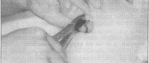
Speaking about the structure of the dental pulp, we will first analyze its structural elements - after which we will move on to a description of its layers, which you could see in Fig. 2. You can see the appearance of the pulp removed from the tooth (wound on a pulp extractor) in the photo below.
1) Structural elements of pulp –
The pulp consists of loose fibrous connective tissue (with a large number of nerve endings, blood and lymphatic vessels). Accordingly, its structure will consist of collagen fibers, the main amorphous substance, as well as a large number of various cellular elements. The total collagen content is from 25 to 30% of the dry mass of the dental pulp, and it is mainly collagen types I and III. In the coronal part of the pulp, collagen fibers are located more loosely, but in its root part they form denser clusters.
As for the main amorphous substance located between the collagen fibers, it consists of water, glycosaminoglycans, as well as glycoproteins and proteoglycans. The intercellular substance has a high ability to diffusion, which allows nutrients from the blood to enter the cellular elements, and metabolic products to be excreted into the venous circulatory system. There is also a wide variety of cellular elements in the pulp - primarily odontoblasts and fibroblasts (fibroblasts are responsible for the formation of intercellular substance and the synthesis of collagen fibrils), as well as dendritic cells, macrophages, lymphocytes, mast cells, etc.
Cellular elements of the pulp -
| Cells | Odontoblasts – form dentin and provide its trophism. Neighboring odontoblasts are interconnected by intercellular connections, which allows the layer of odontoblasts to also perform a barrier function (regulating the movement of molecules and ions between the pulp and predentin). Some authors call these cells “dentinoblasts.” |
| Fibroblasts are the most numerous cells in the pulp (their number decreases with age). Their function is to produce and maintain the composition of the intercellular substance of the pulp, as well as to absorb and digest the components of the intercellular substance. | |
| Macrophages - participate in the renewal of the cellular composition of the pulp, capturing and digesting dead cells and components of the intercellular substance. They phagocytose microorganisms and participate in the development of immune reactions as antigen-presenting effector cells. | |
| Dendritic cells – they are antigen-presenting cells whose function is to absorb various antigens, process them and present them to lymphocytes. They also induce the proliferation of T lymphocytes. The content of dendritic cells increases as the pulp matures, as well as with antigenic stimulation. | |
| T-lymphocytes are contained in the pulp in small quantities, but during inflammation their number increases sharply (of all types of this population of lymphocytes, T-suppressors predominate in the pulp). B-lymphocytes are not normally found in the pulp, and appear only when it is inflamed (they synthesize IgG immunoglobulins and provide humoral immune responses). | |
| Mast cells – they are located around blood vessels; are characterized by the presence in the cytoplasm of a large number of granules with biologically active substances (histamine, heparin, eosinophilic chemotactic factor, etc.). The release of these components causes an increase in vascular permeability. | |
| Poorly differentiated cells - can give rise to odontoblasts and fibroblasts. The content of these cells decreases markedly with age. | |
| Fibers and glycoproteins | Type I and III collagen fibers, reticular fibers, fibronectin. |
| Basic intercellular substance | Glycosaminoglycans, chondroitin, proteoglycan. |