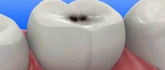
Apical periodontitis of the tooth - what is it?
The diagnosis of periodontitis is made when the patient's periodontal tissues near the apex of the tooth root become inflamed, which is why it is called apical. It affects deep gingival and bone structures, so it is not easy to treat - unlike, for example, caries. Nevertheless, this disease requires immediate intervention, because if the patient is not in a hurry to treat apical periodontitis, then he risks encountering unpleasant consequences of the disease: the appearance of granulomas and cysts, destruction of the alveolar bone, and in the most severe cases, even sepsis.
Apical periodontitis: pathogenesis
Why does inflammation appear in the peri-root zone? Pathogenic bacteria in tissues are the most likely cause of apical periodontitis. It is called an infectious form of periodontitis, it is a logical continuation of untreated caries, which subsequently develops into pulpitis. If the patient continues to ignore the symptoms and does not seek treatment, the pulp dies, allowing pathogenic bacteria to enter the periodontium through the apical foramen.
Another cause of the disease can be mechanical damage - a blow, bruise, displacement or fracture of a tooth. Then the patient is diagnosed with traumatic periodontitis.
The third reason is the penetration of caustic, potent substances into the peri-root tissue, for example, medications. Almost always this is a consequence of a medical error, most often - inaccurate treatment of pulpitis. Such cases are called drug-induced apical periodontitis.
Symptoms
Symptoms of inflammation depend on the cause of development.
- With candidiasis, a white coating appears, resembling cottage cheese.
- Inflammation caused by improper treatment and mechanical damage is characterized by swelling and redness of the tissues, severe pain, and a rise in temperature.
If there are obvious signs of inflammation, you should immediately consult a dentist. Reasons for making an appointment are also:
- decreased sensitivity of mucous membranes,
- pain when chewing and swallowing,
- swelling and bleeding of the gums,
- the appearance of erosions in the sky.
The sooner you contact a specialist, the less time the treatment will take.
Types and symptoms of apical periodontitis
Depending on how the inflammatory process proceeds, two types of disease are distinguished: acute apical periodontitis and chronic apical periodontitis. The second is always a consequence of the first: if acute inflammation is not cured in time, then it has every chance of becoming chronic.
The symptoms of acute apical periodontitis of pulpal origin are difficult to miss - they can cause the patient many unpleasant moments. The acute period is usually accompanied by the following conditions:
- nagging pain in the area of the affected tooth, which intensifies over time, becomes pulsating, and can radiate to other parts of the face;
- swelling of soft tissues, enlarged lymph nodes;
- rise in temperature;
- headache.
The duration of acute apical periodontitis ranges from 2 days to 2 weeks. In the absence of appropriate treatment, two options are possible: either the disease will progress, involving more and more tissue, or it will enter a chronic stage. The clinic of chronic apical periodontitis looks like this:
- pain in the affected area is mild or absent altogether, and can also occur when pressing or biting;
- slight tissue swelling may be observed;
- Possible bad breath;
- the diseased tooth is sensitive to temperature influences.
The chronic course of the disease may be accompanied by acute periods. With exacerbation of chronic apical periodontitis, the same symptoms are observed as in the acute form.
How to choose an apex locator
When choosing an apex locator, the dentist must determine for himself those parameters in the operation of the device that will be convenient for him. These include :
- display parameters (color or black and white);
- type of results on the display: graphic and/or numerical;
- presence or absence of a sound signal;
- ease of use (size of electrodes, length of wires).
In addition to these characteristics, it is necessary to take into account when choosing a device:
- working environment (dry, wet or both dry and wet);
- budget;
- availability of additional functions.
The reputation of the manufacturer is also important . Apex locators from European manufacturers are considered the most stable in operation. Chinese devices break down faster, but they cost less.
Diagnosis of apical periodontitis
It should be remembered that the symptoms of apical periodontitis are similar to the symptoms of some other oral diseases: pulpitis, sinusitis, hilar cyst and others. To make an accurate diagnosis, the following studies are necessary:
- electroodontometry
- helps to assess the degree of pulp necrosis; - radiography
- allows you to see tissue changes in the apical region; for chronic apical periodontitis, x-ray is the best diagnostic method, but it may not detect acute inflammation at an early stage; - A blood test
is an additional diagnostic method. With apical periodontitis, an increase in the level of leukocytes and ESR is observed.
Treatment methods for apical periodontitis of the tooth
How to treat chronic apical periodontitis and how does the treatment method for acute apical periodontitis differ? In both cases, the patient will need several visits to the doctor, since the therapy is carried out in several stages.
- The first stage:
opening the tooth, thoroughly cleaning the dental canals from the remains of necrotic pulp and areas affected by caries. Expansion of channels. - Second stage:
relief of inflammation. It is produced by filling the channels with antiseptic and anti-inflammatory materials. Additionally, the doctor may prescribe medicinal rinses, and in advanced cases, a course of antibiotics. - Third stage:
filling the canals and monitoring the results of treatment using radiography.
In particularly difficult cases, surgical intervention may be required to gain access to the affected root through an opening in the alveolar bone. If none of the treatment methods brings results, the tooth is removed.
Brief reviews of the most popular models
Modern apex locators have many functions . The more there are, the more expensive the device. The leading positions in the market are occupied by manufacturers not only from Germany, Japan (Morita) and Israel, but also from China and Russia (Averon).
NovApex
"NovApex" is an apex locator manufactured by the Israeli company Forum Engineering Technologies Ltd.
Its compact dimensions and low weight allow the device to be placed in any place convenient for the doctor, even on the patient during work. The file allows you to take measurements both in a dry canal and filled with saline, blood or pus. In addition, the device can operate autonomously from a battery . The dentist customizes the alert mode: sound or light. Its cost starts from 22.5 thousand rubles.
Complications after treatment of apical periodontitis
Treatment of acute apical periodontitis (as well as chronic) is a rather complex process that requires accuracy and concentration from the doctor. Like any intervention, it can lead to complications under unfavorable circumstances. What symptoms should you pay attention to after treatment of apical periodontitis?
- A sharp pain that occurs immediately after the end of the anesthesia may indicate that when filling the roots, the filling mass has gone beyond the boundaries of the tooth and is pressing on the gums. There is also a possibility of root perforation and breakage of the thin end of the endoscopic instrument in the root canal.
- After putting the medicine into the canals, the tooth hurts when pressed, and the gums are slightly swollen. These symptoms indicate that the doctor has used a strong medication that irritates periodontal tissue. It could also be a sign of an allergic reaction.
However, in most cases, timely treatment of apical periodontitis ends successfully and does not cause complications.
Extension of filling material beyond the apex: endodontic failure versus clinical success
Many dentists believe that procedural errors in daily practice, such as extrusion beyond the apical foramen and contact of filling material (most often gutta-percha and zinc oxide eugenol sealer) with periapical tissues after treatment, are a direct cause of endodontic failure.
Some authors show the negative impact of extending the filling material beyond the apex on the predictability of endodontic treatment and emphasize the following:
- High incidence of postoperative pain with exacerbation of inflammation
- Toxicity of materials
- Constant irritation of the periapical tissues with manifestations ranging from mild inflammation to a general reaction of the body.
In contrast, other authors have argued that apex extrusion in itself does not directly contribute to endodontic failure, although it should be avoided. Ingle says that endodontic treatment can be successful despite extrusion of material beyond the root apex. Schilder notes that he has not encountered complications specifically associated with excessive filling of the canal. Weine states that periapical tissues, fortunately, tolerate contact with gutta-percha well and therefore endodontic failure due to its removal beyond the apex is rare. In most cases, there is no pathological radiological evidence, and sometimes there is even a decrease in the amount of excreted material due to phagocytosis.
Image 1 – Mandibular first molar with amalgam filling and insufficient root canal obturation.
After retreatment, the radiograph shows three-dimensional filling of the root canals with a slight extension beyond the apex in the mesial canals obturated with Thermafil #25 and in the distal canal obturated by vertical condensation of heated gutta-percha.
After 5 years, the excess material had disappeared and the periapical tissue remained healthy.
These contradictory results can be explained by differences in research methodology and different composition of the derived materials. In general, the most common causes of discrepancy are related to differences in apical anatomy, limiting the effectiveness of endodontic treatment in various clinical situations.
Image 2 - The main factors leading to the removal of material beyond the root apex:
External inflammatory apical root resorption associated with apical damage to a previously treated tooth.
Incompletely formed root.
Excessive instrumentation with deviation from the natural course of the canal and apical perforation.
Inadequate handling of plasticized or unplasticized gutta-percha and sealants during obturation.
It is extremely important to identify and distinguish between the removal of filling material in three-dimensionally cleaned, formed and obturated root canals and in canals with obstructive overflow in combination with incomplete internal obturation.
Image 3 – Typical view of an exposed gutta-percha point: note the scratches on the point.
Image 4 – The apical foramen is completely filled.
Image 5 – Proper expansion for obturation, but insufficient filling of the canal. Note the transport of the apex during canal formation. (Courtesy of Dr. M. Vitullo).
Image 6 - Postoperative radiograph of the mandibular first molar showing a large apical lesion of endodontic origin. The mesial canals were obturated with Thermafil. A follow-up examination after 4 years shows osteoreparation. Excess filling material did not interfere with bone restoration.
Image 7 – Thermafil carrier extended beyond the apex in the distal root. Thermafil is a good method of obturation if the protocol is followed. The media was removed and the canals were refilled. After 6 months, the lesion was smaller than on the preoperative radiograph, and the patient noted the disappearance of pain when chewing.
Image 8 - These images show that even MTA can be released into the periapical tissue. In this case, the rule is the same as in the previous examples: if the canal is well cleaned, excess MTA cannot interfere with tissue restoration. 6-year follow-up kindly provided by Dr. R. Tonini.
Conclusion
Obturation is the last stage of endodontic treatment. Bringing either sealer or gutta-percha beyond the apical foramen will not necessarily be a mistake when performing obturation. However, this may result in an error in canal formation when parallel canal walls were created instead of conical ones, apical resorption or expansion of the apex.
Therefore, it is necessary to determine the direct causes of excessive root canal filling. “Is the excess material remaining after a complete three-dimensional obturation or is the material brought beyond the apex in combination with incomplete internal obturation of the canal?” Excess material beyond the apex is clearly not the goal of endodontic treatment, but rather the result of safely generated hydraulic forces in achieving three-dimensional obturation of the root canal system.
Numerous cases of root apex removal that have been successful, as evidenced by clinical and radiological signs of long-term restoration of periapical tissues, should reassure patients, doctors and insure against any legal charges.
Source: forum.stomatologija.su, styleitaliano.org