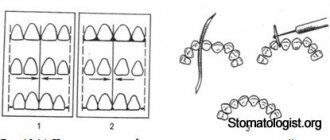
Incisors (dentes incisivi) . There are 8 incisors located in the middle of the dental arches, they are called the front teeth. There are upper and lower incisors, as well as medial and lateral. The incisors have one root and a crown flattened in the vestibulolingual direction with a wide cutting edge. The crowns of the medial incisors are larger than the crowns of the lateral incisors. The dimensions of the lower incisors are smaller than the upper ones; their crown is narrow, the roots are flattened in the mediolateral direction.
Upper incisors. Medial upper incisor. In the vestibular norm, the crown is wide, slightly convex, tapering towards the neck. The enamel of the crown forms a rounded protrusion in the form of an influx in the neck area. The shape of the crown can be different: almost rectangular, trapezoidal with a smaller base at the neck of the tooth, ovoid.
Structure of the medial upper incisor, right
Rice. 1-52. a — vestibular surface; b — medial surface; c - lingual surface; d — vestibular-lingual section; e — mediodistal section (numbers indicate the levels of transverse sections); e - cutting surface, 1, 2, 3 - shapes of cross sections at the level of the crown, middle and upper third of the root, respectively
The medial and incisal edges meet at right angles. The angle between the distal and incisal edges is usually obtuse and rounded. The cutting edge has a slight bevel in the distal direction. On the cutting edge of the teeth in young people, 3 tubercles (rarely 4) are noticeable, which continue in the form of ridges onto the vestibular surface. There are faint grooves between the tubercles and ridges.
The lingual surface of the crown often has medial and lateral marginal ridges (cristae marginales medialis et lateralis), running from the base of the crown to its cutting edge. The degree of expression of the marginal ridges varies. Sometimes they are missing. If the marginal ridges are highly developed, this surface has the appearance of a groove (spatulatate). The marginal ridges converge in the cervical part of the crown, forming the cervical girdle (cingulum cervicale). In the cervical third of the crown, as a rule, the tubercle of the tooth (tuberculum dentale) is clearly visible, the degree of development of which and its shape are different. It can be very developed and divided towards the cutting edge into several teeth (from 2 to 5). More often there are 2 teeth - medial and distal, less often a third, smaller, central tooth is formed between them, and even less often there are 4-5 teeth. The crown of the incisor on the medial side (in the medial norm) is wedge-shaped. Its vestibular contour is convex, with varying curvature of the convexity, and the lingual contour is concave. The enamel border on the medial surface is convex towards the cutting edge. The root of the medial upper incisor is slightly flattened in the mesiodistal direction. The root apex is rounded, and the apical opening of the root canal is clearly defined on it. Along the vestibular surface, the root has a convex contour; along the lingual surface, the contour of the root can be straight, convex or concave. The bend between the crown and the root is greater on the medial edge of the tooth than on the distal edge. This sign of the root, along with the signs of the angle and curvature of the crown, makes it possible to easily determine whether a tooth belongs to the right or left half of the dental arch. the incisor cavity is similar to the external contours of the tooth. Near the cutting edge, the cavity is slit-like, flattened in the vestibulolingual direction. The crown cavity narrows towards the root and passes into the root canal without a sharp boundary. At the apex it is possible to divide the canal into several tubules, each of which can open with an independent opening. The height of the crown of the upper medial incisors along the vestibular surface is 9-12 mm, the width of the cutting edge is 8-9 mm. The mediodistal diameter of the neck is 6-6.9 mm, the vestibulolingual diameter is from 7.1 to 7.5 mm; root length 11.5-15.5 mm. Lateral upper incisor . In all respects, this incisor is very similar to the medial incisor, but significant differences are also noted (Fig. 1-53).
Text of the book “Fundamentals of clinical dental morphology: a textbook”
3.1.3. Mandibular medial incisor
The medial incisor of the mandible is the smallest among the incisors. The mesial-distal size of the crown is significantly less than its height when compared with other incisors. The vestibular-lingual size of the root greatly exceeds its mesial-distal size. Signs of lateralization are weakly expressed.
In vestibular and lingual norms
the shape of the crown is close to an irregular quadrangle with a predominance of the height of the crown over the mesial-distal size (Fig. 15).
Rice. 15. Medial incisor of the lower jaw, right.
a – vestibular norm; b – language norm.
The cutting edge is often relatively smooth. In unworn teeth, there are tubercles on the cutting edge (the medial and distal ones are more pronounced than the middle ones). The crown angles practically do not differ from each other (the sign of the crown angle is not informative).
The approximal contours of the crown weakly converge towards the neck of the tooth. The most prominent points on the contours of the contact surfaces of the crown are located near the border of its occlusal and middle thirds.
The enamel-cementum border is curved towards the root evenly and approximately equally in both the vestibular and lingual norms.
The contact contours of the crown without sharp boundaries transform into the corresponding contours of the cone-shaped root. Often the transition from the medial and distal contours of the crown to the same contours of the root is more noticeable on the distal side. The apical part of the root is slightly curved distally.
The relief of the vestibular surface is weakly expressed. On the vestibular surface of the crown of unworn teeth there are two vertical grooves, separating three vertical ridges that pass into the tubercles of the cutting edge.
On the lingual surface of the crown there are marginal ridges, which are often separated from each other by a median ridge. The marginal ridges, belt and tubercle of the tooth are less developed than those of other incisors. In the lingual norm, both of its contact surfaces are visible on the root.
In medial and distal norms
the shape of the crown, like that of all incisors, is close to a triangle with the base in the area of the neck of the tooth (Fig. 16).
Rice. 16. Medial incisor of the lower jaw, right.
a – medial norm; b – distal norm.
The line of the occlusal contour is straight, short and slightly beveled in the direction from the vestibular to the lingual surface, towards the base of the crown.
The occlusal contour becomes vestibular near the IVS (often on the lingual side of the IVS). The vestibular contour of the crown near the border of the cervical and middle thirds is convex (equator of the tooth).
The lingual contour of the crown at the site of localization of the lingual tubercle is curved in the lingual direction, and along the remaining length to the incisal edge is slightly curved towards the USV.
The line of the enamel-cementum border is convex towards the cutting edge, the most protruding point is located near the USV. The enamel-cement junction in the distal norm has a smaller amplitude of curvature than in the medial one.
The transition of the vestibular and lingual contours of the crown to the corresponding contours of the root is clearly visible. The vestibular contour of the root is usually convex; the lingual contour can be either convex, straight or even concave. The apex of the cone-shaped root is located near the USV.
The approximal surfaces of the tooth have little relief. In the distal norm, the vertical groove of the root is more pronounced than in the medial one. this fact can be used as an additional feature when determining whether a tooth belongs to the right or left side of the dental arch.
In occlusal norm
the shape of the crown is close to an irregular quadrangle with rounded corners in place of the largest convexities of the vestibular and lingual contours and their transitions into each other (Fig. 17, a).
Rice. 17. Medial incisor of the lower jaw, right.
a – occlusal norm and section at the level of the base of the crown; b – tooth cavity.
The vestibular contour forms a weakly defined medial-distal slope. The conical contour of the crown has a greater curvature than the vestibular one.
In horizontal sections, the shape of the root approaches an irregular oval with concave medial and distal contours and a sharp predominance of the vestibular-lingual size over the medial-distal one.
Tooth cavity
resembles its external contours (Fig. 17, b). The cavity of the crown in the upper part is slit-like narrowed in the vestibular-lingual direction, and in the area of the base of the crown, without noticeable boundaries, it passes into the root canal.
In the middle part of the root, the canal often bifurcates into vestibular and lingual, with the subsequent connection of these branches in the apical part. On transverse sections, both channels have a rounded shape. The lingual canal is located close to the lingual surface of the root.
Anatomical options.
In
the vestibular (lingual) norm,
the shape of the crown often looks like a rectangle with a sharp predominance of the height of the crown over the mesial-distal size. There are incisors whose crown shape is close to triangular, oval or trapezoidal.
The cutting edge is often straight, but can be rounded to varying degrees. The severity and number of tubercles of the incisal edge vary. The approximal contours of the crown often converge towards the neck, but can be almost parallel to each other. The crown angle sign is usually uninformative.
The point of greatest convexity of the enamel-cement junction is most often located along the USV, but can be displaced distally. More often, the curvature of the enamel-cementum border in the vestibular and lingual norms is approximately the same, but sometimes it can be more pronounced on the lingual side.
On the vestibular surface of the crown, as a rule, there are three vertical ridges, of which the medial and distal ones are more pronounced. The number of ridges and their severity may vary. There are incisors with a uniformly convex or flattened vestibular surface of the crown.
On the lingual surface of the crown, between the barely noticeable marginal ridges, a median ridge may extend from the tubercle of the tooth. Depending on the severity of the ridge, the central part of the lingual surface of the crown can be concave, flat or convex.
The transition of the approximal contours of the crown to the corresponding contours of the root is often more pronounced on the distal side. In the vestibular (lingual) norm, the direction of the root apex varies.
In the medial (distal) norm
the line of the vestibular contour of the crown varies from slightly convex to straight. The contour of the crown can be smooth, convex or concave. The cutting edge of the crown can be located along the USV or be shifted to the lingual side.
The vestibular contour of the root can be smooth or convex, the lingual contour can be smooth, convex or concave.
The height of the tooth varies from 16.9 to 26.7 mm, while the height of the crown is 6.3-11.6 mm, the height of the root is 7.7-17.9 mm.
The mesial-distal size of the crown between the contact points ranges from 4.4 to 6.7 mm, the neck - from 2.7 to 4.6 mm. The size of the crown in the vestibular-lingual direction in the equator region ranges from 4.8 to 6.8 mm, in the cervical region - from 4.3 to 6.3 mm. 3.1.4.
Lateral incisor of the lower jaw The lateral lower incisor is larger than the medial one, has a wider crown and a more massive root. To determine whether a tooth belongs to the right or left side of the dental arch, all three main signs of lateralization are applicable.
In vestibular and lingual norms
the shape of the crown is close to triangular with a rounded apex at the enamel-cement border (Fig. 18).
Rice. 18. Lateral incisor of the lower jaw, right.
a – vestibular norm; b – language norm.
The occlusal contour of the crown is often uneven and turns into approximal contours, forming crown angles of different sizes. The mesial angle of the crown is less than the distal one (a sign of the angle of the crown).
The approximal contours of the crown converge from the cutting edge towards the neck of the tooth. The mesial contour of the crown slopes more towards the USV than the distal one. The most prominent points of the approximal contours of the crown, through which the equator line passes, are located near the border of the occlusal and middle thirds of the crown.
The enamel-cementum border is curved towards the root, and on the lingual side, as a rule, to a greater extent than on the vestibular side. The points of greatest convexity of the enamel-cementum border in both norms are located near the USV.
The transition of the crown contour to the root contour is more noticeable on the distal side than on the medial side. The root is longer than that of the mandibular medial incisor, with a clearly visible sign of root position.
The relief of the vestibular surface is weakly expressed. In unworn teeth, on the vestibular surface there are two shallow vertical grooves separating the enamel ridges, which turn into the tubercles of the cutting edge. The medial and distal ridges are usually more noticeable than the middle one. The vestibular surface of the root is smooth.
On the lingual surface of the crown, marginal ridges are visible, which are connected to each other near the belt. In the area of the cervical third of the crown there is a tooth tubercle. The lateral surfaces of the root weakly converge towards the lingual side.
In medial and distal norms
the crown has a shape close to the shape of a triangle, the most acute angle of which corresponds to the cutting edge (Fig. 19).
Rice. 19. Lateral incisor of the lower jaw, right.
a – medial norm; b – distal norm.
The occlusal contour has a slight bevel in the lingual direction. The line of the occlusal contour passes into the vestibular and lingual contours of the crown, most often on the lingual side of the USV. The vestibular contour is curved, its most prominent point is located near the border of the cervical and middle thirds of the crown. The lingual contour of the crown is convex towards the lingual side at the site of the lingual tubercle and curved towards the USV from the upper part of the lingual tubercle to the occlusal contour.
The line of the enamel-cementum border is convex towards the occlusal contour. The most prominent point of the enamel-cementum border is located approximately along the USV. The amplitude of the curvature of the enamel-cementum border in the medial norm is, as a rule, greater than in the distal norm.
The transition of the contours of the crown to the corresponding contours of the root is weakly expressed.
The root is conical. The root apex is located near the USV. The vestibular contour is often convex; the lingual contour can be either convex, straight or concave in shape. On the distal surface of the root, the vertical groove is deeper than on the medial surface. This sign can be used as an additional criterion for determining whether a tooth belongs to the right or left side of the dental arch.
In occlusal norm
the shape of the crown, as well as that of the medial incisor of the lower jaw, is close to an irregular quadrangle with rounded corners in the places of greatest curvature of the vestibular and lingual contours and in the places of transition of these contours (Fig. 20, a).
Rice. 20. Lateral incisor of the lower jaw, right.
a – occlusal norm and section at the level of the base of the crown; b – tooth cavity.
The medial contour of the crown is somewhat longer than the distal one.
The degree of curvature of the lingual contour is more pronounced than the vestibular one. The point of greatest convexity of the lingual contour is displaced distally.
The sign of crown curvature is more noticeable than that of the medial incisor of the lower jaw (medial-distal slope of the vestibular contour).
On horizontal sections, the root has the appearance of a laterally “compressed” oval with a more pronounced concavity of the distal contour.
Tooth cavity
resembles its external contours, it is more voluminous than that of the medial lower incisor (Fig. 20, b).
In the upper part, the cavity of the crown is slit-like narrowed in the vestibular-lingual direction and smoothly passes into a narrow root canal.
The root canal, as a rule, is single, flattened in the medial-distal direction.
Anatomical options.
In
the vestibular norm,
the severity of the enamel ridges and the degree of convergence of the contact contours of the crown to its base are variable.
In the language norm
the size of the marginal ridges varies. Sometimes the lingual surface is smooth. The root apex is often directed distally. Less commonly, the root is straight or curved to the medial side.
In contact standards
The vestibular contour of the crown can be either convex or smooth. The lingual contour can be curved to varying degrees.
The root canal in its middle part may bifurcate.
Tooth height varies from 18.5 to 26.6 mm.
In this case, the height of the crown is 7.3-12.6 mm, the height of the root is 9.4-18.1 mm. The mesial-distal size of the crown between the contact points ranges from 4.6 to 8.2 mm, the neck - from 3.0 to 4.9 mm. The size of the crown in the vestibular-lingual direction in the equator region ranges from 5.2 to 7.4 mm, in the cervical region - from 4.3 to 6.8 mm. Test tasks
The largest tooth in the incisor group is:
a – medial incisor of the upper jaw;
b – lateral incisor of the upper jaw;
c – medial incisor of the lower jaw;
d – lateral incisor of the lower jaw.
On the upper jaw, the smaller ones are:
a – medial incisor;
b – lateral incisor.
On the lower jaw, the largest in size are:
a – medial incisor;
b – lateral incisor.
The convexity of the enamel-cement border towards the cutting edge is most pronounced:
a – on the medial surface of the incisors;
b – on the distal surface of the incisors.
Vertical root grooves on both lateral surfaces are pronounced:
a – at the medial incisor of the upper jaw;
b – at the lateral incisor of the upper jaw.
Signs of tooth lateralization are weakly expressed:
a – at the medial incisor of the upper jaw;
b – at the lateral incisor of the upper jaw;
c – at the medial incisor of the lower jaw;
d – at the lateral incisor of the lower jaw.
The lingual tubercle is more developed:
a – at the medial incisor of the upper jaw;
b – at the lateral incisor of the upper jaw.
Bifurcation of the root canal is most typical:
a – for the medial incisor of the upper jaw;
b – for the lateral incisor of the upper jaw;
c – for the medial incisor of the lower jaw.
In the medial incisor of the lower jaw, the vertical root groove is more pronounced:
a – on the medial surface of the root;
b – on the distal surface of the root.
A sign of root position is the deviation of the root apex:
a – to the medial side;
b – to the distal side;
c – to the vestibular side; d – to the lingual side.
A sign of crown curvature is:
a – slope of the vestibular surface of the crown in the medial-distal direction;
b – slope of the vestibular surface of the crown in the distal-medial direction;
c – roundness of the distal corner of the crown.
Answers to test tasks:
1
A;
2
b;
3
b;
4
a;
5
B;
6
in;
7
b;
8
in;
9
b;
10
b;
11
a.
3.2. Group of fangs
Fangs (dentes canini) –
single-rooted teeth with a sharp “tearing cusp” (main cusp) of the occlusal contour. They are located in the dental arch between the incisors and premolars (third position) and are designed to “tear” food.
Humans have four permanent canines:
– canines of the upper jaw
(right and left);
– fangs of the lower jaw
(right and left).
What is common in the anatomy of the fangs is the presence of a cone-shaped crown, pointed on all surfaces, and the longest root.
The canine of the upper jaw is larger than the canine of the lower jaw.
The approximal surfaces of the upper canine converge to a greater extent towards the neck of the tooth, and the lingual cusp is better defined than that of the antagonist of the same name. This makes it easy to determine whether a tooth belongs to the upper or lower jaw. The canines show all the main signs of lateralization. 3.2.1.
Maxillary canine The maxillary canine has a crown pointed on all surfaces, the longest root and well-defined signs of lateralization.
In vestibular and lingual norms
the shape of the crown is close to pentagonal (Fig. 21).
Rice. 21. Maxillary canine, right.
a – vestibular norm; b – language norm.
The occlusal contour of the main tubercle consists of two segments extending from its apex, which is located somewhat mesially from the USV. These segments form an angle between themselves that is close to a right angle. The mesial segment of the occlusal contour is less extensive than the distal one (an important additional sign of tooth lateralization). The place of transition of the occlusal contour to the distal one is closer to the base of the crown than the place of transition of the occlusal contour to the mesial one.
The approximal contours of the crown sharply converge towards the USV, while the distal contour, as a rule, deviates more towards the USV than the mesial contour.
The enamel-cementum border is curved towards the root apex. The degree of curvature of the enamel-cementum border is more pronounced in the lingual norm. The most prominent point of the enamel-cementum boundary in the vestibular norm is located near the USV. On the lingual side, the same point is often displaced mesially from the USV. The transition of the contact contours of the crown to the corresponding contours of the root is more pronounced on the distal side.
The apex of the cone-shaped root of the tooth, as a rule, is located distally from the USV (a sign of root position).
On the vestibular surface of the crown, two vertical depressions are visible, separating three enamel ridges. The most pronounced is the median ridge, which is located from the main tubercle to the neck of the tooth. Of the other two vertical ridges, the mesial one is better expressed than the distal one. The vestibular surface of the root is relatively smooth.
Along the edges of the lingual surface of the crown there are marginal ridges, which are separated by depressions from the median ridge. The depression between the median and distal ridges is more noticeable than the depression between the median and mesial ridges.
The median ridge runs from the main tubercle to the lingual. The prominent tubercle is located near the enamel-cementum border and determines its sharp convexity towards the apex of the tooth root. The marginal ridges converge towards the lingual tubercle. The mesial and distal surfaces of the root converge in the lingual direction and are clearly visible from the lingual side. The distal surface of the root is somewhat convex, the mesial surface is flattened.
In mesial and distal norms
the shape of the crown resembles a triangle with the base located at the neck of the tooth (Fig. 22).
The occlusal contour is rounded and passes into the vestibular contour of the crown on the vestibular side of the IVD.
The vestibular contour of the crown is convex, its most prominent point is located in the cervical third.
Rice. 22. Maxillary canine, right.
a – mesial norm; b – distal norm.
The shape of the lingual contour is determined by the size of the lingual tubercle and the median ridge. In the area of the lingual tubercle, the lingual contour is usually convex. The contour of the median ridge can be convex, straight or concave from the main tubercle to the lingual.
The enamel-cementum border is curved towards the occlusal contour, more so on the mesial side than on the distal side. The point of greatest convexity of the enamel-cement border is located, as a rule, near the USV.
The transition of the vestibular and lingual contours of the crown to the corresponding contours of the root in the mesial and distal norms is well expressed.
The vestibular contour of the root is often convex, and the lingual contour is concave in the apical third and convex throughout the rest. The root apex is located near the USV.
The distal surface of the root is somewhat convex, the mesial surface is flattened. Both approximal root surfaces have vertical grooves. On the distal surface of the root, the groove is less noticeable than on the mesial surface.
In occlusal norm
the shape of the crown resembles an irregular quadrangle with rounded corners (Fig. 23, a).
The points of greatest convexity of the vestibular and lingual contours of the crown are approximately equally distant from the projection of the “tearing tubercle”. The point of greatest convexity of the vestibular contour is shifted to the mesial side. The sign of crown curvature is clearly visible. The curvature of the lingual contour, as a rule, is more pronounced than the vestibular one, with the point of greatest convexity located near the USV.
Rice. 23. Maxillary canine, right.
a – occlusal norm and section at the level of the base of the crown; b – tooth cavity.
In a horizontal section, the shape of the root approaches the shape of an irregular oval, elongated in the vestibular-lingual direction. Along the lateral contours of the root there are depressions, of which the mesial one is more pronounced.
Tooth cavity
matches its external shape. The cavity of the crown is pointed towards the main tubercle and has indentations towards the corners of the crown (Fig. 23, b). From the neck, the cavity of the crown gradually narrows and passes into a relatively wide root canal.
Anatomical options.
The shape of the crown of the upper canine is variable. The shape of the crown of the canine can resemble incisors or premolars.
In the vestibular (lingual) norm
the mesial and distal segments of the occlusal contour (slopes of the “tearing cusp”) often form an angle close to a right angle. There are variants of the tooth crown with an acute angle of the occlusal contour. In cases where the angle formed by the slopes of the main tubercle is close to unfolded, the canine is shaped like an incisor. On the distal slope of the “tearing tubercle” there may be an intermediate (additional) tubercle, from which a weakly defined enamel ridge extends onto the vestibular surface. The degree of convergence of the contact contours of the crown changes towards the neck of the tooth. The amplitude of the curvature of the enamel-cementum boundary is variable.
On the lingual surface
The severity of the median ridge, which largely determines the relief of the lingual surface, varies. In the presence of a wide, rounded and well-developed median ridge, the lingual surface becomes convex, giving the crown a conical shape. With a weak expression of the median ridge, the lingual surface is somewhat concave in the occlusal and middle thirds of the crown and is shaped like the lingual surface of the incisors. The lingual tubercle may be split into two fragments. The grooves separating the ridges and tubercles of the lingual surface are usually well defined. Often the groove separating the distal marginal ridge bifurcates in the middle third of the crown, limiting the so-called triangular fossa. Sometimes the grooves extending from the distal marginal groove (second order) cover a triangular elevation, which passes into the intermediate tubercle of the distal slope of the main tubercle [9].
In contact norm
vary the degree of convexity of the vestibular and lingual contours of the crown. The tip of the lingual tubercle may be close to the enamel-cementum junction or reach the middle of the lingual contour. The point of greatest convexity of the enamel-cementum border can be located along the USV or be shifted vestibular from the USV.
The amount of curvature of the root apex to the distal side is variable. The root sometimes “splits” into vestibular and lingual. The root canal is often deviated distally. There are variants of curvature of the root canal in the vestibular direction.
The height of the tooth ranges from 20.0 to 38.4 mm, while the height of the crown is 8.2-13.6 mm, the height of the root is 10.8-28.5 mm. The mesial-distal size of the crown between the contact points ranges from 6.3 to 9.5 mm, the neck - from 3.6 to 7.3 mm. The size of the crown in the vestibular-lingual direction in the equator region ranges from 6.7 to 10.7 mm, in the cervical region - from 6.1 to 10.4 mm.
Structure of the lateral upper incisor, right:
Rice. 1-53. a — vestibular surface; b — medial surface; c - lingual surface; d — vestibular-lingual section; d — mediodistal section; e - cutting surface; 1, 2, 3 - as in Fig. 1-52
The vestibular surface of the crown is trapezoidal or ovoid. The distal corner of the crown (between the chewing and distal edges) is more rounded than that of the medial incisor. The cutting edge of the lateral incisor is not straight, but rounded. Sometimes the cutting edge is not expressed at all, and on the upper part of the crown there is a pointed tubercle (the so-called peg-shaped tooth). The tubercles and grooves between them are very poorly developed. On the lingual surface of the lateral incisors, the same ridges, tubercles and teeth are noted as on the medial ones, but the shape of the lateral incisors is more variable. The dental tubercle is more pronounced than on the medial incisors, and a deeper fossa is formed under it. Of the tubercle teeth, the distal one is more developed. The lateral incisors are smaller than the medial ones. Crown height 8-10 mm, width - 6-7 mm , mesiodistal size of the crown base 4.8-5.4 mm, vestibulolingual - 5.8-6.2 mm, root length 10.5-14 mm. Lateral incisors may be absent. The row of upper incisors is located in the dental arch either in a slightly curved or even almost straight line. However, there may be deviations from the norm in the position of a number of upper incisors. It is also possible to increase the number of incisors. Between the medial incisors there is rarely an additional middle tooth - a peg-shaped mesiodens. Sometimes the incisors are arranged as if in two rows - the so-called crowding, with one or both lateral incisors located somewhat posterior to the medial ones, the canine thus moving closer to the medial incisor. During crowding, the medial incisors can be rotated around the longitudinal axis with the distal angles anterior or inward. There are enlarged spaces between the incisors - diastemas: more often between the lateral incisor and the canine, less often between the medial incisors. Lower incisors . The dimensions of these incisors are smaller compared to the upper incisors, the crown is narrow, the roots are flattened in the mediolateral direction. Medial lower incisor . The medial lower incisor has a narrow crown, slightly widening towards the cutting edge (Fig. 1-54).
CLINICAL AND ANATOMICAL FEATURES OF THE INCISERS OF THE UPPER AND LOWER JAWS.
⇐ PreviousPage 3 of 3
The group of incisors takes part in the formation of the frontal part of the dentition. These are 8 teeth, 4 of them belong to the upper jaw and 4 to the lower jaw.
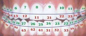
Dental formula.
——-12 11|21 22——-
——-42 41|31 32———
Nature has endowed the group of incisors with very important functions. Basically this is the capture, tearing off of food and partial chewing of it. The surfaces of the teeth with edges located at the appropriate angle to each other perfectly cut off objects in contact with them using the “guillotine” principle. Without making unnecessary movements, with minimal effort, the jaws come into contact with each other through the sharp edges of opposing incisors. The surface cutting mechanism is activated, and then food processing occurs, in which the entire dentofacial apparatus takes part.
Upper central (medial) incisors
- the largest of the group of incisors. They have a crown shaped like a wide shovel or chisel (Fig. 3).
Rice. 3. Upper central incisor.
On the cutting edge of a recently erupted tooth (Fig. 4.) one can distinguish three teeth in the form of tubercles, which are then gradually worn away. The vestibular surface of the crown is not flat, it is noticeably convex and has a trapezoidal shape, the smallest base of which faces the neck, the largest to the cutting edge of the crown. On the vestibular surface there are three ridges (mamelona): longitudinal (1), medial – (2), distal – (3), between which minor depressions can be traced: medial – (4), distal – (5). The longitudinal ridge has gentle slopes, the ridge that unites them is practically not pronounced and ends at the cutting edge with a weakly expressed elevation of the enamel. The differentiation of the surface of the incisors is clearly visible at a young age, when mamelons (enamel-dentin ridges) are clearly visible, which in turn form the cutting edge and also set the contours of the contact surfaces. The location and degree of mamelons vary. These can be completely undefined ridges without indentations, almost flat surfaces of the crowns.
Rice. 4.
The medial angle of the crown (7) is sharp, the distal angle (8) is obtuse, rounded. The cutting edge in the distal section is slightly beveled and raised, due to which the difference between the mesial and distal angles can be seen.
The palatal surface of the incisor is concave (Fig. 5.) and also has the shape of a trapezoid. It has a well-defined dental tubercle (tuberculum dentale), the size of which varies quite a lot. Along the edges of this surface there are two longitudinal ridges (medial and distal), which, gradually thickening towards the neck, merge with the tubercle.
Fig.5.
Sometimes a hole forms at the point where the rollers meet. The contact surfaces have the shape of a curved wedge, tapering towards the cutting edge due to the greater concavity of the lingual surface.
In the cervical region there is a cervical girdle that evenly borders the neck of the tooth.
The contact, medial and distal surfaces of the upper incisors (Fig. 6.) resemble the shape of a wedge, the base of which is located in the area of the tooth neck, the apex is projected along the average longitudinal axis of the root.
Fig.6.
The root of the tooth is quite massive, elongated, cone-shaped. Its vestibular surface is wide, the lateral ones somewhat converge posteriorly in the palatal direction. The root apex is rounded, and the apical foramen of the tooth root is clearly visible on it. The root of the tooth in 100% of cases has one canal, in which lateral canals are observed in 24%. On the upper central incisors, the curvature of the crown is almost always well expressed, the sign of the angle is clearly visible on unworn teeth, the sign of the root is less noticeable. The cavity of a tooth corresponds to its external shape. In the crown, it looks like a compressed triangular gap with three recesses directed towards the cutting edge of the tooth, into which the so-called pulp horns enter. Towards the root, the tooth cavity gradually narrows, turning into a relatively wide and straight root canal. The shape of the canal generally follows the shape of the tooth root.
Upper lateral incisors
relatively smaller in size (Fig. 7). Their shape and size often vary. This is manifested in the existence of a variety of forms of both vestibular and palatal surfaces, the presence of a high degree of differentiation, the appearance of additional morphological elements, an extensive network of fissures and grooves. In terms of the degree of reduction, the medial incisors of the upper jaw occupy 2nd place in the human dental system, inferior to the third permanent molars. They also have a chisel-shaped crown, but with a more convex vestibular surface.
Fig.7.
The vestibular surface of the tooth crown resembles the shape of a pentagon as a result of well-developed longitudinal (1) and two marginal (medial - (2), distal - (3)) ridges, two of which (longitudinal and medial) end in significant elevations on the cutting edge of the enamel. All of the above ridges have gentle slopes and weakly defined ridges; as a result, the vestibular surface, on the one hand, has a high degree of differentiation, but on the other, retains its smooth and rounded shape.
On the palatal surface (Fig. 8.) there are clearly defined two ridges and a dental tubercle, in front of which a blind fossa (foramen caecum) may be located. The palatal surface of the crown has a high degree of differentiation.
Fig.8.
Well-developed medial (1) and distal (2) marginal ridges and slightly defined contours of the main longitudinal ridge give the tooth crown a spade-like shape.
The contact medial and distal surfaces (Fig. 9.) resemble a triangle, the base of which faces the neck of the tooth, and the apex faces the cutting edge.
Fig.9.
The lateral incisors (Fig. 10.) have well-defined signs of curvature of the crown, angle and root. The incisor root is more compressed in the mesial-distal direction, so the contact surfaces are wider and converge towards the palate. The cavity of the lateral incisor copies the shape of the tooth and has a slit-like shape in the crown part. The most voluminous part of the tooth cavity is located in its neck and near the cervical part. Gradually narrowing, it turns into a fairly wide root canal.
Rice. 10. Upper lateral incisor Fig. 11. Lower central incisor
Lower central (medial) incisors
the smallest in the group of incisors. Their crowns are very elongated in the vertical direction and resemble a chisel in shape (Fig. 11). Their vestibular surface is slightly convex, their contact surfaces are triangular in shape, and their lingual surface is concave. The narrow and high vestibular surface of the lower central incisor (Fig. 12.) resembles a chisel shape with three ridges: longitudinal - (1), medial - (2), distal - (3). The marginal ridges are more clearly defined than the longitudinal ones. Each of the rollers ends at the cutting edge with a slight swelling of the enamel. The sign of the angle on the crown of the lower central incisor is not visible, because the medial (8) and distal (9) angles are not much different from each other. The rollers are separated from each other by invisible grooves (medial - (4), distal (5)). In the middle third, all three ridges merge, forming a flat, poorly defined equator (6).
Fig. 12.
The lingual surface of the crown of the lower central incisor is concave (Fig. 13.). There is a small degree of differentiation of the lingual surface of the crown, where the main ridges are visible: weak longitudinal (1), more pronounced medial (2) and distal (3). The recesses separating the ridges are practically undetectable. The cervical middle third is represented by a single, slightly rounded, undifferentiated surface in the cervical area.
Fig. 13. Fig. 14.
The medial and distal contact surfaces resemble a wedge shape. (Fig. 14.)
The incisor root is significantly compressed in the medial-distal direction; there is a longitudinal groove on its lateral surfaces; the sharp apex of the root can deviate in different directions. Signs of angle, curvature of the crown and root are weakly expressed. Belonging to one or another half of the jaw is determined by the presence of a groove, which is more pronounced on the distal surface of the root. The tooth cavity is located more in the frontal plane and has the appearance of a triangular slit, which, narrowing, turns into a narrow slit-like root canal. On a cross section, it often has the shape of a slit elongated in the lingual-vestibular direction. In 20-30% of cases, two canals are observed in the lower central incisors, which gradually turn into one in the apex area.
Lower lateral incisors
They have the same chisel-shaped crowns as the central ones, but are somewhat larger in size (Fig. 15). The vestibular surface is slightly convex, the lingual surface is concave, the contact surfaces are triangular in shape, with the distal one being larger. The cutting edge has angles with a well-defined angular feature. The vestibular surface of the crown resembles the shape of a trapezoid, the smaller base of which faces the cervical region, the larger base faces the cutting edge. On the smoothed, poorly differentiated surface, a weakly defined longitudinal ridge (1) with gentle slopes is visible, as well as marginal ones: medial (2) and distal (3).
Fig. 15.
The lingual surface of the crown has developed marginal ridges and a smoothed longitudinal ridge (1), which creates a spade-like shape (Fig. 16.).
The medial ridge has a pronounced, uniform bend from the cervical region to the cutting edge, where it ends in a slight elevation.
The distal ridge (3) curves in an S-shape from the neck to the distal eminence on the incisal edge of the crown. The mesial angle (6) of the crown is higher and sharper than the distal one (7). In the cervical region, the marginal ridges merge, forming a smoothed lingual tubercle (4). Minor depressions are visible between the main ridges in the area of the middle third and the cutting third.
On the lingual surface in the cervical part of the crown there is an enamel ridge that well contours the neck of the tooth. Signs of crown and root curvature are weakly expressed.
Fig. 16.
The contact medial and distal surfaces of the tooth crown resemble the shape of a triangle, the base of which faces the neck of the tooth, and the apex faces the cutting edge. The root of the lateral incisor, compared to the central one, is less compressed in the mesial-distal direction and also has a longitudinal groove, more pronounced on the distal surface. The cavity of the tooth follows its shape and, narrowing, transforms into a slit-like root canal. In a cross-section, the canal has the shape of a slit elongated in the lingual-buccal direction. Sometimes there is a bifurcation of the canal in the anteroposterior direction, but towards the apex of the root they merge into one. In 56% of cases one channel is noted, in 44% - two.
CONTROL QUESTIONS
:
1. Mechanism of tooth eruption.
2.Number and groups of teeth in the primary and permanent dentition.
3. Average timing of eruption of primary and permanent teeth.
4. Differences in the anatomical structure of milk and permanent teeth.
5.Formula of permanent and primary teeth. The formula is clinical, anatomical and according to WHO.
6. Distinctive features of the structure of the teeth on the right and left sides:
– sign of crown curvature;
– sign of crown angle;
– sign of root deviation;
– a sign of contact surfaces.
7. Clinical and anatomical features of the medial incisors of the upper jaw.
8. Clinical and anatomical features of the lateral incisors of the upper jaw.
9. Clinical and anatomical features of the medial incisors of the lower jaw.
10. Clinical and anatomical features of the lateral incisors of the lower jaw.
HOME TASK:
⇐ Previous3
Recommended pages:
Use the site search:
The structure of the medial lower incisor, right:
Rice. 1-54. a — vestibular surface; b — medial surface; c - lingual surface; d — vestibular-lingual section; d — mediodistal section; e - cutting surface; 1, 2, 3 - shapes of cross sections at the level of the crown, middle and lower third of the root, respectively
The angles between the incisal and medial , as well as lateral edges are almost the same, and the sign of the crown angle is difficult to recognize. The cutting edge of the crown has 3 tubercles, well defined on unworn teeth. On the vestibular surface of the tooth, from the tubercles, the edges go towards the neck of the tooth, three differently pronounced ridges. The medial and distal ridges are usually clearly visible. In the middle third of the crown, the ridges flatten and disappear. The enamel border forms an arc open to the cutting edge of the tooth. The sign of crown curvature is not expressed, so it is not always possible to determine whether a tooth belongs to a specific segment. On the lingual surface, marginal ridges are noticeable, extending from the corners of the cutting edge to the neck of the tooth. On the lower incisors they are less pronounced and sometimes absent. In the cervical part of the crown there is a median dental tubercle, from which a small flattened ridge can sometimes extend to the median tubercle on the cutting surface. The lingual surface of the crown can be concave, flat or slightly convex. The lateral, contact surfaces of the tooth (medial and distal) have a wedge shape. The contour of the vestibular surface of the crown is formed by a convex arch, and the lingual surface is formed by a concave arch. The enamel border is arched, convex towards the cutting edge. The root of the medial lower incisor is flattened in the mesiodistal direction. The contour of the vestibular surface of the root is convex or smooth, while the lingual surface is convex, smooth, or even concave. The root apex quite often deviates vestibularly. Signs of root angle are not expressed. The cavity is similar to the shape of a tooth; the root canal sometimes splits into two. The height of the crown of the medial lower incisor ranges from 7.0 to 9.5 mm, width 5.0-5.7 mm, vestibular-lingual neck size 5.5-6.0 mm, mesiodistal - 3.5-5.0 mm; root length 9.5-14.0 mm. Lateral lower incisor . In the vestibular norm, the crown of the lateral incisors is trapezoidal (Fig. 1-55).
Artistic modeling and restoration of teeth
The cutting edge in the distal section is slightly beveled and raised, due to which the difference between the mesial and distal angles can be seen.The vestibular surface is not flat; there are three ridges (mamelons): longitudinal - (1), medial - (2) distal - (3), between which minor depressions can be traced: medial - (4), distal - (5).
The longitudinal ridge has gentle slopes, the ridge that unites them is practically not pronounced and ends at the cutting edge with a weakly expressed elevation of the enamel.
The marginal ridges forming the contact surfaces converge, with concavity on the medial surface and a slight convexity on the distal surface, which gives the tooth crown a trapezoidal shape.
The ridges themselves are formed by gentle slopes and also end at the cutting edge with slight elevations of enamel. At the border of the middle and cervical thirds, the longitudinal and marginal ridges merge with each other, forming the equator of the tooth (6).
The medial and distal recesses are represented by shallow grooves running almost parallel, ending with notches on the cutting edge. The palatal surface of the crown of the 11th tooth is shown in Fig. 619-621.
The palatal surface of the upper right central incisor also has the shape of a trapezoid. Similar to the vestibular surface, the sharp, lower located medial corner (5) of the crown and the rounded distal corner (6) are visible. However, on this surface
Converging marginal ridges (medial - (1), distal - (2)) reach greater development, uniting with each other in the cervical region; the longitudinal ridge is not traced.
This structure of the tooth crown from the palatal surface gives grounds to talk about the spatulate shape of the central incisor.
In the cervical region there is a cervical girdle (3), which evenly borders the neck of the tooth, however, at the border of the cervical and middle thirds, enamel protrusions appear, tending to the center of the crown, and the distal protrusion occupies a greater extent in relation to the medial one; they are separated by a thin groove.
Thus, the palatal surface of the crown of this 11th tooth is sufficiently differentiated.
Upon closer examination, the presence of several morphological elements is determined; their edges and ridges are insignificantly expressed, and all existing forms have smooth lines and rounded outlines.
The contact medial surface (1) of the upper right central incisor resembles the shape of a wedge, the base of which is located in the area of the tooth neck, the apex is projected along the middle longitudinal line of the root axis (Fig. 622, 623).
| Rice. 623. 1 - medial surface; 2 - vestibular circuit; 3 - palatal contour; 4 - anatomical neck |
The vestibular contour (2) has a uniform, smooth slope towards the incisal edge, the palatal contour (3) is convex in the cervical region due to the development of the cervical girdle, and concave in the middle and incisal third.
The distal contact surface (1), presented in the form of a wedge, is similar to the structure of the anterior contact surface (Fig. 624, 625).
| Rice. 625. 1 - distal surface; 2 - vestibular circuit; 3 - palatal contour; 4 - anatomical neck |
However, in its middle part, a more developed distal ridge is visible than the medial one, giving roundness and a slight convexity to the distal contact surface.