Characteristics of directions
"Distal" means "at a distance from"; “proximally” has the opposite meaning, “close to.” If you find the conditional middle of an organ or limb, then a division will occur into parts: the initial and final, or the farthest and closest from the body, the place of origin or the place of attachment.
Anatomical formations localized to the middle, closer to the beginning, are called distal; after the middle, closer to the end are called proximal.
The description of the location of the structure is given based on the condition that the body is in the position of the anatomical stance of a person: standing on his feet straight, with his arms down and palms facing forward.
The human upper limb is usually considered as an example.
The arm originates from the belt of the upper limbs and is divided into:
- shoulder region;
- elbow area;
- forearm area;
- brush.
The elbow joint can be taken as the conventional middle. The shoulder and forearm are located close to the origin of the arm and are the proximal parts of the arm, the forearm and hand are the opposite: they are located further from the body and are designated as the distal parts.
But if we consider the forearm area as a separate structure, it will have a distal end (further from the shoulder) and a proximal end (closer to the shoulder).
Application in human anatomy.
“Distal” and “proximal” are terms used to indicate the direction in which a particular anatomical structure should be looked for or used to describe it. For example, the anatomical and surgical necks of the humerus are on the proximal part of the bone (located proximally) and are described as “proximal anatomy.”
In addition to anatomical terminology, the concepts are also used in the clinic to designate pathological conditions:
- syndromes: distal Sudeck syndrome (pain in the lower extremities that occurs after injury and is accompanied by multiple disorders);
- diseases: – proximal duodenitis (inflammation of the part of the duodenum, closer to the stomach);
- a symptom of distal arthrogryposis (characteristic abnormal bending in the joints of the hands and feet).
The concepts are widely applied and used in the process of teaching students of medical schools the basics of normal and topographic anatomy, and operative surgery.
When maintaining protocols for surgical interventions and subsequent analysis of standard surgical techniques, one can find a description of the direction of the suture along the wound and clarification of the location of the formation. The location of bone tumors, for example, is described as: “localized at the proximal epiphysis of the humerus.”
Specific examples of the use of the terms “distal” and “proximal”.
| Region | The essence | Example |
| Anatomical terminology | Localization clarification | Location of the distal radioulnar joints. |
| Determining direction | Loss of skin sensation extends distally; the bolus moves from the proximal end of the esophagus to the distal end. | |
| Clinical terminologists | Clarification of diagnostic signs | Symptom of distal attenuation. |
Application in dentistry
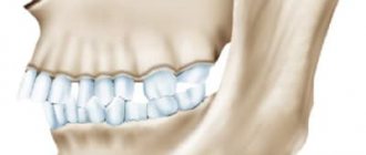
From the point of view of the average person looking at the dentition, the arrangement of the teeth can be described as being “to the side” of each other or one tooth can be said to be “in front” of the other.
To avoid confusion, it is common practice among dentists and oral surgeons to use the adjectives “anterior” and “posterior.” Even if 2 teeth stand in one line, like incisors, the position of the first incisor relative to the second is designated as “front”.
“Proximal” and “distal” as terms when describing the location of a tooth do not describe the tooth itself, but its lateral surfaces or edges. The mesial surface is considered to be facing the previous (behind) tooth. The distal surface is the one that faces the tooth in front.
Both terms are used to understand and discuss the treatment plan. Developing a strategy for dental prosthetics and installation of veneers involves a description of manipulations with the distal and proximal surfaces of the teeth.
Classification of distal occlusion
In orthodontics, several classifications of deep distal bite are used, which are actively used by doctors.
Classification of subclasses according to Engle:
- 1. First subclass . The incisors of the upper row protrude forward, in some cases slightly upward. Because of this, a sagittal gap is formed during closure.
- 2. Second subclass . The incisors of the upper row meet the incisors of the lower row, as they grow slightly backward. This situation is not characterized by the presence of a sagittal fissure.
Types of clinical forms of distal occlusion according to Khoroshilkina:
- 1. Skeletal or gnathic form . The anomaly develops due to uneven development of the jaws and their incorrect position relative to each other.
- 2. Dentoalveolar form . With this form, the alveolar ridge develops incorrectly or individual teeth in the rows are positioned incorrectly.
- 3. Mixed form . A combination of the previous two options.
The method of correcting distal occlusion depends on the type of anomaly and the age at which it was discovered.
Distal and proximal sections
Human internal organs are divided into hollow and parenchymal. Hollow is an organ that has space (cavity) inside. Organs of this type often have a tubular structure. For example, intestines, ureter, trachea, esophagus and others. Parenchymal is an organ without a cavity, consisting only of functional elements.
The use of the words “distal” and “proximal” in relation to parenchymal organs is incorrect, since such organs do not have a beginning, an end or a place of attachment.
They are divided into:
- parts such as head, body and tail;
- right and left parts;
- individual structures such as the vitreous body, chambers and lens of the eye.
Distal and proximal are sections of tubular organs that have a beginning and an end.
One of the functions of the gastrointestinal tract, which is to conduct the food bolus, determines the features of its anatomical structure and makes it possible to imagine the gastrointestinal tract as a long tube consisting of upper (proximal) and lower (distal) sections. The human intestine is divided into thin and thick, the border between which is the ileocecal valve.
The small intestine is the proximal portion of the entire human intestine and is divided into the duodenum, jejunum, and ileum. The cecum, colon, and rectum are the distal sections that make up the large intestine.
The urinary canal, uterus, trachea, larynx, pharynx and other tubular organs have parts according to the areas where they pass and to which they belong. The pharynx is divided into the oropharynx, nasopharynx and hypopharynx; The esophagus has cervical, thoracic and abdominal parts. But all hollow organs begin and end, and thus have proximal and distal ends.
Anatomical classification of teeth
Normally, an adult should have 14-16 teeth on each jaw:
- 4 incisors: 2 central and 2 lateral (right and left);
- 2 fangs;
- 4 premolars;
- 4-6 molars.
Conventionally, all teeth can be divided into central (frontal), usually included in the smile line, that is, up to the fangs.
Their main purpose is to bite food. And chewing (back) teeth, which have a more massive structure, cusps, which provide better grinding of food. Calling chewing teeth molars is incorrect! Any tooth, even the central one, has a root, and, for example, in fangs it is more massive and long in comparison with others. It is the fang that holds the Guinness World Record. The longest canine is 37.2 mm; for comparison, the average tooth length is about 20 mm.
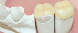
Anatomically, in each tooth one can distinguish:
- Crown
On which the cutting edge of the central and cusps of the chewing teeth are located. The equator can be traced in the crown - the most convex part of the tooth, forming close contact with its neighbors - this is necessary for uniform distribution of the chewing load.
- Neck
The narrowest part of the tooth to which the periodontal ligament is attached, and here the enamel meets the cementum of the root. Normally, most of the cervix is hidden by the gum.
- Root
Depending on the anatomy, a tooth may have 1-3 roots, and this is its longest part.
The anatomical structure of wisdom teeth is not the same, and this is especially expressed in the number of roots. There are cases where a wisdom tooth had as many as 7 roots!
Histological structure of the tooth: enamel
Enamel is the avascular, densest mineralized “tissue” of the human body, covering the crown of the tooth, that is, the part that protrudes above the gum.
The unique structure of enamel allows it to remain relatively unchanged throughout life. Its main structural formation is enamel prisms, which are concentrated into bundles. Retzius lines running in an oblique direction stand out, which can be compared to tree rings - they are formed due to cyclic processes of mineralization, that is, saturation of the enamel with minerals. The main structural unit of an enamel prism is crystals of apatite-like substances that are tightly attached to each other. With age, the size of these crystals increases.
The chemical structure of enamel is as follows:
- 95% inorganic compounds
They are represented by apatites, but the main one is hydroxyapatite Ca10(PO4)6(OH)2 - 75.04%. The condition of the enamel is determined by the ratio of calcium and phosphorus; this coefficient, which depends on the age and group of teeth, is used to determine the condition of the enamel.
- 3.8% water
In addition to bound water, which is represented by the water shell of apatite crystals, free water is also determined in the chemical structure of the tooth. This enamel fluid fills microspaces and participates in ion exchange processes occurring in teeth.
- 1.2% organic compounds
They are represented by lipids, carbohydrates and proteins.
Dentin is the main substance of the tooth
Beneath the enamel lies dentin, a less calcified mineralized tissue. The chemical structure is somewhat different; there are more organic substances here compared to enamel - 28%, but still more inorganic - 72%. Dentin is permeated with tubes, inside which dentinal fluid circulates, performing several functions:
- Nutritious
Delivers substances for metabolic processes from the tooth pulp to dentin and enamel and vice versa - from enamel to pulp.
- Sensitive
Dentinal fluid is a kind of conductor that irritates the nerve endings of the pulp and transmits corresponding impulses.
This is how we feel the temperature of food, pain during irritation, for example, with increased tooth sensitivity. Therefore, the first sensitive zone of the tooth is the dentin-enamel junction.
Root cement
It is also a mineralized tissue, somewhat resembling rough-fibered bone. The chemical composition is dominated by inorganic substances. The root cement is impregnated with inorganic salts, and due to collagen fibers it is tightly welded to the bone tissue of the jaw alveoli.
Dental pulp or "nerve"
This is loose connective tissue, replete with blood and nerve vessels, which fills the endodont of the tooth, that is, its internal part. Anatomically, the coronal pulp and the root pulp are distinguished, which performs the following functions:
- Plastic
Due to the content of odontoblasts in it, it participates in the formation of dentin;
- Trophic
Nutrients and minerals enter the pulp with blood vessels, and the pulp “gives” them to dentin and enamel.
- Sensitive
This is the most sensitive part of the tooth.
- Protective
Due to odontoblasts, it stimulates the production of secondary dentin.
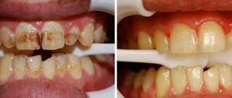
Mineral metabolism
After each meal, tooth enamel is worn down by a few microns, but is restored within 10-40 minutes. Due to what? First of all, due to the protective mechanisms provided by nature. Two processes occur in teeth every day:
- demineralization - the release of minerals, for example, due to mechanical abrasion, deficiency conditions and, of course, the action of acids produced by caries-forming bacteria, etc.;
- remineralization - saturation of enamel and dentin with minerals that are “brought” by saliva, or they come from the pulp.
If these processes are in balance, there are no problems: caries and hypersensitivity do not develop!
But if there is not enough calcium and phosphorus in the diet, and the body simply has nowhere to take them from to deliver them to the teeth, the balance is upset. As a result, the enamel becomes depleted, and sensitivity appears, which only progresses over time. And then the enamel is destroyed, and caries most often begins in these places. Text: Yulia Lapushkina.
Distal and proximal renal tubules
The structural and functional unit of the human kidney is the nephron (a structure involved in the filtration of urine and the reabsorption of microelements, ions and water needed by the body).
To increase the absorption surface area, the nephron has long convoluted proximal and distal tubules, which have different structures and perform a specific function. Proteins, carbohydrates, water and salts are reabsorbed in the proximal convoluted tubules. The distal convoluted tubules are permeable only to ions and water and impermeable to macromolecules.
Also in the structure of nephrons, proximal and distal straight tubules are distinguished. They form the basis of the countercurrent multiplying system: its various sections are permeable to different substances, due to which the urine becomes more concentrated.
Proximal and distal radioulnar joints
The skeleton of the forearms is formed by the ulna and radius bones. To connect the bones and carry out flexion and extension movements of the hand, joints serve as movable joints.
The proximal radioulnar joint is located closer to the elbow area and connects the articular surfaces of the head of the radius and the radial notch of the ulna. The distal one, in turn, is localized closer to the hand. The articular surfaces are the circumference of the head of the radius and the ulnar notch on the radius.
Distal and proximal are the joints of the upper limb, which allow the arm to rotate inward - pronation and outward - supination. The distal radioulnar joint, in combination with the radiocarpal joint, provides flexion and extension of the hand.
Skeletal bones are divided into flat and tubular. Flat bones have edges, surfaces, and sometimes corners. The distal and proximal ends are distinguished only in tubular bones, both long and short. Both ends of the bone have their own specific anatomical formations.
The task of orienting a single bone correctly in space is simplified if there is a clear understanding that the condyle of the humerus belongs to the distal structures.
Diagnosis of distal bite
Only a professional orthodontist can accurately determine the presence of distal occlusion. First, the doctor examines the oral cavity and can first judge what malocclusion the patient has. However, to confirm the diagnosis and draw up a further treatment plan, additional studies are carried out:
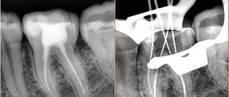
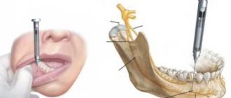
- 1. Computed tomography . Obtaining three-dimensional images of the dentofacial apparatus allows you to determine: the size of the jaws, their relationship, bone structure, the condition of the TMJ, and so on.
- 2. Casts . Taking impressions of the dentition allows you to determine the characteristics of the closure of the teeth. The study helps create a plan for further tooth movement to form a correct bite.
- 3. Photo protocol . Photos are taken with the mouth open and closed, in front and in profile, with a smile and from different angles. This allows you to track the dynamics of bite correction.
Contact our clinic, where diagnostics are performed using modern, precise equipment with a guarantee of reliable results.
Epiphysis bone
The bony epiphysis is an expanded and rounded end section of the tubular bone, which is involved in the formation of articular joints, bone nutrition and hematopoiesis, due to the red bone marrow located inside.
Epiphyses are localized both at the proximal end of the bone and at the distal end.
How to correct a distal bite?
Treatment of distal occlusion in childhood is faster and easier. For therapy, various devices are used to stimulate the growth of the lower jaw and/or inhibit the development of the upper jaw. Orthodontists use:
- - tongue barriers;
- — special trainers;
- - myogymnastics.
If necessary, tooth extraction and canine grinding can be performed for better closure.
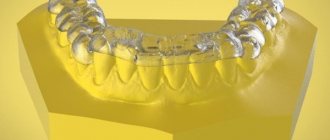
If the problem was diagnosed in adolescence or older, then treatment is performed using braces or removable aligners. Additionally, an articular splint can be used if the function of the temporomandibular joint is impaired.
If there is no effect from conservative treatment, maxillofacial surgery is performed. It's difficult and expensive.
Scheme for surgical correction of bite
The timing of distal bite correction varies from person to person. The exact duration of the correction depends on the severity of the anomaly, the patient’s age, and additional problems with the dentofacial apparatus. In children, treatment usually takes 6-8 months less time than in adults.
Wrist
The 8 carpal bones are divided into distal and proximal rows, respectively. The first row of the wrist, closest to the forearm, includes the scaphoid, lunate triquetrum and pisiform bones.
The first 3 bones of the proximal row articulate to form an articular surface for connection with the distal end of the radius. The distal row of the wrist is formed by the trapezium, trapezoid, capitate and hamate bones, which have articular surfaces for articulation with the proximal parts of the five metacarpal bones.
Phalanges of the fingers.
The feet and hands are the most distal parts of the upper and lower extremities. The bony basis of the toes and wrist are the phalanges. The second, third, fourth and fifth fingers and toes have three phalanges, and the thumbs have two. Each phalanx has a distal and proximal part.
The proximal end of each phalanx is connected to the distal end of the previous bone by a joint:
- interphalangeal on the arms and legs;
- metacarpophalangeal on the hand;
- metatarsophalangeal on the leg.
The last 2 occupy a more proximal position compared to the interphalangeal ones.
Paraparesis
A neurological disorder in which the ability of nerve impulses to travel to the distal parts of both upper or lower extremities is weakened or lost. With severe damage to the nervous system, paraparesis can develop into paralysis. The causes of the disease are injuries, hernias, stenosis of the vertebral arteries, tumors, vascular and neurological pathologies of other origins.
There are different classifications of paraparesis:
- depending on the level at which the nervous system is affected - central (spastic), peripheral (sluggish) or mixed;
- according to localization, it is divided into upper, with the involvement of two upper extremities, and lower, with damage to both lower extremities;
- According to the severity of symptoms, there are complete, deep, moderate and mild.
Symptoms may not extend beyond one limb and may affect a single muscle or group of muscles. Depending on the part of the limb where paraparesis is localized, a division is made into paroxysmal and distal paraparesis.
Damage to the gluteal region, the anterior and posterior surfaces of the thigh, the shoulder and shoulder girdle areas is classified as paroxysmal paresis; distal includes severe symptoms in one of the areas: lower leg, foot, forearm or hand.
Treatment of both distal and proximal paresis is carried out after determining the cause of the pathology. Simultaneously with the treatment of the underlying disease, therapy is carried out aimed at eliminating symptoms, for example, improving blood flow in the distal extremities.
The concepts “distal” and “proximal” in a broad sense can denote directions, parts of organs, ends of bones, and localization of pain syndromes. A clear command of this terminology allows you to avoid mistakes and misunderstandings during contacts within the medical community.
The meaning of the words "distal" and "proximal" is different from the concepts of "anterior", "posterior", "superior" and "inferior". This difference is close to the intuitive level of perception and plays an important role in various fields of medicine.
Author: Storchai O. E.
Distal occlusion (upper macrognathia, prognathia)
Distal bite therapy can be carried out using the following methods and methods:
- orthodontic treatment,
- hardware-surgical,
- surgical,
- prosthetic
- various combined and combined methods.
During treatment, taking into account certain features depending on the clinical form of the anomaly, the age of the patient, the individual characteristics of the structure of the facial skull and the type of its growth, the following tasks must be solved.
- Regulation during jaw growth using a facebow and extraoral traction or a functional apparatus.
- Restriction of growth and shortening of the dentition of the upper jaw due to the distal movement of the upper molars, canines and elimination of protrusion of the anterior teeth
- When treating distal occlusion, it is advisable to transfer form P2 to form II1, which can be achieved by using arches in the traditional sequence, that is, the primary arch, as a rule, is multi-stranded, flexible, allowing the creation of additional bends when teeth are crowded in order to avoid excessive load on them, then a steel one rectangular.
- Distal movement of the upper anterior teeth without or after extraction of individual teeth (most often premolars).
- Stimulation of growth and anterior movement of the lower jaw
- Expansion of the dentition of the upper and/or lower jaw
- Change in interalveolar height and normalization of the Spee curve.
- Normalization of the function of masticatory and facial muscles
- Retention period
It is not always possible to completely correct the anomaly during these manipulations, but a change in position of approximately 45 mm can be achieved. When planning orthodontic treatment of patients with distal occlusion, teleradiological examination data characterizing the type of growth of the maxillofacial complex, the activity of residual growth and their comparison with orthognathic occlusion are very important.
With a distal occlusion, the proportion of the neutral type of growth decreases (from 71% with orthognathic to 50%) in favor of horizontal, namely to 43% versus 15% with orthognathic. This indicates the predominance of development of the facial skeleton in the anteroposterior direction due to intensive growth of the upper jaw, especially in the period of 7-12 years and somewhat less in 12-15 years. That is why the most optimal for modifying jaw growth are the replacement bite and the earliest permanent bite (for 14-15 years, for 12-13).
In patients with a neutral type of growth of the facial skeleton, the main tasks when correcting a distal bite are, first of all, to restrain the growth of the upper jaw and stimulate the growth of the lower jaw. In such patients, mainly removable equipment of functional or combined action should be used.
With the horizontal type, it is necessary, first of all, to restrain the growth of the upper jaw, with simultaneous distal movement of the lateral teeth, using a face bow with a cervical traction. For adult patients, to reduce the upper dentition, treatment is recommended with the removal of the first premolars, followed by distal displacement of the lateral and anterior teeth. This is indicated for prognathia with a reduced or average size of the base of the upper jaw and for prognathia caused by crowding of the upper front teeth, their sharp protrusion, often together with the alveolar process.
In severe forms of distal occlusion with a pronounced horizontal gap, removal of the first premolars is indicated even in a mixed dentition. After this, when treating II1, you can use a flex arch, then a nitinol one, fixing them first on the teeth of the upper jaw. One of the indications for tooth extraction is a reduction in the retromolar space, which enhances the mesial displacement of the lateral teeth and aggravates the close position of the anterior teeth, with insufficient space for the canines of the upper and lower jaw (Zhulev E.N.).
According to WR Proffit (1986), the indication for serial extraction is a discrepancy between the size of the teeth and the dental arch by 10 mm or more, and Ringenberg (1964) believes that the initial value should be smaller, namely 7 mm. According to the point of view of V.P. Norkunaite, with the length of the segment of the dentition “from the distal surfaces of the crowns of the 12th and 22nd teeth to the mesial points of the sixth teeth” equal to 18.5-21.0 mm, and if the sum of the mesiodistal dimensions of the canines and premolars is 22.5-24, 0 mm, then removal of individual permanent teeth is indicated. It should be noted that non-extraction orthodontic treatment is even relatively simpler, since there is no need to move the teeth a significant distance to close the post-extraction gap.
As a last resort, removal of the second molar (sometimes unilateral) is used and distalization of the dentition is performed using a face bow. It is very difficult to move the first molars distally more than 1.5-2.0 mm even after removing the second molars, since distal tooth displacement is much more difficult than mesial displacement. The latter requires more reliable support and stabilization, as E. Engle wrote about. The extraoral thrust should not be low, otherwise extrusion of the molars will occur.
During the period of replacement teeth, when treating distal deep bite, you can use the forgotten but good method of A. Katz, namely, crowns with spikes on the second milk or first permanent molars (the tooth is not prepared) of the lower jaw. When the mandible is advanced forward, the elongated mesial cusps of the artificial crown should fit into the gap between the first and second primary molars of the upper jaw, widened by preparation. In this case, some separation of the bite occurs, which contributes to the dentoalveolar lengthening of the lateral teeth and a decrease in the incisal overlap. Long-term use of such crowns (8-10 months) leads to the formation of an orthognathic bite.
In children with mixed dentition and a clear tendency to develop distal occlusion, McNamara recommends maxillary expansion with overcorrection, usually a rapid maxilla expander. The subsequent use of a retention plate leads to the movement of the lower jaw to a position more convenient for the patient, pushed forward. This eliminates buccal crossbite and, after some time, improves occlusal relationships in the sagittal direction. Somewhat earlier, this phenomenon was explained by H. Taatz and Reichenbach by the fact that the expansion of the upper jaw contributes to the spontaneous displacement of the lower jaw to the anterior position. If such correction does not occur, then RG Alexander recommends the use of a facebow with extraoral traction until the end of the mixed dentition.
In mixed dentition, removable plate devices and a pre-orthodontic trainer are used in the treatment of prognathia.
But in addition to this, when treating anomalies at the dentoalveolar level, especially when combined with a narrowing of the dentition or crowding of teeth, fixed structures can be used. First of all, this is a “2 x 4” device, that is, rings for the first molars and braces for the 4 upper incisors, or utility arches.
Growth can be stimulated using activators, for example Andresen Haüpl or function regulators R.Fränkel. The activator is a removable two-jaw monoblock plastic, functional apparatus, consisting of upper and lower plates connected to each other; a vestibular arch, springs or a screw may be added to them. In addition to the plates adjacent to the inner surface of the alveolar processes, they have a corresponding bed for the oral surfaces of all upper and lower teeth. It is better to fix all types of plates with arrow-shaped clasps and Adams clasps.
The device holds the lower jaw in an extended anterior position (a constructive bite that must be determined by the doctor before treatment), promoting dentoalveolar lengthening in the lateral areas, while the upper anterior teeth are moved posteriorly due to a reciprocal action. On the upper jaw, the plate touches the mesial edges of the surfaces of the teeth, but lags behind the distal ones. On the lower jaw, on the contrary, it fits tightly to the distal edges and lags behind the mesial ones to move the lower jaw.
The clinical laboratory stages of production are as follows.
First clinical impression taking from both jaws; the first laboratory casting of plaster models and making a wax template for the upper jaw with bite ridges to determine the constructive bite, the boundaries of the wax template: in front are the cutting edges of the incisors, behind is a line passing through the middle of the crowns of the last molars, on the side is the chewing surface of the lateral teeth.
The second clinical stage is the determination of a constructive bite: the patient moves the lower jaw forward to a neutral relationship of the first permanent molars (1 class each), and he is asked to close his teeth until they come into contact with the wax. In this case, the separation of the dentition should exceed the “rest height” and it is necessary to monitor the position of the ridge and the coincidence of the midline. If in the position of the constructive bite the neutral closure of the sixth teeth is not achieved and the discrepancy is 45 mm, then this position is fixed. If the sagittal discrepancy exceeds 6 mm, the first activator is first prepared (at 45 mm), and after 6-8 months the second activator is prepared, but with the movement of the lower jaw to the neutral closure of the sixth teeth.
After fixing the constructive bite, plaster models with a wax template are handed over to the dental technician and the doctor gives him instructions:
- make an apparatus with or without a vestibular arch for retrusion of the upper anterior teeth (the shape is being specified),
- install a screw or other additional elements, springs, levers, lingual arches, etc. F.Ya. Khoroshilkina and WR Proffit proposed installing facebow tubes into the activator (bite blocks in the premolar area) in order to be able, along with the functional action of the device, to create additional distal and vertical force using extraoral traction.
Second laboratory stage: the models are plastered in an occluder, the wax template is removed, a plastic base is made, the listed parts or others (as directed by the doctor), the device is polymerized in a special double cuvette or in a regular one, increasing its vertical size
Third clinical stage: fitting the activator in the oral cavity, first to the upper dentition, and then to the lower one; the activator should fit tightly to the teeth, with lips closed; The rules for using and caring for the device are explained to the patient and the next visit is scheduled. During repeated visits, the device is adjusted in the direction of movement of the upper and lower lateral teeth. During the treatment process, the dental bed is polished according to the direction of movement of the teeth, that is, those that need to be moved in the palatal or lingual direction, and vice versa, the plate should fit tightly to those teeth that need to be moved in the vestibular direction. The device can mainly be used while at home or while sleeping. Treatment is especially successful in the early stages of distal and deep bites.
The device helps restore nasal breathing, since the child is forced to breathe more through the nose due to the closure of the oral fissure by the plate. But it is contraindicated if nasal breathing is completely absent. The activator also helps eliminate the habit of sucking fingers, tongue, lips and various objects. The vestibular deviation of the lower teeth can be prevented by the activator hood, which overlaps them by 1/3 of the height of the crowns, so the plastic in it is polished or the hood is completely removed. Similar actions, depending on the progress of treatment, are taken at each visit. You can also stimulate the advancement of the lower jaw using the Balters bionator.
Treatment of distal occlusion (II2) can be carried out in two stages. First, the upper anterior teeth are deviated, eliminating the blocking of the lower jaw, that is, subclass II2 is transferred to II1 using edgewise therapy, rotating the first molar. The latter should be the first step in the treatment of a class II anomaly if there is a tendency for mesial rotation of the molar around the palatal root. If orthodontic treatment II1 is carried out without tooth extraction, then sometimes it is enough to turn the first upper molar with its buccal surface posteriorly, which allows you to create an additional space of 1.5-3.0-4.0 mm and the subclass will move to II1. This can be done with the help of an extraoral yaga, the Gozhgarin palatal clasp, in which the ends of the clasp, curved in two planes, are fixed into the palatal locks on the molars. The device is activated by unbending the loop.
This treatment method can be used when a class II anomaly is combined with an open bite. To illustrate, we give an example from the clinical practice of Dr. P. Ngan et al.: an 8-year-old patient had closure of the molars on both sides according to E. Engle class II, a 5-millimeter sagittal discrepancy in the frontal area, an anterior open bite and lower retrognathia. The main goal of treatment was to delay the anterior growth of the upper jaw, transfer the molar relationship from class II to class I, reduce concomitant skeletal disorders and open bite.
The treatment apparatus consisted of an activator and an extraoral arch attached to it. Because the base plate of the device did not cover the vault of the palate, a connecting arch (diameter 1.2 mm) was used instead, which increased the space for the tongue. For fixation to the extraoral traction activator, a special tube with a diameter of 1.12 mm (0.045 inches) was mounted in the plastic between the upper and lower dentition. The force of extraoral traction was up to 400 grams on each side. Springs for tilting the front teeth were made of elastic steel wire with a diameter of 0.5-0.6 mm, the lower part of which was fixed with horizontal shanks in plastic. The vertical part of the springs had a point contact in the area of the necks of the teeth.
The mandibular part of the apparatus consisted of an incisal platform for advancing the lower jaw. When determining the constructive bite, the lower jaw moved forward until the incisors made direct contact. In patients with hyperactivity of the muscles of the perioral region, to reduce their effect, lip pads in the form of a “tear” according to R. Fränkel were used, which were located in the vestibule of the oral cavity parallel to the alveolar process. The ratio of molars according to class I. was achieved in about a year and at the same time the size of the open bite decreased, which led to an improvement in the lip relationship. The entire treatment lasted about 14 months.
Sometimes symmetrical or unilateral removal of premolars in the upper jaw is performed. At the second stage of treatment, the lower jaw is set in the correct relationship with the upper jaw. To do this, when there is a sharp narrowing of the lower dentition, it is expanded, and then, based on the clinical picture and X-ray data of the temporomandibular joints, sagittal movement of the lower jaw is carried out using plates with an inclined plane. There are a large number of varieties of plates, including those with an inclined plane. Depending on a particular clinical situation, the doctor selects the appropriate design.
A. Katz's bite block is used to treat prognathia in combination with a deep bite. A special feature of its design is an inclined plane and reversible clasps that bend over the cutting edges of the front teeth onto their vestibular surface. The plate does not adhere to the mucous membrane of the anterior part of the palate and the necks of the anterior teeth. When closing with an inclined plane, the lower teeth slide along its surface, trying to return from a forced (constructive) bite to its original position, and the lower jaw moves forward, and the upper teeth tilt towards the palatine side. In the lateral areas, due to the separation of the bite, a vertical restructuring occurs, that is, dentoalveolar elongation.
The fundamental clinical and laboratory stages of making a plate differ little from those described in the manufacture of the activator: obtaining impressions, making a wax composition of the plate with retaining and reversible clasps, determining the constructive bite, polymerization of plastic, fitting and application of the device.
It should be remembered that when treating distal occlusion in patients 15-20 years old, when using bite blocks before stabilization occurs, a double or “wandering” bite may be established, that is, in a position of physiological rest, the lower jaw is fixed in a neutral position, and during function it moves to the former (distal).
The devices proposed by R. Fränkel are called functional regulators, the main parts of which are side shields and pelota, which relieve the dentition from the pressure of the cheeks and lips. As a result, under the influence of the tongue, the growth of the apical base is stimulated in the transversal and sagittal directions. The parts of the apparatus are held together by metal arcs made of elastic wire. This skeletonization made it possible to increase the strength of the regulators, reduce the size of the plastic shields, lighten the apparatus and make it open in the frontal area for better swallowing and speech. Active elements (screws or springs) can be added to the device to accelerate the movement of individual teeth.
R. Fränkel proposed function regulators of three main types: type I (FR I) is used to eliminate protrusion of the anterior teeth and distal occlusion, combined with narrowing of the dentition, fan-shaped arrangement of the upper frontal teeth and with anomalies of 1 class. E. Engla; type II (FR II) for the treatment of distal occlusion of subclass 2 (II2), that is, in combination with deep overlap and retrusion of the upper anterior teeth; type III (FR III) for the treatment of progenia. The principal clinical and laboratory stages of the regulators have been described previously.
The use of this method is effective in early childhood (the period of primary and mixed dentition), that is, when one can count on the growth of the jaw bones and especially the apical base. Treatment with the regulator, especially during the period of its development, is recommended according to the following scheme: for the first two weeks use the device during the day for 1 hour, for the next 2 weeks every day for 2 hours, then during all free time, removing the device only during meals; in 2-3 months around the clock. After correcting the bite with adjusters, retention devices are not required, since already during the active phase of orthodontic treatment, the conditions that contribute to the occurrence of relapse are eliminated.
Sagittal movement of the lower jaw during prognathism should be considered as the last stage of treatment, based on the considerations that the restructuring of muscles, temporomandibular joints, as well as dentoalveolar lengthening in the lateral areas in the vertical direction are not always successful. When treating severe forms of prognathia with deep overlap, the separation between the lateral teeth should be at least 45 mm. With active protraction of the lower jaw, tissue restructuring occurs in the order of activation (stimulation) of functional hypertrophy, mainly of the lateral pterygoid muscle, which is poorly developed during prognathism.
It is necessary to constantly monitor the separation of the bite and, as contact between the lateral teeth is achieved, to re-create the separation of the bite by correcting the inclined plane. It is also necessary to correct the appliance in the area where its base adheres to the palatal surfaces of the anterior teeth. In most cases, a sagittally moved mandible is secured in its new position due to the close contacts of natural teeth or contacts created by dentures.
The use of devices can be combined with active myogymnastics, but they are incompatible with edgewise therapy, although it would be very desirable to correct the dentoalveolar components of the anomaly simultaneously with the correction of jaw growth. This is possible when using non-removable functional devices or when combining braces with a face bow. There is no point in drawing a sharp boundary between the phases of treatment, expecting, for example, leveling of the dentition, because extraoral appliances also contribute to a certain extent to the correction of the dental components of the anomaly.
RG Alexander is a proponent of the use of extraoral traction both for growing jaws (children, adolescents) and for adults. But in the first, with the help of a facial arch, the growth of the upper jaw is suppressed and at the same time its dentition is leveled, the lower jaw is unblocked, providing the opportunity to achieve its genetic potential. In adults, when growth has stopped, the main purpose of extraoral appliances is to hold the upper molars in place, to avoid their displacement forward.
Intermaxillary traction, in the author's opinion, should be used while the dentition of both jaws is aligned and stabilized, rigid end steel archwires are installed (0.17 x 0.25) and torque control is established to prevent incisor tilting. The arches must completely fill the slots of the braces and be in the mouth for at least a month before installing elastic traction according to class II. Having studied the vector of forces in the traditional position of the above-mentioned thrust, that is, from the upper canines to the lower first molars, RG Alexander determined the presence of an undesirable, very significant vertical component of the force. An increase in the horizontal component of the force can be achieved through a different fixation of the traction, namely, from the second lower molar to the spherical hook of the bracket on the upper lateral incisors. This increases the vector of horizontally acting force and reduces the tendency to “open” the bite, for which, by the way, elastics are not used in the “Vari Simplex Discipline” system.
When treating the first subclass (II1) of prognathia, complicated by a deep or open bite, the incorrect position of the teeth and an anomaly in the shape of the dentition are usually eliminated. If it is necessary to expand the lateral areas of the upper dentition, then a rapid palatal expander (rapid maxilla expander) is used in the early stages of treatment, before the installation of fixed appliances. If the teeth have not erupted, then plastic plates with a screw can be used, and only then the lower jaw is moved forward. When treating a distal bite in combination with protrusion of the upper anterior teeth, their close position and narrowing of the dentition or their asymmetry, one should not rush to eliminate the protrusion, since the upper anterior teeth, which have become palatally inclined as a result of treatment, will interfere with the movement of the lower jaw.
According to many clinicians, with early treatment, distal occlusion can be eliminated in approximately 80% of patients with functional devices. The use of edgewise therapy, in particular the direct arch technique, expands the age-related indications for orthodontic correction, but positive dynamics of treatment are observed only at the dentoalveolar level.
In case of distal occlusion (II2), the method of eliminating crowding of teeth must be planned taking into account the structure of the facial skeleton, the age of the patient and the amount of space in the dentition. The correct choice of method allows you to get an optimal result and avoid complications during and after treatment.
Treatment of adult patients is reduced mainly to leveling the position of the teeth and eliminating deep incisal overlap, if any. The process of “detailing” the molars is either prolonged (as a result of the “distalization” of first the second and then the first molars) or is impossible because adults already have erupted second and third molars. In such patients, extraction is more indicated, and a dilemma arises: which tooth should be removed, the first or second premolar? To resolve this issue it is necessary to take into account:
- the magnitude of the space deficit; if, after straightening the teeth, a residual gap of no more than 2.0 mm is predicted, then the first premolar is removed, and if it is more than 2.0 mm, then the second premolar; this choice can be argued by the fact that during the closure of the spaces between the teeth, the tendency for retrusion of the incisors increases, while the removal of the second premolar has a lesser effect on the position of the incisors;
- dental condition, it is preferable to remove the affected teeth (destroyed crown, endodontic treatment, changes in periapical tissues, large filling or severe abrasion);
- after extraction, it is necessary to retract the canines or “first premolar canines”, for which the full arch technique or the segmental arch technique can be used;
- The full-arch technique in its standard implementation is as follows: at the first stage, braces are fixed on all teeth, using for additional support, for example, a Gozhgarin clasp, when indicated in combination with a face bow; the initial arch, as a rule, is nitinol, with simultaneous “distalization” of the canine or first molar using an eight-shaped ligature (within the dentition this can be done using springs, elastic traction, elastomeric power modules); it should, however, be noted that in this case, a protrusive displacement of the incisors occurs, which is very undesirable in adults, since then it is necessary to retract them to eliminate the sagittal discrepancy, therefore it is better to use the segmental arch technique;
- segmental arch technique: braces are fixed only on the teeth of the lateral segment, with additional stabilization of the supporting teeth, as in the previous version; then a steel edge arch with a diameter of 0.40×0.55 mm is passively inserted into the braces; for “distalization” the usual sliding technique; if the canine has an abnormal position initially, then a nitinol arch should first be introduced, fixing it only in braces on the canines and premolars, with simultaneous “distalization” with an 8-shaped ligature; after normalizing the position of the fang, you can move on to a full arch and fixation of braces on the incisors, the leveling of which is carried out according to the traditional method (nitinol arch, steel arches, TMA); This technique allows for canine retraction without side effects on the incisors.
For surgical treatment, write down clear indications:
- justification for the need for such therapy, for example, the magnitude of the sagittal discrepancy of the jaws is 10 mm or more, the angle of the SNSs (SNA) on the teleroentgenogram is greater than normal, which is ~ 82°
- good state of somatic and mental health, the growth of the patient’s facial skeleton must be complete
- pronounced protrusion of the lower incisors (the angle of inclination is less than 70-80°, while the norm is 90-95°); it is well known that the vestibular movement of the lower anterior teeth is very limited and the limit depends on the value of the initial axial angle; the maximum reasonable limit should not be less than 90-95°, therefore, when using equipment for the vestibular movement of the lower front teeth, you need to be very careful; correction of the sagittal position of the lower anterior teeth can be carried out: by changing their vestibular inclination,
- changes in the length of the lower jaw, but this depends on the age and nature of micrognathia, that is, it is of the condylar type (when the articular process, which is the center of longitudinal growth, is affected) or extracondylar,
- changes in the position of the mandibular head, if the anomaly has developed due to distal displacement of the mandible
An approximate set of myogymnastic exercises for the treatment of distal occlusion. Exercises should be selected according to the child’s age and not be too difficult. L.S. Persia recommends first of all determining the level of development of the child and giving the load not to the point of fatigue, but approximately 75% of it. Exercises to correct a specific anomaly should be dosed and performed against the background of general physical exercise, starting 2-3 weeks before orthodontic treatment.
Muscle contractions must be performed with maximum amplitude, their intensity must be within physiological limits, with a gradual increase in speed and duration; between two successive contractions there should be a pause equal to the duration of the contraction itself.
Exercises to normalize breathing function (performed during morning exercises, physical education lessons or while walking); starting position: the state of correct posture: the head and torso are held straight, the shoulders are slightly pulled back and slightly lowered, the chest is turned out, the shoulder blades are adjacent to the back, the stomach is tucked and the knee joints are straightened.
Exercises to normalize lip closure (can be performed during speech development classes). Starting position: sitting in front of a mirror, head held straight, shoulders slightly pulled back and slightly lowered, chest turned out, knee joints bent, legs together, stomach tucked.